
Recomendados
Mais conteúdo relacionado
Semelhante a ANATOMY OF EYE.pdf
Semelhante a ANATOMY OF EYE.pdf (20)
Último
Último (20)
ANATOMY OF EYE.pdf
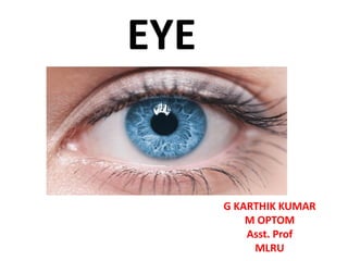
- 1. EYE G KARTHIK KUMAR M OPTOM Asst. Prof MLRU
- 2. ANATOMY OF THE EYE
- 3. THE EYEBALL/GLOBE Each eyeball is a cystic structure kept distended by the pressure inside it. Oblate spheroid. anterior and posterior pole. The equator of the eyeball lies at the mid plane between the two poles.
- 4. Dimensions of an adult eyeball Anteroposterior diameter 24 mm Horizontal diameter 23.5 mm Vertical diameter 23 mm Circumference 75 mm Volume 6.5 ml Weight 7 gm
- 5. Coats of the Eyeball 1. Outer (Fibrous coat) 2. Middle (Vascular coat) 3. Inner (Nervous coat).
- 6. 1.Fibrous coat. It is a dense strong wall which protects the intraocular contents. Anterior 1/6th is transparent called cornea. Posterior 5/6th opaque part is called sclera. Junction of the cornea and sclera is called limbus. Conjunctiva is firmly attached at the limbus.
- 7. 2.Vascular coat (Uveal Tissue). It supplies nutrition to the various structures of the eyeball. It consists of three parts which from anterior to posterior are : Iris Ciliary Body Choroid.
- 8. 3. Nervous coat (Retina). It is concerned with visual functions.
- 9. Segments and chambers of the eyeball 1)Anterior segment: a)Cornea b)Iris c) crystalline lens d)Ciliary body e)Zonules f)Two aqueous humour-filled spaces : Anterior and Posterior chambers. 2. Posterior segment : a) Vitreous humour (a gel like material which fills the space behind the lens). b) Retina c) Choroid d) Optic disc.
- 10. Anterior chamber: It is bounded anteriorly by the back of cornea, and posteriorly by the iris and part of ciliary body. The A/C is about 2.5 mm deep in the centre in normal adults. It contains about 0.25 ml of the Aqueous Humour. It is Shallower in hypermetropes and Deeper in myopes. Posterior chamber: It is a triangular space containing 0.06 ml of aqueous humour. It is bounded anteriorly by the posterior surface of iris and part of ciliary body. Posteriorly by the crystalline lens and its zonules.