Vânia goncalves isbo ng wi nets - accounting interference
Mevic leaflet en 2008
1. MEVIC
Medical Virtual Imaging Chain
Image Quality
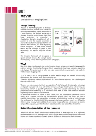
The goal of the MEVIC project is to develop a
software simulation platform which can be used
to reliably determine the clinical performance of Virtual Image
a medical system. The platform will be used to Image Generation
simulate the entire medical image chain: from
image generation, to processing, to Virtual Image Clinical
System Processing Validation
visualization. Using the simulations, it will be
possible to tune the physical parameters of
medical devices from the standpoint of not only Virtual Image
technical measurements, but more importantly, Specialist Visualization
human perception. In other words, medical
devices will be optimized in terms of their Quantification of
performance for specific clinically relevant Clinical Performance
tasks.
The simulation framework will be applied to
specific clinical domains, for example digital
mammography, nuclear medicine, and computed tomography.
Why?
One of the biggest challenges in the medical imaging domain is to accurately and reliably quantify
and then optimize the clinical performance of their acquisition devices, image processing algorithms
and displays. This project seeks to solve two major problems that many companies and academic
institutions are struggling with:
1) As of today, it still is a huge problem to obtain medical images and datasets for validating
developments that are done in the medical imaging area.
2) Reliably determining the clinical performance of a medical system requires a time consuming and
expensive clinical trial.
There are two main reasons why this is such a problem. First of all: people developing the technology
are typically engineers with no or only limited clinical knowledge. Therefore, they will normally use
“engineering metrics” to quantify performance, rather than actually determining the clinical
performance of the technology. It is well known that there is often little correlation between
engineering metrics and clinical performance.
An alternative solution is of course to do a clinical trial. But unfortunately, performing clinical
studies is an extremely expensive and time consuming activity. Even if the money and time is
available, then still there is a huge problem in finding a sufficiently large number of medical images
that can be used for the clinical trial. Not only do patient privacy regulations make it very difficult to
obtain medical images, but in addition these medical images often do not represent a realistic
sample of the population, and therefore there is doubt about the real value of the clinical trial.
Scientific description of the research
The solution that this project consortium will develop consists of three steps. First of all, algorithms
will be developed that can produce realistic medical images, eventually including specific (simulated)
lesions, with known ground truth. This will solve the difficulty of obtaining real medical images.
2. A second step will consist of accurate simulation models of the medical devices that we want to
evaluate. Examples of such devices can be: medical displays and visualization software, acquisition
devices such as scanners and image processing algorithms. Having simulation models available has
the advantage that it is not anymore necessary to develop multiple prototypes of medical systems:
this can be replaced by software simulations.
The third and final step is the development of mathematical observer models. It has been shown that
such mathematical observer models have a very good correlation with the decision process of real
radiologists. In other words: such observer models could replace clinical trials. At the moment there
are still many limitations to these mathematical observer models and it is the goal of this project to
make these models more accurate and reliable. The unique combination of these three steps will give
the project consortium the possibility to perform software simulations that can replace an entire
clinical trial, including the image generation, device prototyping and the actual observer study with
radiologists.
Naturally, the developed algorithms and models are only valuable if we can prove that they
accurately resemble reality and will give the same results as real clinical trials. Therefore, a very
important step in this project will be to compare the developed algorithms and software with real
clinical experiments. For this purpose, a portion of the project will be devoted to validating the
methods with observer studies performed by radiologists.
Valorization of the project
By means of the framework that will be developed in this project, the consortium will be able to
much more easily measure the clinical performance of various medical devices. It will be possible to
simulate clinical trials, for example to compare different technical alternatives, without the need to
make actual prototypes. It is obvious that this will result into higher quality products that will
improve quality of healthcare. Furthermore, the ability to accurately predict the results of human
clinical trials can hopefully improve and simplify the regulatory approval process. Access to
healthcare will also be improved, through quicker time-to-market and reduced research and
development costs.
The benefits of the MEVIC project extend far beyond the borders of Flanders and Belgium: society as
a whole will benefit from reduced healthcare costs – through more cost effective research and
development processes, and an increased quality of healthcare – through higher quality medical
devices.
In cooperation with
IBBT Research Groups
VUB – ETRO http://www.etro.vub.ac.be
UGent – TELIN-IPI http://telin.UGent.be/
UGent – MEDISIP http://medisip.elis.ugent.be/
UZ-KUL http://www.uzleuven.be/