SlideShare uma empresa Scribd logo
1 de 7
Baixar para ler offline
Original Article
Clinical and Radiologic Spectrum of
Septo-optic Dysplasia: Review of 17 Cases
Callie Alt, BSc1
, Michael I. Shevell, MDCM2
, Chantal Poulin, MD2
,
Bernard Rosenblatt, MDCM2
, Christine Saint-Martin, MD3
, and
Myriam Srour, MDCM, PhD2
Abstract
We retrospectively reviewed the clinical and radiologic characteristics of 17 individuals with septo-optic dysplasia (SOD) and
attempted to identify correlations between imaging findings, clinical features, and neurodevelopmental outcome. Surprisingly, only
1 (6%) individual was classified as classic SOD (with septum pellucidum/corpus callosum dysgenesis), 3 (18%) as SOD-like (with
normal septum pellucidum/corpus callosum) and the majority, 13 (76%), as SOD-plus (with cortical brain malformation). Cortical
abnormalities included schizencephaly, polymicrogyria, and gray matter heterotopias. All individuals had optic nerve hypoplasia,
11 (65%) had endocrinologic deficits, and 13 (76%) had abnormal cerebral midlines. Seven individuals (41%) had all 3 features.
Neurodevelopmental outcome was abnormal in 13 (78%), ranging from mild to severe developmental delay. Individuals with
SOD-plus did not have more severe neurologic deficits than individuals with classic or SOD-like subgroups. Thus, SOD is clinically
and radiologically heterogeneous, and cortical abnormalities are very common. Neurodevelopmental deficits are very prevalent,
and of wide-ranging severity.
Keywords
brain, cortical dysplasia, developmental delay, magnetic resonance imaging, malformation, neurodevelopment, neuroradiology,
ophthalmology
Received November 11, 2016. Received revised March 21, 2017. Accepted for publication March 29, 2017.
Septo-optic dysplasia (SOD; OMIM no. 182230), also known
as de Morsier syndrome,1
is a phenotypically variable disor-
der defined by the presence of 2 or more of the following 3
diagnostic features: (1) optic nerve hypoplasia, (2) pituitary
hypofunction, and (3) midline brain abnormalities, typically
dysgenesis of the septum pellucidum and/or corpus callo-
sum.2
Approximately 30% of all SOD patients have the full
manifestation.3
When SOD is associated with cortical
malformations, it is referred to as “SOD-plus.” The most
consistently reported associated cortical abnormality is
schizencephaly.4,5
SOD reflects a disruption in the early stages of forebrain
development, most likely at 4 to 6 weeks of gestation coincid-
ing with the formation of the anterior neural plate.6
The precise
etiology of SOD is not clearly understood, though it is likely
multifactorial, with both environmental and genetic causes.
The majority of cases are sporadic, but on rare accounts, famil-
ial cases have been reported.2
To date, heterozygous mutations
in at least 4 developmental genes, including HESX1, SOX3,
SOX2, and OXT2, have been associated with SOD, though
these only explain less than 1% of all cases.7
Young maternal
age and drug use are also commonly reported.8
Individuals
with SOD can have variable neurodevelopmental outcomes,
with some individuals having normal cognition and develop-
ment and others with developmental delay, autism, and
epilepsy.
In this study, we reviewed the clinical and radiologic fea-
tures of 17 individuals with SOD and attempted to identify
correlations between imaging findings, clinical features, and
developmental outcome.
Methods
We conducted a retrospective chart review of 17 children (9 males and
8 females) diagnosed with SOD between 2010 and 2016 at the Mon-
treal Children’s Hospital. Patients were collected from 2 pediatric
1
McGill University, Montreal, Canada
2
Departments of Pediatrics, Neurology, and Neurosurgery, McGill University,
Montreal, Quebec, Canada
3
Department of Diagnostic Radiology, Montreal Children’s Hospital, McGill
University Health Centre, Montreal, Quebec, Canada
Corresponding Author:
Myriam Srour, MDCM, PhD, Montreal Children’s Hospital, 1001 De´carie Blvd,
EM0.3218, Montre´al, Quebec, H4A 3J1, Canada.
Email: myriam.srour@mcgill.ca
Journal of Child Neurology
2017, Vol. 32(9) 797-803
ª The Author(s) 2017
Reprints and permission:
sagepub.com/journalsPermissions.nav
DOI: 10.1177/0883073817707300
journals.sagepub.com/home/jcn
neurologists and 1 neuroradiologist’s database. A diagnosis was made
when 2 or more of the following features were present: (1) optic nerve
hypoplasia, (2) pituitary hypofunction, and (3) midline brain abnorm-
alities, such as dysgenesis of the septum pellucidum and/or corpus
callosum.2
We systematically collected the following clinical information for
each case: presence of a family history of neurologic disease, prenatal
risk factors (maternal use of tobacco, alcohol, drugs, medications,
maternal vaginal bleeding, hospitalizations), antenatal imaging, and
perinatal factors/complications (labor and delivery complications,
admission to neonatal intensive care unit, prematurity, jaundice).
The following clinical and laboratory features were compiled: the
presence of documented endocrinologic deficits (growth hormone,
adrenocorticotropic hormone, thyroid-stimulating hormone, antidiure-
tic hormone, luteinizing hormone / follicle-stimulating hormone),
delayed or abnormal puberty, developmental delay, seizures, and hear-
ing and visual abnormalities.
Abnormalities on neurologic examination were noted. The pres-
ence of developmental delay in each domain (gross motor, fine motor,
language, and social) was assessed based on clinical records.9
Global
developmental delay was defined as a delay in 2 or more develop-
mental domains. When present, the severity of the delay was stratified
based on the percentage of functional age, noted by a rehabilitation
specialist (eg, physical and/or occupational therapist), using age-
appropriate standardized measures of function and development (such
as the Alberta Infant Motor Scale, the Bayley Scales of Infant Devel-
opment–II and the Denver-II) compared with actual chronologic age
(ie, mild: 67%-100%, moderate: 33%-66%, and severe: <33%).10
Brain magnetic resonance imaging (MRI) was reviewed for all
patients by one pediatric neuroradiologist (CSM) independent of
awareness of clinical features. MRI was performed using 1.5 (in 7
of 17 individuals) or 3 Tesla (10 of 17 individuals) superconduction
systems. The visual pathway, the hypothalamic-pituitary axis, the
cerebral midline, and the cortex were systematically assessed. The
optic nerves were classified as normal, unilateral, or bilateral hypo-
plasia; the optic chiasm as normal or hypoplastic; the septum pelluci-
dum as present or complete agenesis; the corpus callosum as present,
partial, or complete agenesis; the anterior pituitary as normal or hypo-
plastic; the posterior pituitary as normal or ectopic; the pituitary stalk
as normal, hypoplastic, or absent; and the olfactory bulbs as normal or
absent. Presence of cortical malformations was noted (schizencephaly,
polymicrogyria, gray matter heterotopia, transmantle cortical
dysplasia). Based on their MRI and clinical findings, patients were
further classified into one of the 3 subtypes of SOD: classic SOD
(septum pellucidum or corpus callosum abnormality with either optic
nerve hypoplasia and/or pituitary hypofunction in the absence of any
cortical abnormality), SOD-like (optic nerve hypoplasia and pituitary
abnormalities with normal septum pellucidum and corpus callosum, in
the absence of any cortical abnormality), or SOD-plus (in the presence
of any cortical malformation). Note that clinical examination was used
as the criterion standard to establish presence of optic nerve hypoplasia.
Qualitative statistical analysis was performed using the SPSS
Statistics program.
Results
Patient Background Information
A total of 17 patients with a diagnosis of SOD, based on the
presence of 2 of 3 criteria, were included in this study, of whom
9 were males and 8 females. The average age at final
documentation was 6.1 years + 6.3 standard deviations (SDs),
with a median age of 3.0 years and an interquartile interval of
11.8 years (range 2 months-17 years). The average age at pre-
sentation was 5.8 + 6.2 months (range 0-24 months). The mean
maternal age was 22.0 years (+4.4 SDs) with only 1 mother
over the age of 25. This is below the average Canadian maternal
age of 29.8 years in 2012 (Source: Statistics Canada, Canadian
Vital Statistics, Birth Database). Mean gestational age was 38.8
weeks + 1.9 SD, and birth weight was 3.21 kg + 0.51 SD.
None of the patients had another affected family member or was
born to consanguineous parents. Pregnancy risk factors were
noted in 12 of 17 patients. These included maternal smoking
(in 6), urinary tract infection (in 2), epilepsy (in 1), vaginal
bleeding (in 1), malaria (in 1), and treated hypothyroidism (1).
Eleven patients were born by vaginal delivery and 4 by Cesarean
section. Delivery methods for 2 were unknown. Mean gesta-
tional age was 38.8 weeks + 1.9 SD, and birth weight was
3.21 kg + 0.51 SD. Perinatal factors/complications were
reported in 8 patients: 5 had neonatal hypoglycemic events, 4
had neonatal jaundice treated with phototherapy, and 1 was
hospitalized for a cyanotic episode in the context of a hypogly-
cemic episode. Antenatal images were abnormal in 3 patients.
Ventriculomegaly was noted in 2, absent corpus callosum in 1,
and small fetal size in 1 patient.
Diagnostic Classification
Based on MRI and clinical findings, 13 patients (76%) were
classified as SOD-plus (ie, associated with any cortical malfor-
mation) and 3 patients (18%) as SOD-like optic nerve hypopla-
sia and pituitary abnormalities with normal septum pellucidum
and corpus callosum and no cortical abnormality). Only 1 patient
fit the criteria for classic SOD (ie, septum pellucidum or corpus
callosum abnormality with either optic nerve hypoplasia and/or
pituitary hypofunction and no cortical abnormality). Seven
patients (41%) had all 3 diagnostic features. The remaining 10
patients (69%) presented with only 2 features of the triad; 6
(35%) had optic nerve hypoplasia and midline brain abnormal-
ities, and 4 (24%) had optic nerve hypoplasia and pituitary hypo-
function. No patient had pituitary hypofunction or midline brain
abnormalities in the absence of optic nerve hypoplasia. These
findings are summarized in Figure 1.
MRI Findings
The MRI findings are summarized in Table 1. On imaging,
bilateral optic nerve hypoplasia was noted in all (16/17, 94%)
but 1 individual who had normal optic nerves on MRI but
optic nerve hypoplasia on clinical examination. Clinical
examination was used as the criterion standard for determina-
tion of presence of optic nerve hypoplasia. The chiasm was
noted to be hypoplastic in 13 individuals (76%), normal in 2
(12%), and not visualized in the remaining 2 (12%) because of
technical issues.
The cerebral midlines were abnormal in 13 patients (76%):
11 (65%) had agenesis of the septum pellucidum, 2 (12%) had
798 Journal of Child Neurology 32(9)
hypogenesis of the corpus callosum, and 1 had hypoplasia of
the falx cerebri (6%). One patient had both agenesis of the
septum pelucidum and hypogenesis of the corpus callosum.
Thehypothalamic-pituitaryaxiswasabnormalin9individuals
(53%) on imaging. The anterior pituitary was normal in 12 (71%)
and hypoplastic in 5 (29%). The posterior pituitary was normal in
9 (53%) and ectopic in 8 (47%). The pituitary stalk was normal in
10 (59%), absent in 4 (24%), and thin in 3 (18%). Olfactory bulbs
were normal in 13 patients (76%) and absent in 4 (24%).
Cortical malformations were observed in 13 patients (76%)
(see Figure 2 for examples). Isolated polymicrogyria (not asso-
ciated with schizencephaly) was present in 8 patients (47%)
(bilateral, 5; unilateral, 3) and schizencephaly in 5 individuals
(29%) (bilateral, 2; unilateral, 3). Three patients (18%) had
both schizencephaly as well as polymicrogyria at a site distant
from the schizencephaly. Six patients (35%) had gray matter
heterotopia and 1 patient (6%) had transmantle cortical dyspla-
sia. Other cerebral abnormalities were also noted: fusion of the
anterior fornices in 8 patients (47%), arachnoid cyst in 2 (12%),
hydrocephalus in 1 (6%), and Chiari I malformation in 1 (6%).
No patient had white matter abnormalities.
Ophthalmologic Findings
On clinical examination, 14 patients (82%) had bilateral optic
nerve hypoplasia and 3 (18%) had unilateral optic nerve hypo-
plasia. All individuals had either poor vision or blindness,
including the individual with normal optic nerves on imaging.
Eight individuals (47%) were noted to have nystagmus.
Endocrinologic Findings
Endocrinologic deficits were present in 11 children (65%). Six
(35%) had multiple hormone deficiencies and were diagnosed
with panhypopituitarism. Adrenocorticotropic or cortisol defi-
cits were diagnosed in 10 patients (59%), thyroid-stimulating
hormone or thyroid deficits in 7 patients (41%), growth
hormone in 4 (24%), luteinizing hormone in 2 (12%),
follicle-stimulating hormone in 2 (12%), and antidiuretic hor-
mone in 1 (6%). Of the patients with endocrinologic deficits,
64% (7/11) had abnormal pituitary imaging, and 64% (7/11)
had midline abnormalities.
Table 1. Brain MRI Findings in the Study Sample of 17 Children With
Septo-optic Dysplasia.
Finding n
Optic nervea
Normal 1
Unilateral hypoplasia 0
Bilateral hypoplasia 16
Optic chiasm
Normal 2
Hypoplastic 13
Not identified 2
Midline
Normal 4
Abnormal 13
Septum pellucidum
Normal 6
Agenesis 11
Corpus callosum
Normal 15
Partial agenesis 2
Hypothalamic-pituitary axis
Normal 8
Abnormal 9
Anterior pituitary
Normal 12
Hypoplastic 5
Posterior pituitary
Normal 9
Ectopic 8
Pituitary stalk
Normal 10
Thin 3
Absent 4
Olfactory bulbs
Normal 13
Absent 4
Cortical development
Normal 5
Abnormal 13
Polymicrogyria 8
Schizencephaly 5
Unilateral 3
Bilateral 2
Schizencephaly and polymicrogyria 3
Transmantle cortical dysplasia 1
Other cerebral structures
Fusion of anterior fornices 8
Gray matter heterotopia 6
Arachnoid cyst 2
Hydrocephalus 1
Chiari I malformation 1
Abbreviation: MRI, magnetic resonance imaging.
a
Note that 1 individual had normal optic nerves on MRI, but had bilateral optic
nerve hypoplasia on clinical examination, and 2 individuals had bilateral optic
nerve hypoplasia on MRI but unilateral optic nerve hypoplasia on clinical
examination.
Figure 1. Clinical findings in 17 patients with septo-optic dysplasia.
Alt et al 799
Neurologic Findings
Eleven patients (65%) had an abnormal neurologic exam.
Seven (41%) had hypotonia, 2 (12%) had hemiparesis, and 1
(6%) had quadriparesis. Five individuals (29%) had epilepsy,
of which 3 had secondary generalized tonic clonic seizures, 1
had infantile spasms, and 1 had myoclonic epilepsy. Four of the
5 epileptic patients had associated cortical abnormalities: 2 had
schizencephaly and 2 had gray matter heterotopia, including
transmantle cortical dysplasia.
Psychomotor Development
Normal development was reported in 3 (18%) children. Eleven
patients (65%) were globally delayed, 2 (12%) were delayed in
only 1 domain (gross motor), and 1 was lost to follow-up.
Interestingly, all 3 individuals with normal development had
SOD-plus: a 3-year-old boy with bilateral optic nerve hypopla-
sia, hypopituitarism, absence of the septum pellucidum, and
bilateral frontal polymicrogyria; a 17-year-old girl with aca-
demic difficulties, bilateral optic nerve hypoplasia, agenesis of
the septum pellucidum, multiple endocrinologic deficits, right
perisylvian polymicrogyria and gray matter heterotopias, and
fusion of the anterior fornices; and a 6-year-old boy with bilat-
eral optic nerve hypoplasia, agenesis of the septum pellucidum,
and bilateral frontal polymicrogyria. Of those that were glob-
ally delayed, 3 had mild, 4 moderate, and 4 severe delay. Two
of the 3 children that were severely delayed had associated
cortical abnormalities. Two patients (12%) were diagnosed
with autism, and 1 (6%) with autistic features that did not fulfill
the criteria for autism.
Genetic Evaluation
Seven patients had karyotypes or chromosomal microarray
testing and were normal. Only 1 patient had sequencing of
HESX1, which was normal. One patient had a whole exome
sequencing on a clinical basis. No rare variants were noted in
any genes known to be associated with SOD, and no causal
pathogenic mutations were identified in any other known
disease gene.
Figure 2. Brain imaging abnormalities in individuals with SOD. T2-weighted 1.5-Tesla MRI showing (A) left open-lipped schizencephaly (arrow)
and right perisylvian polymicrogyria (arrowhead), (B) fusion of the fornices, (C) transmantle cortical dysplasia, and (D) bilateral frontal
polymicrogyria. (E) T1-weighted coronal 3-Tesla MRI showing small right periventricular heterotopia. A, anterior, L, left; MRI, magnetic
resonance imaging; P, posterior; R, right; SOD, septo-optic dysplasia.
800 Journal of Child Neurology 32(9)
Discussion
We reviewed the clinical and radiologic features and neurode-
velopmental outcomes of 17 patients with SOD. In our cohort,
only 1 (6%) patient was classified as having classical SOD (ie,
with agenesis of septum pellucidum and/or corpus callosum in
the absence of any cortical abnormality), 3 (18%) as SOD-like
(ie, with normal septum pellucidum and corpus callosum in
the absence of any cortical abnormality), and 13 (76%) as
SOD-plus (ie, with cortical malformations).
The most striking finding of our study was the high inci-
dence of cortical malformations within our cohort. Indeed, 76%
(13/17) of individuals had cortical abnormalities on their brain
imaging and thus were categorized as having SOD-plus. Focal
polymicrogyria was noted in 59% (10/17), schizencephaly in
30% (5/17), and heterotopias (subcortical, periventricular, or
transmantle) in 35% (6/17). Three individuals (18%) had both
schizencephaly and polymicrogyria (not associated with schi-
zencephaly). Signorini et al11
reported that 42% of their
patients with SOD have associated cortical malformations. The
reasons for the increased incidence of cortical malformations in
our cohort may be 2-fold. First, the majority (10/17) of our
patients had high-resolution MRIs on a 3-Tesla magnet, allow-
ing detection of more subtle abnormalities. Second, there may
be a selection bias given that many of these patients were
selected from neurologists’ databases and were more likely to
have been referred because of specific neurologic symptoms
such as abnormal development, focal findings on examination,
or epilepsy. Indeed, individuals with SOD having neurologic
symptoms may be more likely to have cortical involvement. In
our cohort, patients with SOD-plus did not seem to have a
higher risk of epilepsy than individuals without cortical
abnormalities (4/13, 31%, vs 1/4, 25%, P ¼ 1.0, on Fisher exact
test); however, studying a larger number of patients would be
required to better assess whether the presence of a cortical
malformation confers a greater risk of epilepsy in SOD. Nev-
ertheless high-resolution brain imaging in patients with SOD is
important to enable search for cortical abnormalities that may
be responsible for focal neurologic findings or epilepsy.
Interestingly, almost all individuals with SOD-plus (12/13,
92%) have a midline abnormality (Table 2), a finding that has
also been noted in other studies.8,11
This suggests the involve-
ment of common or overlapping pathophysiologic mechanisms
that disrupt both midline cerebral development and neuronal
migration such as an in utero vascular event, fetal viral infec-
tion, or common underlying genetic etiology. Indeed, early
vascular disruption has been hypothesized to underlie some
SOD cases12-14
and is also a favored theory in the etiology of
several cortical abnormalities such as perisylvian polymicro-
gyria and schizencephaly.15
Fetal viral infections have been
associated with SOD and various cortical malformations.16
Finally, one can speculate that mutations in genes with an
important function both in midline cerebral development and
neuronal migration may lead to such a phenotype.
All individuals in our cohort had optic nerve hypoplasia on
clinical examination, which is the gold standard for diagnosis.
Optic nerve hypoplasia was bilateral in 82% (14/17) and uni-
lateral in 18% (3/14) of the cohort. Endocrinologic deficits
were present in 65% (11/17) our cohort, and midline abnorm-
alities in 76% (13/17). This is similar to what has been reported
in other studies.6,17
The 3 SOD diagnostic features were present
in 35% of patients (6/17), which is consistent with what has
been previously reported (30%-47%) in other studies.3,11
Endocrinologicdeficiencieswereobservedin11ofourpatients
(65%), of which 7 had associated abnormal pituitary imaging. The
absence of imaging abnormalities of the pituitary does not pre-
cludethepresenceofhormonaldeficits,underlyingtheimportance
of clinical hormonal investigations in any individual with 1 feature
of SOD. In addition, 2 individuals with abnormal pituitaries on
MRI had no endocrinologic deficits when last evaluated.
Developmental impairment was reported in 13 (78%) of our
patients. Poor vision likely contributes to, but does not fully
account for, the patients’ developmental delay. The severity of
the associated developmental impairment is wide-ranging. In
fact, the severity was equally distributed in all severity cate-
gories (see Table 2). SOD-plus patients have a heterogeneous
neurodevelopmental outcome, ranging from normal to severely
delayed development. All 3 individuals with normal develop-
ment had cortical abnormalities and were classified as SOD-
plus. Conversely, SOD-like patients appear to have more severe
delay, with all individuals categorized as moderate-severe delay.
It is difficult to draw clear conclusions given the small number of
patients. It is important to note again that, because of possible
aforementioned selection bias in our study, patients with normal
development are less likely to be referred for neurologic evalua-
tion and thus would not have been included in our cohort. Other
limitations to this present study also include the retrospective
nature of the study and the relatively small number of patients
precluding statistical analysis.
Table 2. Common Clinical Features of the 3 Subtypes of Septo-optic
Dysplasia.
Classic SOD
(1/17)
SOD-like
(3/17)
SOD-plus
(13/17)
Vision
Normal 0 0 1
Abnormal 1 3 12
Developmental level
Normal 0 0 3
Mildly delayed 1 0 3
Moderately delayed 0 1 3
Severely delayed 0 2 3
Not classified 0 0 1
Autistic 0 1 2
Endocrine function
Normal 1 1 4
Abnormal 0 2 9
Presence of midline defect
No 0 3 1
Yes 1 0 12
Abbreviation: SOD, septo-optic dysplasia.
Alt et al 801
Few studies such as ours have focused on the neurologic and
developmental features in SOD,8,11,18,19
with the majority of
studies concentrating on endocrinologic and ophthalmologic
aspects.20-25
Riedl et al8
looked at 68 patients with optic nerve
hypoplasia, of which 42 had SOD. In their study, unilateral
optic nerve hypoplasia and septum pellucidum remnants were
associated with a milder endocrinologic and neurologic SOD
phenotype, and abnormalities of the falx and hippocampus
were associated with worse endocrinologic and neurodevelop-
mental outcomes. Signori et al11
reviewed 17 SOD patients and
observed a heterogeneous clinical spectrum, with nervous sys-
tem involvement as a key feature of the syndrome: develop-
mental/cognitive delay present in nearly one-half of the study
sample. In this study, patients classified as SOD or SOD-like
had more marked neurodevelopmental delay than patients in
the SOD-plus category. Parr et al19
systematically studied the
presence of autistic features in a cohort of 83 patients with optic
nerve hypoplasia or SOD, and found a 36% prevalence of aut-
ism spectrum disorder in SOD patients compared to 26% in
optic nerve hypoplasia patients. A cross-sectional study by
Jutley-Neilson et al18
looking at the occurrence of autism spec-
trum disorder in 42 children with SOD or optic nerve hypopla-
sia reported an autism spectrum disorder diagnosis in 33%,
which is greater than that observed in our cohort.
The underlying etiology of SOD remains overwhelmingly
unknown. In our cohort, 7 patients (41%) had chromosomal
studies (karyotype or microarray), only 1 had molecular
genetic testing for known SOD genes, and no patient had an
identified etiology. All our cases were sporadic. With the
exception of one, all mothers were under the age of 25 years.
This is in line with other studies that suggest that SOD patients
are the children of mothers who are younger than average.26
In summary, our study reveals that SOD is clinically hetero-
geneous. Cortical abnormalities are frequently associated with
SOD, and should be systematically looked into for using high-
resolution MRI. Neurodevelopmental deficits are very preva-
lent, and of wide-ranging severity. Larger studies are needed to
better assess clinical and radiologic predictors of outcome in
SOD.
Acknowledgments
MS holds a clinician-scientist award from the Fonds de Recherche en
Sante´ du Quebec (FRQ-S).
Declaration of Conflicting Interests
The authors declared no potential conflicts of interest with respect to
the research, authorship, and/or publication of this article.
Funding
The authors received no financial support for the research, authorship,
and/or publication of this article.
References
1. De Morsier G. Studies on malformation of cranio-encephalic
sutures. III. Agenesis of the septum lucidum with malformation
of the optic tract [in French]. Schweiz Arch Neurol Psychiatr.
1956;77:267-292.
2. Kelberman D, Dattani MT. Genetics of septo-optic dysplasia.
Pituitary. 2007;10:393-407.
3. Morishima A, Aranoff GS. Syndrome of septo-optic-pituitary
dysplasia: the clinical spectrum. Brain Dev. 1986;8:233-239.
4. Kuban KC, Teele RL, Wallman J. Septo-optic-dysplasia-
schizencephaly. Radiographic and clinical features. Pediatr
Radiol. 1989;19:145-150.
5. Miller SP, Shevell MI, Patenaude Y, Poulin C, O’Gorman AM.
Septo-optic dysplasia plus: a spectrum of malformations of cor-
tical development. Neurology. 2000;54:1701-1703.
6. Webb EA, Dattani MT. Septo-optic dysplasia. Eur J Hum Genet.
2010;18:393-397.
7. Saranac L, Gucev Z. New insights into septo-optic dysplasia.
Prilozi. 2014;35:123-128.
8. Riedl S, Vosahlo J, Battelino T, et al. Refining clinical phenotypes
in septo-optic dysplasia based on MRI findings. Eur J Pediatr.
2008;167:1269-1276.
9. Srour M, Shevell M. Genetics and the investigation of developmen-
tal delay/intellectual disability. Arch Dis Child. 2014;99:386-389.
10. Srour M, Mazer B, Shevell MI. Analysis of clinical features pre-
dicting etiologic yield in the assessment of global developmental
delay. Pediatrics. 2006;118:139-145.
11. Signorini SG, Decio A, Fedeli C, et al. Septo-optic
dysplasia in childhood: the neurological, cognitive and neuro-
ophthalmological perspective. Dev Med Child Neurol. 2012;54:
1018-1024.
12. Lubinsky MS. Hypothesis: septo-optic dysplasia is a vascular
disruption sequence. Am J Med Genet. 1997;69:235-236.
13. Chiaramonte I, Cappello G, Uccello A, et al. Vascular cerebral
anomalies associated with septo-optic dysplasia. A case report.
Neuroradiol J. 2013;26:66-70.
14. Mitchell LA, Thomas PQ, Zacharin MR, Scheffer IE. Ectopic
posterior pituitary lobe and periventricular heterotopia: cerebral
malformations with the same underlying mechanism? AJNR Am J
Neuroradiol. 2002;23:1475-1481.
15. Guerrini R, Dobyns WB. Malformations of cortical development:
clinicalfeaturesandgeneticcauses.LancetNeurol.2014;13:710-726.
16. Barkovich AJ, Lindan CE. Congenital cytomegalovirus infection
of the brain: imaging analysis and embryologic considerations.
AJNR Am J Neuroradiol. 1994;15:703-715.
17. McCabe MJ, Alatzoglou KS, Dattani MT. Septo-optic dysplasia
and other midline defects: the role of transcription factors:
HESX1 and beyond. Best Pract Res Clin Endocrinol Metab.
2011;25:115-124.
18. Jutley-Neilson J, Harris G, Kirk J. The identification and
measurement of autistic features in children with septo-optic
dysplasia, optic nerve hypoplasia and isolated hypopituitarism.
Res Dev Disabil. 2013;34:4310-4318.
19. Parr JR, Dale NJ, Shaffer LM, Salt AT. Social communication
difficulties and autism spectrum disorder in young children with
optic nerve hypoplasia and/or septo-optic dysplasia. Dev Med
Child Neurol. 2010;52:917-921.
20. Cemeroglu AP, Coulas T, Kleis L. Spectrum of clinical presenta-
tions and endocrinological findings of patients with septo-optic
802 Journal of Child Neurology 32(9)
dysplasia: a retrospective study. J Pediatr Endocrinol Metab.
2015;28:1057-1063.
21. Bachmann-Gagescu R, Phelps IG, Dempsey JC, et al. KIAA0586
is mutated in Joubert syndrome. Hum Mutat. 2015;36:831-835.
22. Antonini SR, Grecco Filho A, Elias LL, Moreira AC, Castro M.
Cerebral midline developmental anomalies: endocrine, neurora-
diographic and ophthalmological features. J Pediatr Endocrinol
Metab. 2002;15:1525-1530.
23. Atapattu N, Ainsworth J, Willshaw H, et al. Septo-optic dysplasia:
antenatal risk factors and clinical features in a regional study.
Horm Res Paediatr. 2012;78:81-87.
24. Ahmad T, Garcia-Filion P, Borchert M, Kaufman F, Burkett L,
Geffner M. Endocrinological and auxological abnormalities in
young children with optic nerve hypoplasia: a prospective study.
J Pediatr. 2006;148:78-84.
25. Garcia-Filion P, Borchert M. Optic nerve hypoplasia syndrome: a
review of the epidemiology and clinical associations. Curr Treat
Options Neurol. 2013;15:78-89.
26. Murray PG, Paterson WF, Donaldson MD. Maternal age in
patients with septo-optic dysplasia. J Pediatr Endocrinol Metab.
2005;18:471-476.
Alt et al 803

Mais conteúdo relacionado

Mais procurados

Bardet- biedl syndrome/ dental courses
Bardet- biedl syndrome/ dental coursesBardet- biedl syndrome/ dental courses
Bardet- biedl syndrome/ dental coursesIndian dental academy
 
Fetal alcohol spectrum disorders: an overview from the glia perspective
Fetal alcohol spectrum disorders: an overview from the glia perspectiveFetal alcohol spectrum disorders: an overview from the glia perspective
Fetal alcohol spectrum disorders: an overview from the glia perspectiveBARRY STANLEY 2 fasd
 
Neuropsychiatric Manifestation In 8 Years Old Girl Presented With Obstructive...
Neuropsychiatric Manifestation In 8 Years Old Girl Presented With Obstructive...Neuropsychiatric Manifestation In 8 Years Old Girl Presented With Obstructive...
Neuropsychiatric Manifestation In 8 Years Old Girl Presented With Obstructive...AR Muhamad Na'im
 
IOSR Journal of Pharmacy (IOSRPHR)
IOSR Journal of Pharmacy (IOSRPHR)IOSR Journal of Pharmacy (IOSRPHR)
IOSR Journal of Pharmacy (IOSRPHR)iosrphr_editor
 
barded bidel syndrome
barded bidel syndromebarded bidel syndrome
barded bidel syndromeJani Mehul
 
Plgf, a placental marker of fetal brain defects after in utero alcohol exposu...
Plgf, a placental marker of fetal brain defects after in utero alcohol exposu...Plgf, a placental marker of fetal brain defects after in utero alcohol exposu...
Plgf, a placental marker of fetal brain defects after in utero alcohol exposu...BARRY STANLEY 2 fasd
 
Genotype Phenotype Correlation In Prader Willi Syndrome
Genotype Phenotype Correlation In Prader Willi SyndromeGenotype Phenotype Correlation In Prader Willi Syndrome
Genotype Phenotype Correlation In Prader Willi SyndromeMihaiela Fazacas
 
Duchenne muscular dystrophy -Prenatal Diagnosis & Genetic Counseling- Dr.Padmesh
Duchenne muscular dystrophy -Prenatal Diagnosis & Genetic Counseling- Dr.PadmeshDuchenne muscular dystrophy -Prenatal Diagnosis & Genetic Counseling- Dr.Padmesh
Duchenne muscular dystrophy -Prenatal Diagnosis & Genetic Counseling- Dr.PadmeshDr Padmesh Vadakepat
 
Mucopolysccharidoses : A Rare Cause For Bilateral Cloudy Cornea.
Mucopolysccharidoses : A Rare Cause For Bilateral Cloudy Cornea.Mucopolysccharidoses : A Rare Cause For Bilateral Cloudy Cornea.
Mucopolysccharidoses : A Rare Cause For Bilateral Cloudy Cornea.Dr. Jagannath Boramani
 
Brain Imaging Abnormalities in Autism Disorders
Brain Imaging Abnormalities in Autism DisordersBrain Imaging Abnormalities in Autism Disorders
Brain Imaging Abnormalities in Autism Disordersasclepiuspdfs
 
Neurometabolic Disorders
Neurometabolic  DisordersNeurometabolic  Disorders
Neurometabolic DisordersAmr Hassan
 
Clinical Assessment & Management of Prion Disease
Clinical Assessment & Management of Prion DiseaseClinical Assessment & Management of Prion Disease
Clinical Assessment & Management of Prion Diseaseapplebyb
 

Mais procurados (20)

Bardet- biedl syndrome/ dental courses
Bardet- biedl syndrome/ dental coursesBardet- biedl syndrome/ dental courses
Bardet- biedl syndrome/ dental courses
 
Fetal alcohol spectrum disorders: an overview from the glia perspective
Fetal alcohol spectrum disorders: an overview from the glia perspectiveFetal alcohol spectrum disorders: an overview from the glia perspective
Fetal alcohol spectrum disorders: an overview from the glia perspective
 
Neuropsychiatric Manifestation In 8 Years Old Girl Presented With Obstructive...
Neuropsychiatric Manifestation In 8 Years Old Girl Presented With Obstructive...Neuropsychiatric Manifestation In 8 Years Old Girl Presented With Obstructive...
Neuropsychiatric Manifestation In 8 Years Old Girl Presented With Obstructive...
 
Congenital malformations
Congenital malformationsCongenital malformations
Congenital malformations
 
IOSR Journal of Pharmacy (IOSRPHR)
IOSR Journal of Pharmacy (IOSRPHR)IOSR Journal of Pharmacy (IOSRPHR)
IOSR Journal of Pharmacy (IOSRPHR)
 
barded bidel syndrome
barded bidel syndromebarded bidel syndrome
barded bidel syndrome
 
Plgf, a placental marker of fetal brain defects after in utero alcohol exposu...
Plgf, a placental marker of fetal brain defects after in utero alcohol exposu...Plgf, a placental marker of fetal brain defects after in utero alcohol exposu...
Plgf, a placental marker of fetal brain defects after in utero alcohol exposu...
 
Genotype Phenotype Correlation In Prader Willi Syndrome
Genotype Phenotype Correlation In Prader Willi SyndromeGenotype Phenotype Correlation In Prader Willi Syndrome
Genotype Phenotype Correlation In Prader Willi Syndrome
 
Duchenne muscular dystrophy -Prenatal Diagnosis & Genetic Counseling- Dr.Padmesh
Duchenne muscular dystrophy -Prenatal Diagnosis & Genetic Counseling- Dr.PadmeshDuchenne muscular dystrophy -Prenatal Diagnosis & Genetic Counseling- Dr.Padmesh
Duchenne muscular dystrophy -Prenatal Diagnosis & Genetic Counseling- Dr.Padmesh
 
O.I. RESEARCH POSTER
O.I. RESEARCH POSTERO.I. RESEARCH POSTER
O.I. RESEARCH POSTER
 
Mucopolysccharidoses : A Rare Cause For Bilateral Cloudy Cornea.
Mucopolysccharidoses : A Rare Cause For Bilateral Cloudy Cornea.Mucopolysccharidoses : A Rare Cause For Bilateral Cloudy Cornea.
Mucopolysccharidoses : A Rare Cause For Bilateral Cloudy Cornea.
 
Dokotorat_abstract_MJMS
Dokotorat_abstract_MJMSDokotorat_abstract_MJMS
Dokotorat_abstract_MJMS
 
Brain Imaging Abnormalities in Autism Disorders
Brain Imaging Abnormalities in Autism DisordersBrain Imaging Abnormalities in Autism Disorders
Brain Imaging Abnormalities in Autism Disorders
 
Tarzana_Providence_031809_final
Tarzana_Providence_031809_finalTarzana_Providence_031809_final
Tarzana_Providence_031809_final
 
Neurometabolic Disorders
Neurometabolic  DisordersNeurometabolic  Disorders
Neurometabolic Disorders
 
Clinical Assessment & Management of Prion Disease
Clinical Assessment & Management of Prion DiseaseClinical Assessment & Management of Prion Disease
Clinical Assessment & Management of Prion Disease
 
Fetal surgery for neural tube defects
Fetal surgery for neural tube defectsFetal surgery for neural tube defects
Fetal surgery for neural tube defects
 
Approach to leukodystrophy
Approach to leukodystrophyApproach to leukodystrophy
Approach to leukodystrophy
 
Achondroplasia
AchondroplasiaAchondroplasia
Achondroplasia
 
2018 BDSRA Gayko
2018 BDSRA Gayko2018 BDSRA Gayko
2018 BDSRA Gayko
 

Semelhante a Displasia septooptica

5 la genetica clínica en pediatria
5 la genetica clínica en pediatria5 la genetica clínica en pediatria
5 la genetica clínica en pediatriaapepasm
 
1 s2.0-s0887899413001999-main
1 s2.0-s0887899413001999-main1 s2.0-s0887899413001999-main
1 s2.0-s0887899413001999-mainllilia
 
Smith-Lemli-Opitz Syndrome2
Smith-Lemli-Opitz Syndrome2Smith-Lemli-Opitz Syndrome2
Smith-Lemli-Opitz Syndrome2llilia
 
Autism spectrum disorder
Autism spectrum disorderAutism spectrum disorder
Autism spectrum disorderEnoch R G
 
Adrenolukodystrphy dr Musa Atarzadeh
Adrenolukodystrphy   dr Musa AtarzadehAdrenolukodystrphy   dr Musa Atarzadeh
Adrenolukodystrphy dr Musa AtarzadehMusa Atazadeh
 
Pt assesment BY PRASANTH PS
Pt assesment BY PRASANTH PSPt assesment BY PRASANTH PS
Pt assesment BY PRASANTH PSPrasanth Ps
 
Phenotypic Analysis of a Case of “3MC Syndrome” with Review of Literature
Phenotypic Analysis of a Case of “3MC Syndrome” with Review of LiteraturePhenotypic Analysis of a Case of “3MC Syndrome” with Review of Literature
Phenotypic Analysis of a Case of “3MC Syndrome” with Review of LiteratureBIJCROO
 
Fahr Syndrome- A Rare Case Report
Fahr Syndrome- A Rare Case ReportFahr Syndrome- A Rare Case Report
Fahr Syndrome- A Rare Case ReportDr Gauri Kapila
 
Down syndrome and Physiotherapy Management
Down syndrome and Physiotherapy Management Down syndrome and Physiotherapy Management
Down syndrome and Physiotherapy Management Anumeha Sharma
 
Trisomy 8, Turners syndrome, Triple X: A Case Report
Trisomy 8, Turners syndrome, Triple X: A Case ReportTrisomy 8, Turners syndrome, Triple X: A Case Report
Trisomy 8, Turners syndrome, Triple X: A Case ReportDominick Maino
 
Cereberal palsy dr hussein abass 2019 ppt
Cereberal palsy dr hussein abass  2019  pptCereberal palsy dr hussein abass  2019  ppt
Cereberal palsy dr hussein abass 2019 pptHosin Abass
 
Dental management downs syndrome, fetal alcohol syndrome
Dental management downs syndrome, fetal alcohol syndromeDental management downs syndrome, fetal alcohol syndrome
Dental management downs syndrome, fetal alcohol syndromeDr Ravneet Kour
 
Congenital epilepsi
Congenital epilepsiCongenital epilepsi
Congenital epilepsiSanu Sphere
 
Ipoglicemia da iperinsulinemia
Ipoglicemia da iperinsulinemiaIpoglicemia da iperinsulinemia
Ipoglicemia da iperinsulinemiaMerqurio
 

Semelhante a Displasia septooptica (20)

5 la genetica clínica en pediatria
5 la genetica clínica en pediatria5 la genetica clínica en pediatria
5 la genetica clínica en pediatria
 
1 s2.0-s0887899413001999-main
1 s2.0-s0887899413001999-main1 s2.0-s0887899413001999-main
1 s2.0-s0887899413001999-main
 
Smith-Lemli-Opitz Syndrome2
Smith-Lemli-Opitz Syndrome2Smith-Lemli-Opitz Syndrome2
Smith-Lemli-Opitz Syndrome2
 
Autism spectrum disorder
Autism spectrum disorderAutism spectrum disorder
Autism spectrum disorder
 
Biological aspects of schizophrenia
Biological aspects of schizophreniaBiological aspects of schizophrenia
Biological aspects of schizophrenia
 
Adrenolukodystrphy dr Musa Atarzadeh
Adrenolukodystrphy   dr Musa AtarzadehAdrenolukodystrphy   dr Musa Atarzadeh
Adrenolukodystrphy dr Musa Atarzadeh
 
Pt assesment BY PRASANTH PS
Pt assesment BY PRASANTH PSPt assesment BY PRASANTH PS
Pt assesment BY PRASANTH PS
 
Meningiomyleocele
MeningiomyleoceleMeningiomyleocele
Meningiomyleocele
 
Phenotypic Analysis of a Case of “3MC Syndrome” with Review of Literature
Phenotypic Analysis of a Case of “3MC Syndrome” with Review of LiteraturePhenotypic Analysis of a Case of “3MC Syndrome” with Review of Literature
Phenotypic Analysis of a Case of “3MC Syndrome” with Review of Literature
 
Fahr Syndrome- A Rare Case Report
Fahr Syndrome- A Rare Case ReportFahr Syndrome- A Rare Case Report
Fahr Syndrome- A Rare Case Report
 
Down syndrome and Physiotherapy Management
Down syndrome and Physiotherapy Management Down syndrome and Physiotherapy Management
Down syndrome and Physiotherapy Management
 
1750 1172-6-36
1750 1172-6-361750 1172-6-36
1750 1172-6-36
 
Trisomy 8, Turners syndrome, Triple X: A Case Report
Trisomy 8, Turners syndrome, Triple X: A Case ReportTrisomy 8, Turners syndrome, Triple X: A Case Report
Trisomy 8, Turners syndrome, Triple X: A Case Report
 
Cereberal palsy dr hussein abass 2019 ppt
Cereberal palsy dr hussein abass  2019  pptCereberal palsy dr hussein abass  2019  ppt
Cereberal palsy dr hussein abass 2019 ppt
 
Dental management downs syndrome, fetal alcohol syndrome
Dental management downs syndrome, fetal alcohol syndromeDental management downs syndrome, fetal alcohol syndrome
Dental management downs syndrome, fetal alcohol syndrome
 
4133 pb8-8420-r3
4133 pb8-8420-r34133 pb8-8420-r3
4133 pb8-8420-r3
 
4133 pb8-8420-r3
4133 pb8-8420-r34133 pb8-8420-r3
4133 pb8-8420-r3
 
Congenital epilepsi
Congenital epilepsiCongenital epilepsi
Congenital epilepsi
 
Ipoglicemia da iperinsulinemia
Ipoglicemia da iperinsulinemiaIpoglicemia da iperinsulinemia
Ipoglicemia da iperinsulinemia
 
Prader Willi Syndrome ppt1
Prader Willi Syndrome ppt1Prader Willi Syndrome ppt1
Prader Willi Syndrome ppt1
 

Último

College Call Girls Pune Mira 9907093804 Short 1500 Night 6000 Best call girls...
College Call Girls Pune Mira 9907093804 Short 1500 Night 6000 Best call girls...College Call Girls Pune Mira 9907093804 Short 1500 Night 6000 Best call girls...
College Call Girls Pune Mira 9907093804 Short 1500 Night 6000 Best call girls...Miss joya
 
Call Girls Kanakapura Road Just Call 7001305949 Top Class Call Girl Service A...
Call Girls Kanakapura Road Just Call 7001305949 Top Class Call Girl Service A...Call Girls Kanakapura Road Just Call 7001305949 Top Class Call Girl Service A...
Call Girls Kanakapura Road Just Call 7001305949 Top Class Call Girl Service A...narwatsonia7
 
Book Call Girls in Yelahanka - For 7001305949 Cheap & Best with original Photos
Book Call Girls in Yelahanka - For 7001305949 Cheap & Best with original PhotosBook Call Girls in Yelahanka - For 7001305949 Cheap & Best with original Photos
Book Call Girls in Yelahanka - For 7001305949 Cheap & Best with original Photosnarwatsonia7
 
Artifacts in Nuclear Medicine with Identifying and resolving artifacts.
Artifacts in Nuclear Medicine with Identifying and resolving artifacts.Artifacts in Nuclear Medicine with Identifying and resolving artifacts.
Artifacts in Nuclear Medicine with Identifying and resolving artifacts.MiadAlsulami
 
Call Girls ITPL Just Call 7001305949 Top Class Call Girl Service Available
Call Girls ITPL Just Call 7001305949 Top Class Call Girl Service AvailableCall Girls ITPL Just Call 7001305949 Top Class Call Girl Service Available
Call Girls ITPL Just Call 7001305949 Top Class Call Girl Service Availablenarwatsonia7
 
Call Girls Jayanagar Just Call 7001305949 Top Class Call Girl Service Available
Call Girls Jayanagar Just Call 7001305949 Top Class Call Girl Service AvailableCall Girls Jayanagar Just Call 7001305949 Top Class Call Girl Service Available
Call Girls Jayanagar Just Call 7001305949 Top Class Call Girl Service Availablenarwatsonia7
 
Russian Call Girl Brookfield - 7001305949 Escorts Service 50% Off with Cash O...
Russian Call Girl Brookfield - 7001305949 Escorts Service 50% Off with Cash O...Russian Call Girl Brookfield - 7001305949 Escorts Service 50% Off with Cash O...
Russian Call Girl Brookfield - 7001305949 Escorts Service 50% Off with Cash O...narwatsonia7
 
Low Rate Call Girls Pune Esha 9907093804 Short 1500 Night 6000 Best call girl...
Low Rate Call Girls Pune Esha 9907093804 Short 1500 Night 6000 Best call girl...Low Rate Call Girls Pune Esha 9907093804 Short 1500 Night 6000 Best call girl...
Low Rate Call Girls Pune Esha 9907093804 Short 1500 Night 6000 Best call girl...Miss joya
 
College Call Girls Vyasarpadi Whatsapp 7001305949 Independent Escort Service
College Call Girls Vyasarpadi Whatsapp 7001305949 Independent Escort ServiceCollege Call Girls Vyasarpadi Whatsapp 7001305949 Independent Escort Service
College Call Girls Vyasarpadi Whatsapp 7001305949 Independent Escort ServiceNehru place Escorts
 
Call Girls Jp Nagar Just Call 7001305949 Top Class Call Girl Service Available
Call Girls Jp Nagar Just Call 7001305949 Top Class Call Girl Service AvailableCall Girls Jp Nagar Just Call 7001305949 Top Class Call Girl Service Available
Call Girls Jp Nagar Just Call 7001305949 Top Class Call Girl Service Availablenarwatsonia7
 
VIP Call Girls Pune Vrinda 9907093804 Short 1500 Night 6000 Best call girls S...
VIP Call Girls Pune Vrinda 9907093804 Short 1500 Night 6000 Best call girls S...VIP Call Girls Pune Vrinda 9907093804 Short 1500 Night 6000 Best call girls S...
VIP Call Girls Pune Vrinda 9907093804 Short 1500 Night 6000 Best call girls S...Miss joya
 
Call Girls Hosur Just Call 7001305949 Top Class Call Girl Service Available
Call Girls Hosur Just Call 7001305949 Top Class Call Girl Service AvailableCall Girls Hosur Just Call 7001305949 Top Class Call Girl Service Available
Call Girls Hosur Just Call 7001305949 Top Class Call Girl Service Availablenarwatsonia7
 
Kolkata Call Girls Services 9907093804 @24x7 High Class Babes Here Call Now
Kolkata Call Girls Services 9907093804 @24x7 High Class Babes Here Call NowKolkata Call Girls Services 9907093804 @24x7 High Class Babes Here Call Now
Kolkata Call Girls Services 9907093804 @24x7 High Class Babes Here Call NowNehru place Escorts
 
Call Girls Frazer Town Just Call 7001305949 Top Class Call Girl Service Avail...
Call Girls Frazer Town Just Call 7001305949 Top Class Call Girl Service Avail...Call Girls Frazer Town Just Call 7001305949 Top Class Call Girl Service Avail...
Call Girls Frazer Town Just Call 7001305949 Top Class Call Girl Service Avail...narwatsonia7
 
Sonagachi Call Girls Services 9907093804 @24x7 High Class Babes Here Call Now
Sonagachi Call Girls Services 9907093804 @24x7 High Class Babes Here Call NowSonagachi Call Girls Services 9907093804 @24x7 High Class Babes Here Call Now
Sonagachi Call Girls Services 9907093804 @24x7 High Class Babes Here Call NowRiya Pathan
 
VIP Call Girls Mumbai Arpita 9910780858 Independent Escort Service Mumbai
VIP Call Girls Mumbai Arpita 9910780858 Independent Escort Service MumbaiVIP Call Girls Mumbai Arpita 9910780858 Independent Escort Service Mumbai
VIP Call Girls Mumbai Arpita 9910780858 Independent Escort Service Mumbaisonalikaur4
 
Call Girls Service Chennai Jiya 7001305949 Independent Escort Service Chennai
Call Girls Service Chennai Jiya 7001305949 Independent Escort Service ChennaiCall Girls Service Chennai Jiya 7001305949 Independent Escort Service Chennai
Call Girls Service Chennai Jiya 7001305949 Independent Escort Service ChennaiNehru place Escorts
 
call girls in Connaught Place DELHI 🔝 >༒9540349809 🔝 genuine Escort Service ...
call girls in Connaught Place  DELHI 🔝 >༒9540349809 🔝 genuine Escort Service ...call girls in Connaught Place  DELHI 🔝 >༒9540349809 🔝 genuine Escort Service ...
call girls in Connaught Place DELHI 🔝 >༒9540349809 🔝 genuine Escort Service ...saminamagar
 
Call Girl Koramangala | 7001305949 At Low Cost Cash Payment Booking
Call Girl Koramangala | 7001305949 At Low Cost Cash Payment BookingCall Girl Koramangala | 7001305949 At Low Cost Cash Payment Booking
Call Girl Koramangala | 7001305949 At Low Cost Cash Payment Bookingnarwatsonia7
 

Último (20)

College Call Girls Pune Mira 9907093804 Short 1500 Night 6000 Best call girls...
College Call Girls Pune Mira 9907093804 Short 1500 Night 6000 Best call girls...College Call Girls Pune Mira 9907093804 Short 1500 Night 6000 Best call girls...
College Call Girls Pune Mira 9907093804 Short 1500 Night 6000 Best call girls...
 
Call Girls Kanakapura Road Just Call 7001305949 Top Class Call Girl Service A...
Call Girls Kanakapura Road Just Call 7001305949 Top Class Call Girl Service A...Call Girls Kanakapura Road Just Call 7001305949 Top Class Call Girl Service A...
Call Girls Kanakapura Road Just Call 7001305949 Top Class Call Girl Service A...
 
Book Call Girls in Yelahanka - For 7001305949 Cheap & Best with original Photos
Book Call Girls in Yelahanka - For 7001305949 Cheap & Best with original PhotosBook Call Girls in Yelahanka - For 7001305949 Cheap & Best with original Photos
Book Call Girls in Yelahanka - For 7001305949 Cheap & Best with original Photos
 
Artifacts in Nuclear Medicine with Identifying and resolving artifacts.
Artifacts in Nuclear Medicine with Identifying and resolving artifacts.Artifacts in Nuclear Medicine with Identifying and resolving artifacts.
Artifacts in Nuclear Medicine with Identifying and resolving artifacts.
 
Call Girls ITPL Just Call 7001305949 Top Class Call Girl Service Available
Call Girls ITPL Just Call 7001305949 Top Class Call Girl Service AvailableCall Girls ITPL Just Call 7001305949 Top Class Call Girl Service Available
Call Girls ITPL Just Call 7001305949 Top Class Call Girl Service Available
 
Call Girls Jayanagar Just Call 7001305949 Top Class Call Girl Service Available
Call Girls Jayanagar Just Call 7001305949 Top Class Call Girl Service AvailableCall Girls Jayanagar Just Call 7001305949 Top Class Call Girl Service Available
Call Girls Jayanagar Just Call 7001305949 Top Class Call Girl Service Available
 
Russian Call Girl Brookfield - 7001305949 Escorts Service 50% Off with Cash O...
Russian Call Girl Brookfield - 7001305949 Escorts Service 50% Off with Cash O...Russian Call Girl Brookfield - 7001305949 Escorts Service 50% Off with Cash O...
Russian Call Girl Brookfield - 7001305949 Escorts Service 50% Off with Cash O...
 
Low Rate Call Girls Pune Esha 9907093804 Short 1500 Night 6000 Best call girl...
Low Rate Call Girls Pune Esha 9907093804 Short 1500 Night 6000 Best call girl...Low Rate Call Girls Pune Esha 9907093804 Short 1500 Night 6000 Best call girl...
Low Rate Call Girls Pune Esha 9907093804 Short 1500 Night 6000 Best call girl...
 
College Call Girls Vyasarpadi Whatsapp 7001305949 Independent Escort Service
College Call Girls Vyasarpadi Whatsapp 7001305949 Independent Escort ServiceCollege Call Girls Vyasarpadi Whatsapp 7001305949 Independent Escort Service
College Call Girls Vyasarpadi Whatsapp 7001305949 Independent Escort Service
 
Call Girls Jp Nagar Just Call 7001305949 Top Class Call Girl Service Available
Call Girls Jp Nagar Just Call 7001305949 Top Class Call Girl Service AvailableCall Girls Jp Nagar Just Call 7001305949 Top Class Call Girl Service Available
Call Girls Jp Nagar Just Call 7001305949 Top Class Call Girl Service Available
 
VIP Call Girls Pune Vrinda 9907093804 Short 1500 Night 6000 Best call girls S...
VIP Call Girls Pune Vrinda 9907093804 Short 1500 Night 6000 Best call girls S...VIP Call Girls Pune Vrinda 9907093804 Short 1500 Night 6000 Best call girls S...
VIP Call Girls Pune Vrinda 9907093804 Short 1500 Night 6000 Best call girls S...
 
Call Girls Hosur Just Call 7001305949 Top Class Call Girl Service Available
Call Girls Hosur Just Call 7001305949 Top Class Call Girl Service AvailableCall Girls Hosur Just Call 7001305949 Top Class Call Girl Service Available
Call Girls Hosur Just Call 7001305949 Top Class Call Girl Service Available
 
Kolkata Call Girls Services 9907093804 @24x7 High Class Babes Here Call Now
Kolkata Call Girls Services 9907093804 @24x7 High Class Babes Here Call NowKolkata Call Girls Services 9907093804 @24x7 High Class Babes Here Call Now
Kolkata Call Girls Services 9907093804 @24x7 High Class Babes Here Call Now
 
Escort Service Call Girls In Sarita Vihar,, 99530°56974 Delhi NCR
Escort Service Call Girls In Sarita Vihar,, 99530°56974 Delhi NCREscort Service Call Girls In Sarita Vihar,, 99530°56974 Delhi NCR
Escort Service Call Girls In Sarita Vihar,, 99530°56974 Delhi NCR
 
Call Girls Frazer Town Just Call 7001305949 Top Class Call Girl Service Avail...
Call Girls Frazer Town Just Call 7001305949 Top Class Call Girl Service Avail...Call Girls Frazer Town Just Call 7001305949 Top Class Call Girl Service Avail...
Call Girls Frazer Town Just Call 7001305949 Top Class Call Girl Service Avail...
 
Sonagachi Call Girls Services 9907093804 @24x7 High Class Babes Here Call Now
Sonagachi Call Girls Services 9907093804 @24x7 High Class Babes Here Call NowSonagachi Call Girls Services 9907093804 @24x7 High Class Babes Here Call Now
Sonagachi Call Girls Services 9907093804 @24x7 High Class Babes Here Call Now
 
VIP Call Girls Mumbai Arpita 9910780858 Independent Escort Service Mumbai
VIP Call Girls Mumbai Arpita 9910780858 Independent Escort Service MumbaiVIP Call Girls Mumbai Arpita 9910780858 Independent Escort Service Mumbai
VIP Call Girls Mumbai Arpita 9910780858 Independent Escort Service Mumbai
 
Call Girls Service Chennai Jiya 7001305949 Independent Escort Service Chennai
Call Girls Service Chennai Jiya 7001305949 Independent Escort Service ChennaiCall Girls Service Chennai Jiya 7001305949 Independent Escort Service Chennai
Call Girls Service Chennai Jiya 7001305949 Independent Escort Service Chennai
 
call girls in Connaught Place DELHI 🔝 >༒9540349809 🔝 genuine Escort Service ...
call girls in Connaught Place  DELHI 🔝 >༒9540349809 🔝 genuine Escort Service ...call girls in Connaught Place  DELHI 🔝 >༒9540349809 🔝 genuine Escort Service ...
call girls in Connaught Place DELHI 🔝 >༒9540349809 🔝 genuine Escort Service ...
 
Call Girl Koramangala | 7001305949 At Low Cost Cash Payment Booking
Call Girl Koramangala | 7001305949 At Low Cost Cash Payment BookingCall Girl Koramangala | 7001305949 At Low Cost Cash Payment Booking
Call Girl Koramangala | 7001305949 At Low Cost Cash Payment Booking
 

Displasia septooptica

  • 1. Original Article Clinical and Radiologic Spectrum of Septo-optic Dysplasia: Review of 17 Cases Callie Alt, BSc1 , Michael I. Shevell, MDCM2 , Chantal Poulin, MD2 , Bernard Rosenblatt, MDCM2 , Christine Saint-Martin, MD3 , and Myriam Srour, MDCM, PhD2 Abstract We retrospectively reviewed the clinical and radiologic characteristics of 17 individuals with septo-optic dysplasia (SOD) and attempted to identify correlations between imaging findings, clinical features, and neurodevelopmental outcome. Surprisingly, only 1 (6%) individual was classified as classic SOD (with septum pellucidum/corpus callosum dysgenesis), 3 (18%) as SOD-like (with normal septum pellucidum/corpus callosum) and the majority, 13 (76%), as SOD-plus (with cortical brain malformation). Cortical abnormalities included schizencephaly, polymicrogyria, and gray matter heterotopias. All individuals had optic nerve hypoplasia, 11 (65%) had endocrinologic deficits, and 13 (76%) had abnormal cerebral midlines. Seven individuals (41%) had all 3 features. Neurodevelopmental outcome was abnormal in 13 (78%), ranging from mild to severe developmental delay. Individuals with SOD-plus did not have more severe neurologic deficits than individuals with classic or SOD-like subgroups. Thus, SOD is clinically and radiologically heterogeneous, and cortical abnormalities are very common. Neurodevelopmental deficits are very prevalent, and of wide-ranging severity. Keywords brain, cortical dysplasia, developmental delay, magnetic resonance imaging, malformation, neurodevelopment, neuroradiology, ophthalmology Received November 11, 2016. Received revised March 21, 2017. Accepted for publication March 29, 2017. Septo-optic dysplasia (SOD; OMIM no. 182230), also known as de Morsier syndrome,1 is a phenotypically variable disor- der defined by the presence of 2 or more of the following 3 diagnostic features: (1) optic nerve hypoplasia, (2) pituitary hypofunction, and (3) midline brain abnormalities, typically dysgenesis of the septum pellucidum and/or corpus callo- sum.2 Approximately 30% of all SOD patients have the full manifestation.3 When SOD is associated with cortical malformations, it is referred to as “SOD-plus.” The most consistently reported associated cortical abnormality is schizencephaly.4,5 SOD reflects a disruption in the early stages of forebrain development, most likely at 4 to 6 weeks of gestation coincid- ing with the formation of the anterior neural plate.6 The precise etiology of SOD is not clearly understood, though it is likely multifactorial, with both environmental and genetic causes. The majority of cases are sporadic, but on rare accounts, famil- ial cases have been reported.2 To date, heterozygous mutations in at least 4 developmental genes, including HESX1, SOX3, SOX2, and OXT2, have been associated with SOD, though these only explain less than 1% of all cases.7 Young maternal age and drug use are also commonly reported.8 Individuals with SOD can have variable neurodevelopmental outcomes, with some individuals having normal cognition and develop- ment and others with developmental delay, autism, and epilepsy. In this study, we reviewed the clinical and radiologic fea- tures of 17 individuals with SOD and attempted to identify correlations between imaging findings, clinical features, and developmental outcome. Methods We conducted a retrospective chart review of 17 children (9 males and 8 females) diagnosed with SOD between 2010 and 2016 at the Mon- treal Children’s Hospital. Patients were collected from 2 pediatric 1 McGill University, Montreal, Canada 2 Departments of Pediatrics, Neurology, and Neurosurgery, McGill University, Montreal, Quebec, Canada 3 Department of Diagnostic Radiology, Montreal Children’s Hospital, McGill University Health Centre, Montreal, Quebec, Canada Corresponding Author: Myriam Srour, MDCM, PhD, Montreal Children’s Hospital, 1001 De´carie Blvd, EM0.3218, Montre´al, Quebec, H4A 3J1, Canada. Email: myriam.srour@mcgill.ca Journal of Child Neurology 2017, Vol. 32(9) 797-803 ª The Author(s) 2017 Reprints and permission: sagepub.com/journalsPermissions.nav DOI: 10.1177/0883073817707300 journals.sagepub.com/home/jcn
  • 2. neurologists and 1 neuroradiologist’s database. A diagnosis was made when 2 or more of the following features were present: (1) optic nerve hypoplasia, (2) pituitary hypofunction, and (3) midline brain abnorm- alities, such as dysgenesis of the septum pellucidum and/or corpus callosum.2 We systematically collected the following clinical information for each case: presence of a family history of neurologic disease, prenatal risk factors (maternal use of tobacco, alcohol, drugs, medications, maternal vaginal bleeding, hospitalizations), antenatal imaging, and perinatal factors/complications (labor and delivery complications, admission to neonatal intensive care unit, prematurity, jaundice). The following clinical and laboratory features were compiled: the presence of documented endocrinologic deficits (growth hormone, adrenocorticotropic hormone, thyroid-stimulating hormone, antidiure- tic hormone, luteinizing hormone / follicle-stimulating hormone), delayed or abnormal puberty, developmental delay, seizures, and hear- ing and visual abnormalities. Abnormalities on neurologic examination were noted. The pres- ence of developmental delay in each domain (gross motor, fine motor, language, and social) was assessed based on clinical records.9 Global developmental delay was defined as a delay in 2 or more develop- mental domains. When present, the severity of the delay was stratified based on the percentage of functional age, noted by a rehabilitation specialist (eg, physical and/or occupational therapist), using age- appropriate standardized measures of function and development (such as the Alberta Infant Motor Scale, the Bayley Scales of Infant Devel- opment–II and the Denver-II) compared with actual chronologic age (ie, mild: 67%-100%, moderate: 33%-66%, and severe: <33%).10 Brain magnetic resonance imaging (MRI) was reviewed for all patients by one pediatric neuroradiologist (CSM) independent of awareness of clinical features. MRI was performed using 1.5 (in 7 of 17 individuals) or 3 Tesla (10 of 17 individuals) superconduction systems. The visual pathway, the hypothalamic-pituitary axis, the cerebral midline, and the cortex were systematically assessed. The optic nerves were classified as normal, unilateral, or bilateral hypo- plasia; the optic chiasm as normal or hypoplastic; the septum pelluci- dum as present or complete agenesis; the corpus callosum as present, partial, or complete agenesis; the anterior pituitary as normal or hypo- plastic; the posterior pituitary as normal or ectopic; the pituitary stalk as normal, hypoplastic, or absent; and the olfactory bulbs as normal or absent. Presence of cortical malformations was noted (schizencephaly, polymicrogyria, gray matter heterotopia, transmantle cortical dysplasia). Based on their MRI and clinical findings, patients were further classified into one of the 3 subtypes of SOD: classic SOD (septum pellucidum or corpus callosum abnormality with either optic nerve hypoplasia and/or pituitary hypofunction in the absence of any cortical abnormality), SOD-like (optic nerve hypoplasia and pituitary abnormalities with normal septum pellucidum and corpus callosum, in the absence of any cortical abnormality), or SOD-plus (in the presence of any cortical malformation). Note that clinical examination was used as the criterion standard to establish presence of optic nerve hypoplasia. Qualitative statistical analysis was performed using the SPSS Statistics program. Results Patient Background Information A total of 17 patients with a diagnosis of SOD, based on the presence of 2 of 3 criteria, were included in this study, of whom 9 were males and 8 females. The average age at final documentation was 6.1 years + 6.3 standard deviations (SDs), with a median age of 3.0 years and an interquartile interval of 11.8 years (range 2 months-17 years). The average age at pre- sentation was 5.8 + 6.2 months (range 0-24 months). The mean maternal age was 22.0 years (+4.4 SDs) with only 1 mother over the age of 25. This is below the average Canadian maternal age of 29.8 years in 2012 (Source: Statistics Canada, Canadian Vital Statistics, Birth Database). Mean gestational age was 38.8 weeks + 1.9 SD, and birth weight was 3.21 kg + 0.51 SD. None of the patients had another affected family member or was born to consanguineous parents. Pregnancy risk factors were noted in 12 of 17 patients. These included maternal smoking (in 6), urinary tract infection (in 2), epilepsy (in 1), vaginal bleeding (in 1), malaria (in 1), and treated hypothyroidism (1). Eleven patients were born by vaginal delivery and 4 by Cesarean section. Delivery methods for 2 were unknown. Mean gesta- tional age was 38.8 weeks + 1.9 SD, and birth weight was 3.21 kg + 0.51 SD. Perinatal factors/complications were reported in 8 patients: 5 had neonatal hypoglycemic events, 4 had neonatal jaundice treated with phototherapy, and 1 was hospitalized for a cyanotic episode in the context of a hypogly- cemic episode. Antenatal images were abnormal in 3 patients. Ventriculomegaly was noted in 2, absent corpus callosum in 1, and small fetal size in 1 patient. Diagnostic Classification Based on MRI and clinical findings, 13 patients (76%) were classified as SOD-plus (ie, associated with any cortical malfor- mation) and 3 patients (18%) as SOD-like optic nerve hypopla- sia and pituitary abnormalities with normal septum pellucidum and corpus callosum and no cortical abnormality). Only 1 patient fit the criteria for classic SOD (ie, septum pellucidum or corpus callosum abnormality with either optic nerve hypoplasia and/or pituitary hypofunction and no cortical abnormality). Seven patients (41%) had all 3 diagnostic features. The remaining 10 patients (69%) presented with only 2 features of the triad; 6 (35%) had optic nerve hypoplasia and midline brain abnormal- ities, and 4 (24%) had optic nerve hypoplasia and pituitary hypo- function. No patient had pituitary hypofunction or midline brain abnormalities in the absence of optic nerve hypoplasia. These findings are summarized in Figure 1. MRI Findings The MRI findings are summarized in Table 1. On imaging, bilateral optic nerve hypoplasia was noted in all (16/17, 94%) but 1 individual who had normal optic nerves on MRI but optic nerve hypoplasia on clinical examination. Clinical examination was used as the criterion standard for determina- tion of presence of optic nerve hypoplasia. The chiasm was noted to be hypoplastic in 13 individuals (76%), normal in 2 (12%), and not visualized in the remaining 2 (12%) because of technical issues. The cerebral midlines were abnormal in 13 patients (76%): 11 (65%) had agenesis of the septum pellucidum, 2 (12%) had 798 Journal of Child Neurology 32(9)
  • 3. hypogenesis of the corpus callosum, and 1 had hypoplasia of the falx cerebri (6%). One patient had both agenesis of the septum pelucidum and hypogenesis of the corpus callosum. Thehypothalamic-pituitaryaxiswasabnormalin9individuals (53%) on imaging. The anterior pituitary was normal in 12 (71%) and hypoplastic in 5 (29%). The posterior pituitary was normal in 9 (53%) and ectopic in 8 (47%). The pituitary stalk was normal in 10 (59%), absent in 4 (24%), and thin in 3 (18%). Olfactory bulbs were normal in 13 patients (76%) and absent in 4 (24%). Cortical malformations were observed in 13 patients (76%) (see Figure 2 for examples). Isolated polymicrogyria (not asso- ciated with schizencephaly) was present in 8 patients (47%) (bilateral, 5; unilateral, 3) and schizencephaly in 5 individuals (29%) (bilateral, 2; unilateral, 3). Three patients (18%) had both schizencephaly as well as polymicrogyria at a site distant from the schizencephaly. Six patients (35%) had gray matter heterotopia and 1 patient (6%) had transmantle cortical dyspla- sia. Other cerebral abnormalities were also noted: fusion of the anterior fornices in 8 patients (47%), arachnoid cyst in 2 (12%), hydrocephalus in 1 (6%), and Chiari I malformation in 1 (6%). No patient had white matter abnormalities. Ophthalmologic Findings On clinical examination, 14 patients (82%) had bilateral optic nerve hypoplasia and 3 (18%) had unilateral optic nerve hypo- plasia. All individuals had either poor vision or blindness, including the individual with normal optic nerves on imaging. Eight individuals (47%) were noted to have nystagmus. Endocrinologic Findings Endocrinologic deficits were present in 11 children (65%). Six (35%) had multiple hormone deficiencies and were diagnosed with panhypopituitarism. Adrenocorticotropic or cortisol defi- cits were diagnosed in 10 patients (59%), thyroid-stimulating hormone or thyroid deficits in 7 patients (41%), growth hormone in 4 (24%), luteinizing hormone in 2 (12%), follicle-stimulating hormone in 2 (12%), and antidiuretic hor- mone in 1 (6%). Of the patients with endocrinologic deficits, 64% (7/11) had abnormal pituitary imaging, and 64% (7/11) had midline abnormalities. Table 1. Brain MRI Findings in the Study Sample of 17 Children With Septo-optic Dysplasia. Finding n Optic nervea Normal 1 Unilateral hypoplasia 0 Bilateral hypoplasia 16 Optic chiasm Normal 2 Hypoplastic 13 Not identified 2 Midline Normal 4 Abnormal 13 Septum pellucidum Normal 6 Agenesis 11 Corpus callosum Normal 15 Partial agenesis 2 Hypothalamic-pituitary axis Normal 8 Abnormal 9 Anterior pituitary Normal 12 Hypoplastic 5 Posterior pituitary Normal 9 Ectopic 8 Pituitary stalk Normal 10 Thin 3 Absent 4 Olfactory bulbs Normal 13 Absent 4 Cortical development Normal 5 Abnormal 13 Polymicrogyria 8 Schizencephaly 5 Unilateral 3 Bilateral 2 Schizencephaly and polymicrogyria 3 Transmantle cortical dysplasia 1 Other cerebral structures Fusion of anterior fornices 8 Gray matter heterotopia 6 Arachnoid cyst 2 Hydrocephalus 1 Chiari I malformation 1 Abbreviation: MRI, magnetic resonance imaging. a Note that 1 individual had normal optic nerves on MRI, but had bilateral optic nerve hypoplasia on clinical examination, and 2 individuals had bilateral optic nerve hypoplasia on MRI but unilateral optic nerve hypoplasia on clinical examination. Figure 1. Clinical findings in 17 patients with septo-optic dysplasia. Alt et al 799
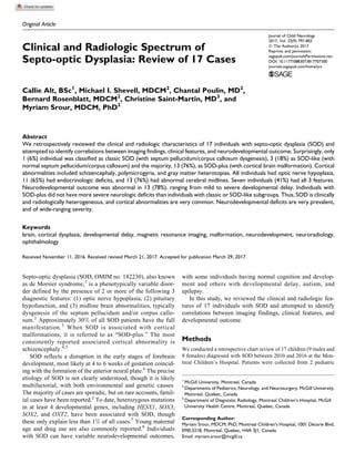
  • 4. Neurologic Findings Eleven patients (65%) had an abnormal neurologic exam. Seven (41%) had hypotonia, 2 (12%) had hemiparesis, and 1 (6%) had quadriparesis. Five individuals (29%) had epilepsy, of which 3 had secondary generalized tonic clonic seizures, 1 had infantile spasms, and 1 had myoclonic epilepsy. Four of the 5 epileptic patients had associated cortical abnormalities: 2 had schizencephaly and 2 had gray matter heterotopia, including transmantle cortical dysplasia. Psychomotor Development Normal development was reported in 3 (18%) children. Eleven patients (65%) were globally delayed, 2 (12%) were delayed in only 1 domain (gross motor), and 1 was lost to follow-up. Interestingly, all 3 individuals with normal development had SOD-plus: a 3-year-old boy with bilateral optic nerve hypopla- sia, hypopituitarism, absence of the septum pellucidum, and bilateral frontal polymicrogyria; a 17-year-old girl with aca- demic difficulties, bilateral optic nerve hypoplasia, agenesis of the septum pellucidum, multiple endocrinologic deficits, right perisylvian polymicrogyria and gray matter heterotopias, and fusion of the anterior fornices; and a 6-year-old boy with bilat- eral optic nerve hypoplasia, agenesis of the septum pellucidum, and bilateral frontal polymicrogyria. Of those that were glob- ally delayed, 3 had mild, 4 moderate, and 4 severe delay. Two of the 3 children that were severely delayed had associated cortical abnormalities. Two patients (12%) were diagnosed with autism, and 1 (6%) with autistic features that did not fulfill the criteria for autism. Genetic Evaluation Seven patients had karyotypes or chromosomal microarray testing and were normal. Only 1 patient had sequencing of HESX1, which was normal. One patient had a whole exome sequencing on a clinical basis. No rare variants were noted in any genes known to be associated with SOD, and no causal pathogenic mutations were identified in any other known disease gene. Figure 2. Brain imaging abnormalities in individuals with SOD. T2-weighted 1.5-Tesla MRI showing (A) left open-lipped schizencephaly (arrow) and right perisylvian polymicrogyria (arrowhead), (B) fusion of the fornices, (C) transmantle cortical dysplasia, and (D) bilateral frontal polymicrogyria. (E) T1-weighted coronal 3-Tesla MRI showing small right periventricular heterotopia. A, anterior, L, left; MRI, magnetic resonance imaging; P, posterior; R, right; SOD, septo-optic dysplasia. 800 Journal of Child Neurology 32(9)
  • 5. Discussion We reviewed the clinical and radiologic features and neurode- velopmental outcomes of 17 patients with SOD. In our cohort, only 1 (6%) patient was classified as having classical SOD (ie, with agenesis of septum pellucidum and/or corpus callosum in the absence of any cortical abnormality), 3 (18%) as SOD-like (ie, with normal septum pellucidum and corpus callosum in the absence of any cortical abnormality), and 13 (76%) as SOD-plus (ie, with cortical malformations). The most striking finding of our study was the high inci- dence of cortical malformations within our cohort. Indeed, 76% (13/17) of individuals had cortical abnormalities on their brain imaging and thus were categorized as having SOD-plus. Focal polymicrogyria was noted in 59% (10/17), schizencephaly in 30% (5/17), and heterotopias (subcortical, periventricular, or transmantle) in 35% (6/17). Three individuals (18%) had both schizencephaly and polymicrogyria (not associated with schi- zencephaly). Signorini et al11 reported that 42% of their patients with SOD have associated cortical malformations. The reasons for the increased incidence of cortical malformations in our cohort may be 2-fold. First, the majority (10/17) of our patients had high-resolution MRIs on a 3-Tesla magnet, allow- ing detection of more subtle abnormalities. Second, there may be a selection bias given that many of these patients were selected from neurologists’ databases and were more likely to have been referred because of specific neurologic symptoms such as abnormal development, focal findings on examination, or epilepsy. Indeed, individuals with SOD having neurologic symptoms may be more likely to have cortical involvement. In our cohort, patients with SOD-plus did not seem to have a higher risk of epilepsy than individuals without cortical abnormalities (4/13, 31%, vs 1/4, 25%, P ¼ 1.0, on Fisher exact test); however, studying a larger number of patients would be required to better assess whether the presence of a cortical malformation confers a greater risk of epilepsy in SOD. Nev- ertheless high-resolution brain imaging in patients with SOD is important to enable search for cortical abnormalities that may be responsible for focal neurologic findings or epilepsy. Interestingly, almost all individuals with SOD-plus (12/13, 92%) have a midline abnormality (Table 2), a finding that has also been noted in other studies.8,11 This suggests the involve- ment of common or overlapping pathophysiologic mechanisms that disrupt both midline cerebral development and neuronal migration such as an in utero vascular event, fetal viral infec- tion, or common underlying genetic etiology. Indeed, early vascular disruption has been hypothesized to underlie some SOD cases12-14 and is also a favored theory in the etiology of several cortical abnormalities such as perisylvian polymicro- gyria and schizencephaly.15 Fetal viral infections have been associated with SOD and various cortical malformations.16 Finally, one can speculate that mutations in genes with an important function both in midline cerebral development and neuronal migration may lead to such a phenotype. All individuals in our cohort had optic nerve hypoplasia on clinical examination, which is the gold standard for diagnosis. Optic nerve hypoplasia was bilateral in 82% (14/17) and uni- lateral in 18% (3/14) of the cohort. Endocrinologic deficits were present in 65% (11/17) our cohort, and midline abnorm- alities in 76% (13/17). This is similar to what has been reported in other studies.6,17 The 3 SOD diagnostic features were present in 35% of patients (6/17), which is consistent with what has been previously reported (30%-47%) in other studies.3,11 Endocrinologicdeficiencieswereobservedin11ofourpatients (65%), of which 7 had associated abnormal pituitary imaging. The absence of imaging abnormalities of the pituitary does not pre- cludethepresenceofhormonaldeficits,underlyingtheimportance of clinical hormonal investigations in any individual with 1 feature of SOD. In addition, 2 individuals with abnormal pituitaries on MRI had no endocrinologic deficits when last evaluated. Developmental impairment was reported in 13 (78%) of our patients. Poor vision likely contributes to, but does not fully account for, the patients’ developmental delay. The severity of the associated developmental impairment is wide-ranging. In fact, the severity was equally distributed in all severity cate- gories (see Table 2). SOD-plus patients have a heterogeneous neurodevelopmental outcome, ranging from normal to severely delayed development. All 3 individuals with normal develop- ment had cortical abnormalities and were classified as SOD- plus. Conversely, SOD-like patients appear to have more severe delay, with all individuals categorized as moderate-severe delay. It is difficult to draw clear conclusions given the small number of patients. It is important to note again that, because of possible aforementioned selection bias in our study, patients with normal development are less likely to be referred for neurologic evalua- tion and thus would not have been included in our cohort. Other limitations to this present study also include the retrospective nature of the study and the relatively small number of patients precluding statistical analysis. Table 2. Common Clinical Features of the 3 Subtypes of Septo-optic Dysplasia. Classic SOD (1/17) SOD-like (3/17) SOD-plus (13/17) Vision Normal 0 0 1 Abnormal 1 3 12 Developmental level Normal 0 0 3 Mildly delayed 1 0 3 Moderately delayed 0 1 3 Severely delayed 0 2 3 Not classified 0 0 1 Autistic 0 1 2 Endocrine function Normal 1 1 4 Abnormal 0 2 9 Presence of midline defect No 0 3 1 Yes 1 0 12 Abbreviation: SOD, septo-optic dysplasia. Alt et al 801
  • 6. Few studies such as ours have focused on the neurologic and developmental features in SOD,8,11,18,19 with the majority of studies concentrating on endocrinologic and ophthalmologic aspects.20-25 Riedl et al8 looked at 68 patients with optic nerve hypoplasia, of which 42 had SOD. In their study, unilateral optic nerve hypoplasia and septum pellucidum remnants were associated with a milder endocrinologic and neurologic SOD phenotype, and abnormalities of the falx and hippocampus were associated with worse endocrinologic and neurodevelop- mental outcomes. Signori et al11 reviewed 17 SOD patients and observed a heterogeneous clinical spectrum, with nervous sys- tem involvement as a key feature of the syndrome: develop- mental/cognitive delay present in nearly one-half of the study sample. In this study, patients classified as SOD or SOD-like had more marked neurodevelopmental delay than patients in the SOD-plus category. Parr et al19 systematically studied the presence of autistic features in a cohort of 83 patients with optic nerve hypoplasia or SOD, and found a 36% prevalence of aut- ism spectrum disorder in SOD patients compared to 26% in optic nerve hypoplasia patients. A cross-sectional study by Jutley-Neilson et al18 looking at the occurrence of autism spec- trum disorder in 42 children with SOD or optic nerve hypopla- sia reported an autism spectrum disorder diagnosis in 33%, which is greater than that observed in our cohort. The underlying etiology of SOD remains overwhelmingly unknown. In our cohort, 7 patients (41%) had chromosomal studies (karyotype or microarray), only 1 had molecular genetic testing for known SOD genes, and no patient had an identified etiology. All our cases were sporadic. With the exception of one, all mothers were under the age of 25 years. This is in line with other studies that suggest that SOD patients are the children of mothers who are younger than average.26 In summary, our study reveals that SOD is clinically hetero- geneous. Cortical abnormalities are frequently associated with SOD, and should be systematically looked into for using high- resolution MRI. Neurodevelopmental deficits are very preva- lent, and of wide-ranging severity. Larger studies are needed to better assess clinical and radiologic predictors of outcome in SOD. Acknowledgments MS holds a clinician-scientist award from the Fonds de Recherche en Sante´ du Quebec (FRQ-S). Declaration of Conflicting Interests The authors declared no potential conflicts of interest with respect to the research, authorship, and/or publication of this article. Funding The authors received no financial support for the research, authorship, and/or publication of this article. References 1. De Morsier G. Studies on malformation of cranio-encephalic sutures. III. Agenesis of the septum lucidum with malformation of the optic tract [in French]. Schweiz Arch Neurol Psychiatr. 1956;77:267-292. 2. Kelberman D, Dattani MT. Genetics of septo-optic dysplasia. Pituitary. 2007;10:393-407. 3. Morishima A, Aranoff GS. Syndrome of septo-optic-pituitary dysplasia: the clinical spectrum. Brain Dev. 1986;8:233-239. 4. Kuban KC, Teele RL, Wallman J. Septo-optic-dysplasia- schizencephaly. Radiographic and clinical features. Pediatr Radiol. 1989;19:145-150. 5. Miller SP, Shevell MI, Patenaude Y, Poulin C, O’Gorman AM. Septo-optic dysplasia plus: a spectrum of malformations of cor- tical development. Neurology. 2000;54:1701-1703. 6. Webb EA, Dattani MT. Septo-optic dysplasia. Eur J Hum Genet. 2010;18:393-397. 7. Saranac L, Gucev Z. New insights into septo-optic dysplasia. Prilozi. 2014;35:123-128. 8. Riedl S, Vosahlo J, Battelino T, et al. Refining clinical phenotypes in septo-optic dysplasia based on MRI findings. Eur J Pediatr. 2008;167:1269-1276. 9. Srour M, Shevell M. Genetics and the investigation of developmen- tal delay/intellectual disability. Arch Dis Child. 2014;99:386-389. 10. Srour M, Mazer B, Shevell MI. Analysis of clinical features pre- dicting etiologic yield in the assessment of global developmental delay. Pediatrics. 2006;118:139-145. 11. Signorini SG, Decio A, Fedeli C, et al. Septo-optic dysplasia in childhood: the neurological, cognitive and neuro- ophthalmological perspective. Dev Med Child Neurol. 2012;54: 1018-1024. 12. Lubinsky MS. Hypothesis: septo-optic dysplasia is a vascular disruption sequence. Am J Med Genet. 1997;69:235-236. 13. Chiaramonte I, Cappello G, Uccello A, et al. Vascular cerebral anomalies associated with septo-optic dysplasia. A case report. Neuroradiol J. 2013;26:66-70. 14. Mitchell LA, Thomas PQ, Zacharin MR, Scheffer IE. Ectopic posterior pituitary lobe and periventricular heterotopia: cerebral malformations with the same underlying mechanism? AJNR Am J Neuroradiol. 2002;23:1475-1481. 15. Guerrini R, Dobyns WB. Malformations of cortical development: clinicalfeaturesandgeneticcauses.LancetNeurol.2014;13:710-726. 16. Barkovich AJ, Lindan CE. Congenital cytomegalovirus infection of the brain: imaging analysis and embryologic considerations. AJNR Am J Neuroradiol. 1994;15:703-715. 17. McCabe MJ, Alatzoglou KS, Dattani MT. Septo-optic dysplasia and other midline defects: the role of transcription factors: HESX1 and beyond. Best Pract Res Clin Endocrinol Metab. 2011;25:115-124. 18. Jutley-Neilson J, Harris G, Kirk J. The identification and measurement of autistic features in children with septo-optic dysplasia, optic nerve hypoplasia and isolated hypopituitarism. Res Dev Disabil. 2013;34:4310-4318. 19. Parr JR, Dale NJ, Shaffer LM, Salt AT. Social communication difficulties and autism spectrum disorder in young children with optic nerve hypoplasia and/or septo-optic dysplasia. Dev Med Child Neurol. 2010;52:917-921. 20. Cemeroglu AP, Coulas T, Kleis L. Spectrum of clinical presenta- tions and endocrinological findings of patients with septo-optic 802 Journal of Child Neurology 32(9)
  • 7. dysplasia: a retrospective study. J Pediatr Endocrinol Metab. 2015;28:1057-1063. 21. Bachmann-Gagescu R, Phelps IG, Dempsey JC, et al. KIAA0586 is mutated in Joubert syndrome. Hum Mutat. 2015;36:831-835. 22. Antonini SR, Grecco Filho A, Elias LL, Moreira AC, Castro M. Cerebral midline developmental anomalies: endocrine, neurora- diographic and ophthalmological features. J Pediatr Endocrinol Metab. 2002;15:1525-1530. 23. Atapattu N, Ainsworth J, Willshaw H, et al. Septo-optic dysplasia: antenatal risk factors and clinical features in a regional study. Horm Res Paediatr. 2012;78:81-87. 24. Ahmad T, Garcia-Filion P, Borchert M, Kaufman F, Burkett L, Geffner M. Endocrinological and auxological abnormalities in young children with optic nerve hypoplasia: a prospective study. J Pediatr. 2006;148:78-84. 25. Garcia-Filion P, Borchert M. Optic nerve hypoplasia syndrome: a review of the epidemiology and clinical associations. Curr Treat Options Neurol. 2013;15:78-89. 26. Murray PG, Paterson WF, Donaldson MD. Maternal age in patients with septo-optic dysplasia. J Pediatr Endocrinol Metab. 2005;18:471-476. Alt et al 803