SlideShare uma empresa Scribd logo
1 de 8
Baixar para ler offline
JOURNAL OF VIROLOGY, Oct. 2004, p. 10213–10220                                                                              Vol. 78, No. 19
0022-538X/04/$08.00 0 DOI: 10.1128/JVI.78.19.10213–10220.2004
Copyright © 2004, American Society for Microbiology. All Rights Reserved.


                                                       MINIREVIEW

            Pathogenesis of Intestinal and Systemic Rotavirus Infection
                                                           Robert F. Ramig*
                   Department of Molecular Virology and Microbiology, Baylor College of Medicine, Houston, Texas


   Rotaviruses are responsible for significant gastrointestinal           posed of 11 segments of double-stranded RNA. There are six
disease, primarily in children 5 years of age and the young of           structural proteins and six nonstructural proteins, each en-
other mammalian species. Each year rotaviruses cause approx-             coded in a unique genome segment except for nonstructural
imately 111 million episodes of gastroenteritis in children,             proteins 5 and 6 (NSP5 and NSP6), which are encoded in
which result in 25 million visits to clinics, 2 million hospital-        overlapping reading frames of a single segment. The rotavirus
izations, and 352,000 to 592,000 deaths. On a worldwide basis,           genus is divided into serological groups (A to E). Groups A to
nearly every child experiences rotavirus gastroenteritis by age          C infect humans, and all groups infect animals. All the infor-
5, 1 in 5 visits a clinic, 1 in 65 is hospitalized, and 1 in 293 dies.   mation presented here is in regard to group A virus infections.
Children in the poorest countries account for 82% of rotavirus              Pathophysiology of rotavirus diarrhea. The enterocytes lin-
deaths (60). This disease burden underscores a need for inter-           ing the small intestine are generally divided into two types:
ventions such as vaccines. A vaccine was developed and ap-               enterocytes and crypt cells (Fig. 1). Villus enterocytes are ma-
proved, but recommendation for its use was withdrawn be-                 ture, nonproliferating cells covering the villi that are differen-
cause of vaccination-associated adverse events (57). Additional          tiated to digestive and absorptive functions. The absorptive
information about the molecular biology, immunology, and                 enterocytes synthesize a number of disaccharidases, pepti-
pathogenesis of rotavirus infection will inform ongoing vaccine          dases, and other enzymes that are expressed on the apical
development efforts.                                                     surface, where they carry out their digestive functions. Absorp-
   Our understanding of rotavirus-induced diarrheal disease is           tion across the enterocyte barrier occurs both by passive dif-
incomplete compared to that of several other pathogens (e.g.,            fusion of solutes along electrochemical or osmotic gradients
cholera). Rotavirus diarrhea has been attributed to several              and by active transport. While the majority of water transport
different mechanisms, including malabsorption secondary to               is passive along osmotic gradients, transporters such as the
enterocyte destruction, a virus-encoded toxin, stimulation of            sodium-glucose cotransporter 1 (SGLT1) transport water
the enteric nervous system (ENS), and villus ischemia (13, 45).          along with solute (42). The crypt epithelium lines the crypts
Over the past several years, numerous studies have addressed             and is the progenitor of the villus enterocytes. Crypt cells lack
mechanisms of diarrhea induction at the cellular and tissue              the well-defined microvilli and absorptive functions of the en-
levels, and a new understanding of the mechanisms is begin-              terocyte and actively secrete Cl ions into the intestinal lumen.
ning to emerge. Here I will briefly outline the new data and              In the normal animal, the combined activity of the enterocytes
present our current understanding of how rotaviruses induce              and crypt cells results in a constant bidirectional flux of elec-
diarrhea in the infected host.                                           trolytes and water across the epithelium. On the villi, the bal-
   Recent studies confirm sporadic case reports that rotavirus            ance is toward absorption, and in the crypts, the balance favors
infection is not confined to the intestine as was generally as-           secretion (51).
sumed. Although systemic sequellae to rotavirus infection are               Our understanding of rotavirus pathophysiology comes pri-
apparently rare, reports have continually appeared in the lit-           marily from animal models. Rotaviruses replicate in the non-
erature, and recent work with animal models has begun to shed            dividing mature enterocytes near the tips of the villi, suggesting
light on how the virus spreads to extraintestinal sites. Our             that differentiated enterocytes express factors required for ef-
current understanding of extraintestinal spread and infection            ficient infection and replication (13). The severity and local-
will also be considered below.                                           ization of rotavirus intestinal infection vary among animal spe-
                                                                         cies and between studies; however, the pathological changes
   GENERAL CHARACTERISTICS OF ROTAVIRUS                                  are almost exclusively limited to the small intestine. In various
INFECTION AND DISEASE IN HUMANS AND ANIMALS                              animal models, rotavirus infection is associated with virtually
                                                                         no visible lesions; slight lesions, such as enterocyte vacuoliza-
  Rotavirus. Rotaviruses comprise a genus within the family              tion and loss; or larger changes, such as villus blunting and
Reoviridae. The rotavirion has a nonenveloped, complex, tri-             crypt hyperplasia. Inflammation is generally mild compared to
ple-layered capsid structure that surrounds a genome com-                that for other intestinal pathogens. This picture of pathology
                                                                         suggests that there is no absolute correlation between histo-
                                                                         logical lesions and disease symptoms.
  * Mailing address: Department of Molecular Virology and Micro-
biology, Baylor College of Medicine, One Baylor Plaza, Houston, TX
                                                                            Rotavirus infection alters the function of the small intestinal
77030. Phone: (713) 798-4830. Fax: (713) 798-3586. E-mail: rramig        epithelium, resulting in diarrhea. The diarrhea was generally
@bcm.tmc.edu.                                                            considered to be malabsorptive, secondary to enterocyte de-

                                                                     10213
10214      MINIREVIEW                                                                                                                    J. VIROL.


                                                                           struction (34). In addition to enterocyte destruction, absorp-
                                                                           tion of Na , water, and mucosal disaccharidases are decreased
                                                                           (10, 28), while mucosal cyclic AMP appears not to be altered
                                                                           (16). Malabsorption results in the transit of undigested mono-
                                                                           and disaccharides, carbohydrates, fats, and proteins into the
                                                                           colon. The undigested bolus is osmotically active, and the co-
                                                                           lon is unable to absorb sufficient water, leading to an osmotic
                                                                           diarrhea (27). Another study suggested that the diarrhea was
                                                                           malabsorptive and resulted from epithelial damage caused by
                                                                           villus ischemia (58). A secretory component of the diarrhea
                                                                           was suggested, based on elevated levels of prostaglandin E2
                                                                           (PGE2) in the infected gut and the stimulation of secretion by
                                                                           PGE2 (79). The fact that gut lesions often do not correlate with
                                                                           the presence of diarrhea stimulated the search for other mech-
                                                                           anisms of diarrhea induction. The viral nonstructural protein
                                                                           NSP4, a secreted fragment of NSP4, or certain NSP4 peptides
                                                                           were found to have toxin-like activity and to induce diarrhea
                                                                           when inoculated into mice (3, 23, 77). The NSP4 enterotoxin
                                                                           activity provides a way to mediate diarrheagenic changes in the
                                                                           absence of significant damage or to mediate changes at unin-
                                                                           fected sites. Recently, it was shown that several drugs that
                                                                           block the action of the ENS attenuate rotavirus-induced secre-
                                                                           tion in the intestine, suggesting a role for the ENS in rotavirus
                                                                           diarrhea (44, 45, 46). It was estimated that 67% of the fluid
                                                                           and electrolyte secretion in rotavirus diarrhea in experiments
                                                                           with mice was due to activation of the ENS (46). Thus, it is
                                                                           clear that rotavirus diarrhea is multifactoral, resulting from the
                                                                           direct effects of virus infection and the indirect effects of in-
                                                                           fection and the host response.
                                                                              Some rotavirus infections are asymptomatic (10), which sug-
                                                                           gests that both viral and host factors can affect disease severity
                                                                           (13). Among the viral factors are the following. (i) Some alleles
                                                                           of VP4 may be associated with asymptomatic disease (24). (ii)
                                                                           Virus strains can be attenuated, particularly by passage in cell
                                                                           culture. Attenuation generally results in a restricted ability to
                                                                           replicate and cause disease in the host (32). (iii) Virus strains


                                                                           plasmic reticulum (blue), increasing [Ca2 ]i. (ii) A cell is secondarily
                                                                           infected after virus release from the initial cell. NSP4 produced by the
                                                                           infection disrupts the tight junctions, allowing paracellular flow of
                                                                           water and electrolytes (green arrow). (iii) NSP4 binds to a specific
                                                                           receptor on a cell and triggers a signaling cascade through PLC and IP3
                                                                           that results in release of Ca2 and an increase in [Ca2 ]i. Intracellular
                                                                           expression of NSP4 does not stimulate PLC. The increase in [Ca2 ]i
                                                                           acts to disrupt the microvillar cytoskeleton. (iv) The brown cell repre-
                                                                           sents a crypt cell. It can be acted on directly by NSP4, or NSP4 can
                                                                           stimulate the ENS, which in turn signals an increase in [Ca2 ]i that
                                                                           induces Cl secretion. Panel B shows the normal architecture of the
                                                                           small intestine, with the circulatory system removed for clarity. This
                                                                           panel emphasizes the ENS and its ganglia in the different submucosal
                                                                           levels. (Panel B is adapted from reference 25, with the permission of
                                                                           M. D. Gershon and the publisher.) Panel C shows a reflex arc in the
                                                                           ENS that can receive signals from the villus epithelium and activate the
                                                                           crypt epithelium. (Adapted from reference 44, with the permission of
   FIG. 1. Model of rotavirus-induced diarrhea. Panel A depicts            L. Svensson and the publisher.) Inset 1 shows a whole mount of an
events in the infected epithelium. For clarity, not all events are shown   adult mouse small intestinal villus, stained with antibody to protein
in each cell. The following processes are shown in order from left to      gene product 9.5 to reveal the rich innervation (yellow stain). (Inset 1
right across the four cells. (i) The initial cell is infected by luminal   is used by courtesy of R. O. Heuckeroth and with permission.) Inset 2
virus, with virus entry and uncoating, formation of a viroplasm (Vi),      shows that infected villus enterocytes may stimulate the ENS by the
and the release of virus and viral proteins. NSP4 (red triangles) may be   basolateral release of NSP4 or other effector molecules. (Inset 2 is
released via a nonclassical secretory pathway. Intracellular NSP4 also     adapted from reference 45 with permission of L. Svensson and the
induces release of Ca2 from the internal stores, primarily the endo-       publisher.)
VOL. 78, 2004                                                                                                   MINIREVIEW         10215


seem to be adapted for growth in particular host species (host       triggers a phospholipase C–inositol 1,3,5-triphosphate (PLC-
range) (5). Several host factors have also been shown to affect      IP3) cascade that culminates in the release of Ca2 from the
the severity of rotavirus disease, including the following. (i)      endoplasmic reticulum, increasing [Ca2 ]i. If NSP4 acts on
Malnutrition is documented to increase the severity of rotavi-       enterocytes, one of the results is the disruption of tight junc-
rus diarrhea (71), where it delays small intestinal recovery (78)    tions, resulting in paracellular permeability. If NSP4 acts on
and modifies intestinal inflammatory responses (79). (ii) Ro-          crypt cells, the resulting increase in [Ca2 ]i leads to secretion
tavirus symptomatic infections are generally age restricted          in the crypt, mediated by activation of a Cl transporter, re-
(13). The age dependence appears to be unrelated to receptor         sulting in an increased secretory component of the diarrhea.
expression, as both the viral and the NSP4 receptors are ex-         Secreted NSP4, or other effector molecules released from in-
pressed in adult animals. However, signaling downstream of           fected cells, may also stimulate the ENS (Fig. 1C). Indeed,
the NSP4 receptor does appear to be age dependent (53), but          experiments with agents that block function of the ENS
age restriction may be related to immunity, as neutralizing          showed that rotavirus infection induced secretion via stimula-
antibodies increase with age and virus exposure. (iii) Rotavirus     tion of the ENS. This information begins to shed light on the
disease may be related to age-dependent protease expression,         mechanism(s) by which relatively few infected cells, causing
as viral infectivity requires protease cleavage of VP4 and new-      little visible damage to the mucosa, can elicit a diarrheal re-
borns have low levels of protease in the gut (29). (iv) The          sponse.
expression of intestinal mucins and the rate of epithelial cell
replacement and fluid absorption are both age dependent and
                                                                      THE MOLECULAR BASIS OF DIARRHEA INDUCTION
have been shown to affect rotavirus infection and disease in the
host (51, 76).                                                          Rotavirus diarrhea is multifactoral, has malabsorption and
                                                                     secretion components, and may have other components sug-
  THE CURRENT MODEL OF DIARRHEA INDUCTION                            gested to be related to villus ischemia and intestinal motility.
               BY ROTAVIRUS                                          Here I present the relevant data on the induction of each of
                                                                     these components (references are selective and for illustrative
   Many of the salient events in rotavirus induction of diarrhea     purposes).
are shown in Fig. 1. Most of the data used to construct this            Malabsorption. A malabsorptive component of rotavirus di-
figure were derived from model animal and cell culture sys-           arrhea appears to be related to the primary infection with the
tems, although some items were demonstrated in humans as             virus. Infection of villus enterocytes leads to a cascade of
well. Numerous recent reviews have dealt with specific aspects        events involving Ca2 (Fig. 1A). This disruption of Ca2 ho-
of diarrhea induction (21–23, 43–45, 50, 52, 67). From these         meostasis appears to be mediated by synthesis of viral proteins
reviews and the discussion above, it is clear that a multitude of    (17, 49). Increased Ca2 permeability at both the plasma mem-
viral and host factors have the potential to influence rotavirus      brane and the endoplasmic reticulum leads to an increase in
infection and disease production. Below, I have assembled            [Ca2 ]i, triggering a chain of events that leads to cell lysis (61).
what we currently understand about the interplay of these            The fact that NSP4 expressed in cells also leads to increases in
factors in the induction of rotavirus diarrhea.                      [Ca2 ]i implicates it as the mediator of virus-induced [Ca2 ]i
   The process leading to diarrhea is initiated when rotavirus       dysregulation (74). A fragment of NSP4 (amino acids 112 to
binds to and infects enterocytes in the small intestine (Fig. 1A).   175) is secreted via a nonclassical pathway early after infection
Binding is mediated by sequential interaction with a series of       (77), and this fragment added exogenously to cells also causes
sialic acid-containing and nonsialylated receptor molecules.         an increase in [Ca2 ]i (73). The increase in [Ca2 ]i follows
The virus is internalized by an unknown mechanism, and the           NSP4 binding to a specific apical receptor (22) that triggers a
outer capsid is lost, activating the virion-associated transcrip-    PLC-IP3 cascade resulting in release of Ca2 from intracellular
tase and viral macromolecular syntheses. Viral proteins and          stores (19). In contrast, the increase in [Ca2 ]i induced by
RNAs concentrate in cytoplasmic structures called viroplasms,        intracellular NSP4 is independent of PLC stimulation (73).
where RNA replication and packaging take place. Intracellular        The NSP4-mediated effects may amplify the diarrheagenic ef-
events, probably involving NSP4, cause release of Ca2 from           fect of infection in the absence of significant visible tissue
the endoplasmic reticulum. The increase in intracellular Ca2         damage. However, the ability of inactivated rotavirus particles
concentration ([Ca2 ]i) triggers a number of cellular processes,     to induce diarrhea (68) suggests that viral structural proteins
including disruption of the microvillar cytoskeletal network,        may also play a role in the dysregulation leading to diarrhea.
lowered expression of disaccharidases and other enzymes at              Rotavirus infection has other effects on enterocytes that may
the apical surface, general inhibition of the Na -solute co-         contribute to malabsorption (Fig. 1A). Infection leads to an
transport systems, and necrosis. NSP4 appears to be released         increase in [Na ]i and a decrease in [K ]i, which appear to be
specifically by a Ca2 -dependent, nonclassical secretion path-        related to increased plasma membrane permeability and not
way prior to cell lysis. These events lead to a malabsorption        inhibition of the Na /K pump (17). Changes in intracellular
component of the diarrhea through reduction in absorptive            levels of Na and K could impair electroneutral NaCl ab-
capacity of the epithelium, reduced activity of Na -solute co-       sorption and Na -linked nutrient absorption, resulting in a loss
transporters, and reduction of digestive enzyme expression on        of fluid (50). [Na ]i dysregulation may be related to a general
the epithelial surface.                                              inhibition of the Na -solute cotransport systems (30). NSP4
   The release of NSP4 from infected cells allows paracrine          may also be involved, as the NSP4114-135 peptide is a specific
effects to occur on uninfected cells (Fig. 1A). NSP4 binds to        and noncompetitive inhibitor of SGLT1 (31). Infection also
these cells, using specific, unidentified receptor(s) (22), and        reduces the expression of digestive enzymes at the apical sur-
10216     MINIREVIEW                                                                                                          J. VIROL.


face of infected enterocytes. For example, the activities of         agents that block ENS stimulation (lidocaine, tetrodotoxin,
alkaline phosphatase, lactase, sucrase, and maltase are reduced      and mecamylamide) significantly lowered the transmembrane
(6, 12, 16). The expression of sucrase and isomaltase in cul-        potential difference in a virus dose-dependent manner in
tured human intestinal epithelium was also reduced, probably         Ussing chamber or organ bath experiments using rotavirus-
as a result of perturbation of protein targeting and the mi-         infected intestinal tissues. In organ bath experiments, blocking
crovillar cytoskeleton (33). Rotavirus infection alters the struc-   the ENS could change net secretion to net absorption (45). In
ture of polarized enterocytes in a number of ways. The increase      live infected animals, repeated administration of lidocaine sig-
in [Ca2 ]i induced by rotavirus infection affects the Ca2 -          nificantly prevented fluid losses. Thus, it is clear that the ENS
sensitive proteins F-actin, villin, and tubulin, damaging the        is activated during rotavirus infection, and this activation could
microvillar cytoskeleton, whereas rearrangements of other cy-        explain how relatively few infected cells at the villus tips could
toskeletal proteins (cytokeratin-18) are independent of              stimulate crypt cells to secrete electrolytes and water (45).
changes in [Ca2 ]i (6, 7). Both rotavirus infection and NSP4         While it is unknown if NSP4 directly stimulates the ENS, the
promote functional changes in tight junctions between entero-        ENS is known to respond to a number of molecules released
cytes that maintain the epithelial barrier (18, 72). The drop in     from enterocytes. Cholera toxin induces the release of 5-hy-
transepithelial resistance induced by either the virus or NSP4       droxytryptamine (5-HT) from enterochromaffin cells in the
suggests that infection can cause paracellular leakage. Rotavi-      villus epithelium, and 5-HT is a stimulator of the ENS (44). It
rus also induces intestinal epithelial cells to secrete CXC and      is possible that secreted NSP4 binds to enterochromaffin cells,
CC chemokines, suggesting that enterocyte chemokine secre-           inducing a release of 5-HT and stimulation of the ENS (44)
tion plays a role in initiating the immune response to infection     (Fig. 1C, inset 2). Likewise, the secretion of chemokines and
(9, 66, 69). Interleukin-8 (IL-8), GRO- , RANTES, interferon         prostaglandins by infected enterocytes may serve to stimulate
(IFN)-stimulated protein 10, and granulocyte-macrophage col-         the ENS. Currently, the ENS and NSP4 appear to have the
ony-stimulating factor (GM-CSF) are stimulated, whereas              major roles in the secretory response to infection.
other chemokines (tumor necrosis factor alpha, IL-1 , IFN- ,            Villus ischemia. Although damage to the intestinal epithe-
IFN- , MIP- , MCP-1, and IL-6) are unchanged. Induction of           lium is minimal in rotavirus-infected mice, villus ischemia was
IL-8 and RANTES is noteworthy because these are the most             observed in some studies (58, 70). It was proposed that diar-
potent chemoattractants for intestinal intraepithelial lympho-       rhea could result from virus-induced release of an unknown
cytes (20). It is unclear if virus replication is required for       vasoactive agent from infected epithelium, causing a local vil-
induction of chemokine secretion (9, 66). In addition, the lev-      lus ischemia and subsequent functional damage to enterocytes
els of PGE2 are increased in infected intestine (79). The che-       (59). However, villus ischemia has not been observed in other
mokines may activate the immune response rather than di-             animal models, so the significance of this observation remains
rectly contribute to diarrhea. Thus, rotavirus infection causes a    unknown.
number of changes in the villus epithelium that contribute to           Intestinal motility. In some diarrheal infections, intestinal
malabsorption.                                                       motility is significantly increased. The intestinal transit time is
   Secretion. The secretory component of rotavirus diarrhea          decreased in rotavirus infection, implying increased motility
appears to be secondary to virus-induced functional changes at       (50). The ENS generally controls motility, but the molecular
the villus epithelium. The central players in secretion appear to    stimulator of motility is not known. It could be any of the ENS
be NSP4 and the ENS. The precise role and targets of secreted        stimulators discussed above.
NSP4 are unknown. NSP4 may simply amplify the effects of                To summarize, rotavirus diarrhea is clearly a multicompo-
infection in the enterocyte epithelium. However, NSP4 may            nent disease. Good evidence exists for malabsorptive and se-
also act at the crypt epithelium (Fig. 1A), where it would           cretory components. The mediators of these disease compo-
induce increases in crypt cell [Ca2 ]i, activate Cl secretion,       nents range from primary cellular damage to a secreted viral
and lead to an outflow of water. This Cl secretion is known to        enterotoxic peptide and a virus-induced interaction with the
be unrelated to the cAMP-dependent Cl channel of crypt               ENS.
cells because CFTR-knockout mice are susceptible to rotavirus
or NSP4-induced diarrhea (2, 53). The identity of the Cl                    SYSTEMIC INFECTION WITH ROTAVIRUS
channel involved in rotavirus diarrhea in CFTR-knockout mice
remains unknown. It is proposed that NSP4 itself may form a             L. M. Kraft performed some of the early studies of rotavirus
channel or that NSP4 activates a dormant Ca2 -activated an-          infection, using epidemic diarrhea of infant mice virus (EDIM)
ion channel (53). Interestingly, these studies also showed that      before it was recognized as a rotavirus (1). An important but
the age dependence of rotavirus diarrhea is not due to age-          under-appreciated aspect of this work is that rotavirus spread
dependent expression of a NSP4 receptor or age-dependent             throughout the bodies of the infected mice following oral in-
Ca2 mobilization but rather to the age dependence of Cl              fection (39, 40, 41). By 72 h postinfection, infectious EDIM
permeability (53). Another possible target of secreted NSP4 is       was found in the lungs, liver, spleen, kidney, bladder, brain,
the ENS, which is also a target in classic cholera toxin-induced     and blood. Since most of the organs where EDIM was detected
diarrhea (45). Indeed, the ENS is rich immediately under the         are highly vascular, it was generally assumed that virus in the
villus epithelium, and it is situated to receive stimuli from the    tissues reflected the presence of blood.
rotavirus-damaged epithelium (Fig. 1C, inset 1). Although               Subsequently, a large number of clinical case reports sug-
NSP4 stimulation of the ENS has not been shown experimen-            gested that rotaviruses could be found at extraintestinal sites
tally, it has been shown that the ENS is involved in rotavirus       following infection. It was not clear if the virus was replicating
diarrhea (46). The application of a number of pharmacologic          and infectious at these sites or passively present in the blood or
VOL. 78, 2004                                                                                                  MINIREVIEW        10217


if it resulted from contamination during sample collection.           replicate to spread, but it need not replicate to high titer to
Examples include the finding of virus in the liver following fatal     spread to the liver. A follow-up study showed that virus es-
disease (8), the finding of elevated liver enzymes associated          caped the intestine via a lymphatic route, appearing sequen-
with virus infection (38), and the demonstration of viral repli-      tially in the Peyer’s patch, the mesenteric lymph node, and,
cation in the liver and kidneys of an immunodeficient child            finally, the peripheral tissues (55). These studies also showed
(26). Rotavirus involvement in biliary atresia was suggested,         that genome segment 6, encoding VP6, was a secondary de-
although the best data involve group C rotaviruses (65). Neu-         terminant of spread. The combination of segment 6 from a
rological involvement was suggested by several reports of chil-       spreading virus with segment 7 from a nonspreading virus
dren with concomitant convulsions and rotavirus diarrhea (for         allowed escape from the intestine but only to the mesenteric
examples, see references 35 and 56). Whether putative central         lymph node and no farther (55). The association of a nonstruc-
nervous system (CNS) infection results from contamination is          tural protein, NSP3, with escape of virus from the intestine
not known, but one study strongly suggested infection of the          suggested that virus transits in infected cells. Indeed, prelimi-
CNS (48). In recent studies, rotavirus antigens were detected         nary experiments indicate that virus in the mesenteric lymph
in myocardium from patients who died unexpectedly (11), and           node is internalized by some cells of the lymphocytic or myloid
in vitro studies demonstrated the ability of rotavirus to repli-      lineage (E. C. Mossel and R. F. Ramig, unpublished data).
cate in primary islet cells, an observation that correlates with      NSP3 is described as a protein that functions in the regulation
the temporal association of infection with development of pan-        of protein synthesis by binding to a specific sequence at the 3
creatic islet autoantibodies (14). These case reports strongly        termini of the nonpolyadenylated viral mRNAs and interacts
indicate that rotavirus infection may have rare systemic sequel-      with the translation initiation complex to favor viral protein
lae, although one must bear in mind that infectious viruses           synthesis in the infected cell (62). How NSP3 functions as the
were not isolated and replication was shown only once (26).           primary determinant of spread to the liver is not understood.
   In animal models, rotaviruses have also been documented to            Potential role for viremia in rotavirus spread and patho-
spread beyond the intestine after oral infection. In the mouse        genesis. The numerous clinical reports of rotavirus at systemic
model, sites of spread include the lamina propria, Peyer’s            sites and work with mice that showed systemic spread
patches, mesenteric lymph nodes, lung, liver, kidney, and bile        prompted a systematic search for evidence of widespread ro-
duct (13). Group A rotaviruses have been shown to induce              tavirus viremia in children and animal model systems (4). In a
biliary atresia in mice, but this system requires intraperitoneal,    retrospective study, 22 of 33 (66%) serum samples from rota-
not oral, inoculation of virus (15). The most detailed studies of     virus-infected children were positive for rotavirus antigen by
extraintestinal spread in the mouse examined spread to the            enzyme immunoassay (EIA), whereas 0 of 35 rotavirus-nega-
liver following oral inoculation.                                     tive children had antigens in their sera. In a subset of six
   The liver as a site of systemic infection. Spread of virus in      children with paired acute and convalescent-phase sera, viral
orally infected mice was shown in persistently infected SCID          antigen was present in all acute-phase sera and none of the
mice where a diffuse hepatitis was noted. Infectious virus was        convalescent-phase sera, indicating that the antigenemia is
isolated from the liver, and surviving mice developed chronic         transient (4). Only three of six serum samples from rotavirus-
liver disease (64, 75). Infectious virus was also found in the        infected children were positive by PCR, the lower positivity
livers of normal mice, although the hepatitis was less severe         probably reflecting the lower sensitivity of the PCR assay used
and resolved spontaneously (75). Spread of virus to the liver         (4). In another study, three children who died of rotavirus-
and development of hepatitis were described as virus strain           associated disease were examined retrospectively. Two of the
dependent. These results were supported by studies of cultured        three children had rotavirus RNA in extraintestinal tissues
human hepatoma (HepG2) cells, where some viruses were                 (spleen, heart, lung, kidney, testes, bladder, adrenal gland, and
capable of complete infectious cycles and others were not (37,        pancreas), as determined by reverse transcription-PCR. Con-
63). The strain-specific ability to infect HepG2 cells segregated      firmation of positive PCR products by hybridization with se-
with viral genome segment 4, encoding the outer capsid spike          rotype-specific probes indicated that the two children with
protein VP4 (36, 63). Subsequent studies indicated that viruses       extraintestinal rotavirus were infected with different G sero-
unable to replicate in HepG2 cells entered less efficiently, but       types (47). These findings show that viral antigen and/or virus
the absolute block to infection occurred at a late step in the        particles enter the circulation.
intracellular replication cycle (S. Jafar and R. F. Ramig, un-           Prospective studies with animal models confirmed the anti-
published data).                                                      genemia seen in humans and also demonstrated viremia. Ro-
   Recent studies utilized the strain difference in extraintestinal   tavirus antigen could be detected by EIA in 100% of the sera
spread to the liver as the basis for a genetic approach to            from infant and adult mice, rats, rabbits, and calves that were
identifying viral determinants of the spread phenotype. A col-        experimentally infected (4). Three of three infected infant
lection of reassortants made from virus strains RRV (spread           mouse serum samples yielded infectious virus when assayed by
competent) and SA11-Cl4 (spread incompetent) were inocu-              oral inoculation of naive mice with sera, and 9 of 11 sera from
lated orally into suckling mice, and the presence of infectious       adult mice yielded infectious virus in the same assay (4). Stud-
virus in the liver was used as a proxy for extraintestinal spread.    ies were subsequently carried out with infant rats infected with
A statistical analysis of the data indicated that genome seg-         the heterologous virus strains rhesus RRV and human
ment 7, encoding the nonstructural protein NSP3, was signif-          HAL1166 (S. E. Crawford, D. G. Patel, E. Cheng, Z. Berkova,
icantly linked to virus spread to the liver (54). However, rep-       J. M. Hyser, A. A. McKinstry, and M. K. Estes, Abstr. 8th
lication of virus to high titer in the intestine was not              International Symposium on Double-Stranded RNA Viruses,
significantly linked to the ability to spread. Thus, a virus must      abstr. W6.4, 2003). In these studies, the sacrificed animals were
10218        MINIREVIEW                                                                                                                       J. VIROL.


   TABLE 1. Rotavirus antigenemia and viremia in various hosts                      about 25% of mice experience liver infection after oral infec-
                                               % Infected animals with:
                                                                                    tion with the same virus strain (54, 55).
      Host                              Serum                           Serum
                                     antigenemia                      infectivity                      CLOSING COMMENTS
Childrena                                 66                              NTd          Rotaviruses naturally infect the enteric tract and cause di-
Mice (infant)a                           100                              100       arrheal disease in children and young animals. The pathophys-
Mice (adult)a                            100                              NTd
                                                                                    iology of rotavirus diarrhea is clearly multifactoral. There is a
Rat (infant)a,b                          100                              100
Rabbit (adult)a                          100                              NTd       malabsorptive component of the diarrhea that seems related to
Bovine (adult)a                          100                              NTd       primary damage to intestinal epithelium by virus infection and
Pig (infant)c                            100                              100       the action of a secreted viral enterotoxin (NSP4). The effects
   a
     The children had natural infections, the mice were challenged with homol-      observed on the epithelium can be traced primarily to dysregu-
ogous or heterologous virus, the rats were challenged with heterologous virus,      lation of Ca2 in the epithelium. A secretory component of
the rabbits were challenged with homologous virus, and the bovines were natu-       rotavirus diarrhea appears to result from stimulation of the
rally infected.
   b
     Identical results were obtained with rhesus or human challenge virus.          ENS. The mode of activation of the ENS is not clear, but it
   c
     Challenged with virulent human virus (Saif et al., 8th International Sympo-    may be through secreted NSP4 or the chemokines and other
sium on Double-Stranded RNA Viruses).
   d
     NT, not tested.
                                                                                    factors released from infected epithelial cells. The secretory
                                                                                    component of the diarrhea appears to result from Ca2 dys-
                                                                                    regulation of the secretory crypt cells. We have learned a great
perfused with buffer to wash blood out of the tissues. Rotavirus                    deal of the molecular biology of rotavirus diarrheal disease
antigen was detected in small intestine, stomach, liver, kidney,                    components, but a great deal of work remains to be done
and spleen in some animals after perfusion, suggesting that the                     before rotavirus diarrhea induction is completely understood.
virus present was specifically associated with those tissues and                        In the past several years, interest in rotavirus spread to
not passively present in the circulation. Infectious virus could                    extraintestinal tissues has been renewed. It appears that some
be detected in 100% of sera from infected rat pups by fluores-                       of this spread depends on viral factors, but other components
cent focus assay on MA104 cells, at titers up to 103 focus-                         of the spread may be related to viremic hosts. It remains
forming units/ml (Crawford et al., 8th International Symposium                      unclear if viremia is, in any way, related to diarrheal disease. It
on Double-Stranded RNA Viruses). Finally, in a study with                           is important to determine if systemic infection with rotavirus is
pigs, no viremia was detected in gnotobiotic piglets infected                       responsible for, or plays a role in, clinical syndromes not cur-
with attenuated human rotavirus Wa, but viremia was detected                        rently associated with rotavirus.
in 100% of gnotobiotic piglets infected with virulent human                            The studies reviewed here indicate that we have made sig-
strain Wa. Inoculating additional gnotobiotic piglets with the                      nificant progress in understanding the molecular basis for ro-
viremic sera and observing diarrhea confirmed the presence of                        tavirus intestinal pathogenesis and spread to peripheral sites.
infectious virus in the serum (L. J. Saif, M. S. P. Azevedo, L.                     However, there remain a large number of questions to be
Yuan, K. I. Nguyen, S. M. Pouly, and M. Gochnauer, Abstr. 8th                       answered. For example, we need to know if NSP4 is the key
International Symposium on Double-Stranded RNA Viruses,                             viral protein or if other viral structural and/or nonstructural
abstr. W6.3, 2003). It is interesting that viremia was not ob-                      proteins play a role in induction of diarrhea. How is a NSP4
served routinely in the studies of rotavirus intestinal escape to                   fragment specifically secreted from infected cells early in in-
the liver (55). In these studies, infectious virus was detected in                  fection? How is the ENS activated by rotavirus infection, and
the blood of only 2 of 60 (3%) animals infected with strain                         what are the roles of NSP4, chemokines, or other stimulatory
RRV, the strain that induced antigenemia in 100% of mice (4)                        molecules released by infected cells? Do rotavirus strains with
and viremia in 100% of rats (Crawford et al., 8th International                     NSP4 polymorphisms differ in activation of the ENS? What is
Symposium on Double-Stranded RNA Viruses). The reason                               the nature of the age-dependent, Ca2 -regulated Cl channel
for this difference is unclear, although it may indicate that virus                 activated by infection? Does the host innate or adaptive im-
moves from the intestine to the liver by a mechanism that does                      mune response play any role in the development of intestinal
not involve viremia.                                                                disease or systemic spread? How does a viral nonstructural
   The results presented here indicate that antigenemia is a                        protein known to regulate translation play a central role in the
nearly universal event in children and animal models (Table 1).                     spread of virus from the intestine to the liver? How does
In animals, antigenemia was accompanied by a transient vire-                        rotavirus gain access to the circulation, and does it leave cir-
mia, as demonstrated by the presence of infectious virus in the                     culation to cause clinically significant infections at systemic
sera of virtually all animals tested. While viremia is not yet                      sites? Finally, it will be important to determine if viremia
proven in children for lack of demonstration of infectious virus                    and/or systemic spread contribute to pathogenesis in the aver-
in the serum, it seems likely that viremia also occurs in chil-                     age rotavirus infection where extraintestinal sequellae are not
dren. This viremia may provide a mechanism for seeding pe-                          seen. Answers to these and other questions are exciting to
ripheral tissues with virus during rotavirus infection and may                      anticipate and will increase our understanding of this ubiqui-
account for the reports of systemic sequellae associated with                       tous disease of childhood.
rotavirus infection. However, these data also suggest that sys-
temic sequellae to viremia are rare, since, for example, 100%                                            ACKNOWLEDGMENTS
of mice and rats are viremic (4; Crawford et al., 8th Interna-                        I thank Robert O. Heuckeroth for allowing use of his unpublished
tional Symposium on Double-Stranded RNA Viruses) but only                           work (Fig. 1C, inset 1) and M. D. Gershon (Fig. 1B) and L. Svensson
VOL. 78, 2004                                                                                                                              MINIREVIEW             10219

(Fig. 1C and inset 2) for use of their published figures. I also thank my              24. Flores, J., K. Midthun, Y. Hoshino, K. Green, M. Gorziglia, A. Z. Kapikian,
colleagues at Baylor College of Medicine, S. Crawford, S. Blutt, M.                       and R. M. Chanock. 1986. Conservation of the fourth gene among rotavi-
Conner, M. Estes, E. Mossel, and B. V. V. Prasad, for helpful discus-                     ruses recovered from asymptomatic newborn infants and its possible role in
sions and for critically reviewing the manuscript.                                        attenuation. J. Virol. 60:972–979.
                                                                                      25. Gershon, M. D. 1999. The enteric nervous system: a second brain. Hosp.
   The work from the author’s laboratory was supported by National
                                                                                          Pract. 34:31–39.
Institutes of Health grants RO1-AI16687 and T32-AI07471.                              26. Gilger, M. A., D. O. Matson, M. E. Conner, H. M. Rosenblatt, M. J. Fine-
                                                                                          gold, and M. K. Estes. 1992. Extraintestinal rotavirus infections in children
                                 REFERENCES                                               with immunodeficiency. J. Pediatr. 120:912–917.
 1. Adams, W. R., and L. M. Kraft. 1963. Epizootic diarrheas of infant mice:          27. Graham, D. Y. and M. K. Estes. 1988. Viral infections of the intestine, p.
    identification of the etiologic agent. Science 141:359–360.                            566–578. In G. Gitnick (ed.), Gastroenterology. Medical Examination Pub-
 2. Angel, J., B. Tang, N. Feng, H. B. Greenberg, and D. Bass. 1998. Studies of           lishing Company, New Hyde Park, N.Y.
    the role for NSP4 in the pathogenesis of homologous murine rotavirus              28. Graham, D. Y., J. W. Sackman, and M. K. Estes. 1984. Pathogenesis of
    diarrhea. J. Infect. Dis. 177:455–458.                                                rotavirus-induced diarrhea: preliminary studies in miniature swine piglet.
 3. Ball, J. M., P. Tian, C. Q-Y. Zeng, A. P. Morris, and M. K. Estes. 1996.              Dig. Dis. Sci. 29:1028–1035.
    Age-dependent diarrhea induced by a rotavirus nonstructural glycoprotein.         29. Greenberg, H. B., H. F. Clark, and P. A. Offit. 1994. Rotavirus pathology and
    Science 272:101–104.                                                                  pathophysiology. Curr. Top. Microbiol. Immunol. 185:255–283.
 4. Blutt, S. E., C. D. Kirkwood, V. Parreno, K. L. Warfield, M. Ciarlet, M. K.        30. Halaihel, N., V. Lievin, F. Alvarado, and M. Vasseur. 2000. Rotavirus infec-
    Estes, K. Bok, R. F. Bishop, and M. E. Conner. 2003. Rotavirus antigenae-             tion impairs intestinal brush-border membrane Na -solute cotransport ac-
    mia and viraemia: a common event? Lancet 362:1445–1449.                               tivities in young rabbits. Am. J. Physiol. Gastrointest. Liver Physiol. 279:
 5. Broome, R. L., P. T. Vo, R. L. Ward, H. F. Clark, and H. B. Greenberg. 1993.          G587–G596.
    Murine rotavirus genes encoding outer capsid proteins VP4 and VP7 are not         31. Halaihel, N., V. Lievin, J. M. Ball, M. K. Estes, F. Alvarado, and M. Vasseur.
    major determinants of host range restriction and virulence. J. Virol. 67:2448–        2000. Direct inhibitory effect of rotavirus NSP4(114–135) peptide on the
    2455.                                                                                 Na -D-glucose symporter of rabbit intestinal brush border membrane. J. Vi-
 6. Brunet, J. P., J. Cotte-Laffitte, C. Linxe, A. M. Quero, M. Geniteau-Leg-              rol. 74:9464–9470.
    endre, and A. L. Servin. 2000. Rotavirus infection induces an increase in         32. Hall, G. A., J. C. Bridger, K. R. Parsons, and R. Cook. 1993. Variation in
    intracellular calcium concentration in human intestinal epithelial cells: role        rotavirurus virulence: a comparison of pathogenesis in calves between two
    in microvillar actin alteration. J. Virol. 74:2323–2332.                              rotaviruses of different virulence. Vet. Pathol. 30:223–233.
 7. Brunet, J. P., N. Jourdan, J. Cotte-Laffitte, C. Linxe, M. Geniteau-Legendre,      33. Jourdan, N., J. P. Brunet, C. Sapin, A. Blais, J. Cotte-Laffitte, F. Forestier,
    A. L. Servin, and A. M. Quero. 2000. Rotavirus infection induces cytoskel-            A. M. Quero, G. Trugnan, and A. L. Servin. 1998. Rotavirus infection
    eton disorganization in human intestinal epithelial cells: implication of an          reduces sucrase-isomaltase expression in human intestinal epithelial cells by
    increase in intracellular calcium concentration. J. Virol. 74:10801–10806.            perturbing protein targeting and organization of microvillar cytoskeleton.
 8. Carlson, J. A., P. J. Middleton, M. T. Szymanski, J. Huber, and M. Petric.            J. Virol. 72:7228–7236.
    1978. Fatal rotavirus gastroenteritis: an analysis of 21 cases. Am. J. Dis.       34. Kapikian, A. Z., Y. Hoshino, and R. M. Chanock. 2001. Rotaviruses, p.
    Child. 132:477–479.                                                                   1787–1833. In D. M. Knipe, P. M. Howley, D. E. Griffin, R. A. Lamb, M. A.
 9. Casola, A., M. K. Estes, S. E. Crawford, P. L. Ogra, P. B. Ernst, R. P.               Martin, B. Roizman, and S. E. Straus, Fields virology, 4th ed. Lippincott
    Garfalo, and S. E. Crowe. 1998. Rotavirus infection of cultured intestinal            Williams & Wilkins, Philadelphia, Pa.
    epithelial cells induces secretion of CXC and CC chemokines. Gastroenter-         35. Keidan, I., I. Shif, G. Keren, and J. H. Passwell. 1992. Rotavirus encepha-
    ology 114:947–955.                                                                    lopathy: evidence of central nervous system involvement during rotavirus
10. Chrystie, I. L., B. M. Totterdell, and J. E. Banatvala. 1978. Asymptomatic            infection. Pediatr. Infect. Dis. J. 11:773–775.
    endemic rotavirus infections of the newborn. Lancet i:1176–1178.                  36. Kitamoto, N., N. M. Mattion, and M. K. Estes. 1993. Alterations in the
11. Cioc, A. M., and G. J. Nuovo. 2002. Histologic and in situ viral findings in the       sequence of the gene 4 from a human rotavirus after multiple passages in
    myocardium in cases of sudden unexpected death. Mod. Pathol. 9:914–922.               HepG2 cells. Arch. Virol. 130:179–185.
12. Collins, J., W. G. Starkey, T. S. Wallis, G. J. Clarke, K. J. Worton, A. J.       37. Kitamoto, N., R. F. Ramig, D. O. Matson, and M. K. Estes. 1991. Compar-
    Spencer, S. J. Haddon, M. P. Osborne, C. D. Candy, and J. Stephen. 1988.              ative growth of different rotavirus strains in differentiated cells (MA104,
    Intestinal enzyme profiles in normal and rotavirus-infected mice. J. Pediatr.          HepG2, and CaCo-2). Virology 184:729–737.
    Gastroenterol. Nutr. 7:264–272.                                                   38. Kovacs, A., L. Chan, C. Hotrakitya, G. Overturf, and B. Portnoy. 1986.
13. Conner, M. E., and R. F. Ramig. 1997. Viral enteric diseases, p. 713–743. In          Serum transaminase elevations in infants with rotavirus gastroenteritis. J. Pe-
    N. Nathanson (ed.), Viral pathogenesis. Lippincott-Raven Publishers, Phil-            diatr. Gastroenterol. Nutr. 5:873–877.
    adelphia, Pa.                                                                     39. Kraft, L. M. 1958. Observations on the control and natural history of epi-
14. Coulson, B. S., P. D. Witterick, Y. Tan, M. J. Hewish, J. N. Mountford, L. C.         demic diarrhea of infant mice (EDIM). Yale J. Biol. Med. 31:121–137.
    Harrison, and M. C. Honeyman. 2002. Growth of rotaviruses in primary              40. Kraft, L. M. 1962. Two viruses causing diarrhea in infant mice, p.115–127. In
    pancreatic cells. J. Virol. 76:9537–9544.                                             R. J. C. Harris (ed.), The problems of laboratory animal disease. Academic
15. Czech-Schmidt, G., W. Verhagen, P. Szavay, J. Leonhardt, and C. Petersen.             Press, New York, N.Y.
    2001. Immunological gap in the infectious animal model for biliary atresia.       41. Kraft, L. M. 1982. Viral diseases of the digestive system, p. 159–191. In H. L.
    J. Surg. Res. 101:62–67.                                                              Foster, J. G. Fox and D. J. Small (ed.), The mouse in biomedical research,
16. Davidson, G. P., D. G. Gall, M. Petric, D. G. Butler, and J. R. Hamilton.             vol. 2. Academic Press, New York, N.Y.
    1977. Human rotavirus enteritis induced in conventional piglets: intestinal       42. Loo, D. D. F., E. M. Wright, and T. Zeuthen. 2002. Water pumps. J. Physiol.
    structure and transport. J. Clin. Investig. 60:1402–1409.                             542:53–60.
17. del Castillo, J. R., J. E. Ludert, A. Sanchez, M. C. Ruiz, F. Michelangeli, and   43. Lopez, S., and C. F. Arias. 2003. Attachment and post-attachment receptors
    F. Liprandi. 1991. Rotavirus infection alters Na and K homeostasis in                 for rotavirus, p. 143–163. In U. Desselberger and J. Gray (ed.), Viral gas-
    MA104 cells. J. Gen. Virol. 72:541–547.                                               troenteritis. Elsevier Science BV, Amsterdam, The Netherlands.
18. Dickman, K. G., S. J. Mempson, J. Anderson, S. Lippe, L. Shao, and R. D.          44. Lundgren, O., and L. Svensson. 2001. Pathogenesis of rotavirus diarrhea.
    Shaw. 2000. Rotavirus alters paracellular permeability and energy metabo-             Microbes Infect. 3:1145–1156.
    lism in Caco-2 cells. Am. J. Physiol. 279:G757–G766.                              45. Lundgren, O., and L. Svensson. 2003. The enteric nervous system and in-
19. Dong, Y., C. Q. Zeng, J. M. Ball, M. K. Este, and A. P. Morris. 1997. The             fectious diarrhea, p. 51–68. In U. Desselberger and J. Gray (ed.), Viral
    rotavirus enterotoxin NSP4 mobilizes intracellular calcium in human intes-            gastroenteritis. Elsevier Science BV, Amsterdam, The Netherlands.
    tinal cells by stimulating phospholipase C-mediated inositol 1,4,5-trisphos-      46. Lundgren, O., A. Timar-Peregrin, K. Persson, S. Kordasti, I. Uhnoo, and L.
    phate production. Proc. Natl. Acad. Sci. USA 94:3960–3965.                            Svensson. 2000. Role of the enteric nervous system in the fluid and electro-
20. Ebert, E. 1995. Human intestinal intraepithelial lymphocyres have potent              lyte secretion of rotavirus diarrhea. Science 287:491–495.
    chemotactic activity. Gastroenterology 109:1154–1159.                             47. Lynch, M., W.-J. Sheih, K. Tatti, J. R. Gentsch, T. Ferebee-Harris, B. Jiang,
21. Estes, M. K., G. Kang, C. Q. Zeng, S. E. Crawford, and M. Ciarlet. 2001.              J. Guarner, J. S. Bresee, M. Greenwald, S. Cullen, H. D. Davies, C. Trevens,
    Pathogenesis of rotavirus gastroenteritis, p. 82–96. In D. J. Chadwick and            S. R. Zaki, and R. I. Glass. 2003. The pathology of rotavirus-associated
    J. A. Goode, (ed.), Gastroenteritis viruses. Novartis Foundation Symposium            deaths using new molecular diagnostics. Clin. Infect. Dis. 37:1327–1333.
    238. John Wiley & Sons, Inc., New York, N.Y.                                      48. Lynch, M., B. Lee, P. Azimi, J. Gentsch, C. Glaser, S. Gilliam, H. G-H.
22. Estes, M. K. 2003. The rotavirus NSP4 enterotoxin: current status and                 Chang, R. Ward, and R. I. Glass. 2001. Rotavirus and central nervous system
    challenges, p. 207–224. In U. Desselberger and J. Gray (ed.), Viral gastro-           symptoms: cause or contaminant? Case reports and review. Clin. Infect. Dis.
    enteritis. Elsevier Science BV, Amsterdam, The Netherlands.                           33:932–938.
23. Estes, M. K. and A. P. Morris. 1999. A viral enterotoxin: a new mechanism         49. Michelangeli, F., M. C. Ruiz, J. R. del Castillo, J. E. Ludert, and F. Liprandi.
    of virus induced pathogenesis, p. 73–82. In P. S. Paul and D. H. Francis (ed.),       1991. Effect of rotavirus infection on intracellular calcium homeostasis in
    Mechanisms in the pathogenesis of enteric diseases 2. Kluwer Academic/                cultured cells. Virology 181:520–527.
    Plenum Publishers, New York, N.Y.                                                 50. Michelangeli, F., and M. C. Ruiz. 2003. Physiology and pathophysiology of
10220          MINIREVIEW                                                                                                                                       J. VIROL.

      the gut in relation to viral diarrhea, p. 23–50. In U. Desselberger and J. Gray     65. Riepenhoff-Talty, M., V. Gouvea, M. J. Evans, L. Svensson, E. Hoffenberg,
      (ed.), Viral gastroenteritis. Elsevier Science BV, Amsterdam, The Nether-               R. J. Sokol, I. Uhnoo, S. J. Greenberg, K. Schakel, G. Zhaori, J. Fitzgerald,
      lands.                                                                                  S. Chong, M. El-Yousef, A. Nemeth, M. Brown, D. Piccoli, J. Hyans, D.
51.   Moon, H. W. 1994. Pathophysiology of viral diarrhea, p. 27–52. In A. Z.                 Ruffin, and T. Rossi. 1996. Detection of group C rotavirus in infants with
      Kapikian (ed.), Viral infections of the gastrointestinal tract. Marcel Dekker,          extrahepatic biliary atresia. J. Infect. Dis. 174:8–15.
      Inc., New York.                                                                     66. Rollo, E. E., K. P. Kumar, N. C. Reich, J. Cohen, J. Angel, H. B. Greenberg,
52.   Morris, A. P., and M. K. Estes. 2001. Microbes and microbial toxins: para-              R. Sheth, J. Anderson, B. Oh, S. J. Hempson, E. R. Mackow, and R. D. Shaw.
      digms for microbial-mucosal interactions. VIII. Pathological consequences               1999. The epithelial cell response to rotavirus infection. J. Immunol. 163:
      of rotavirus infection and its enterotoxin. Am. J. Physiol. Gastrointest. Liver         4442–4452.
      Physiol. 281:G303–G310.                                                             67. Ruiz, M., J. Cohen, and F. Michelangeli. 2000. Role of Ca2 in the replica-
53.   Morris, A. P., J. K. Scott, J. M. Ball, C. Q. Zeng, W. K. O’Neal, and M. K.             tion and pathogenesis of rotavirus and other viral infections. Cell Calcium
      Estes. 1999. NSP4 elicits age-dependent diarrhea and Ca2 mediated I                     28:137–149.
      influx into intestinal crypts of CF mice. Am. J. Physiol. 277:G431–G444.             68. Shaw, R. D., S. J. Hempson, and E. R. Mackow. 1995. Rotavirus diarrhea is
54.   Mossel, E. C., and R. F. Ramig. 2002. Rotavirus genome segment 7 (NSP3)                 caused by nonreplicating viral particles. J. Virol. 69:5946–5950.
      is a determinant of extraintestinal spread in the neonatal mouse. J. Virol.         69. Sheth, R., J. Anderson, T. Sato, B. Oh, S. J. Hempson, E. Rollo, E. R.
      76:6502–6509.                                                                           Mackow, and R. D. Shaw. 1996. Rotavirus stimulates IL-8 secretion from
55.   Mossel, E. C., and R. F. Ramig. 2003. A lymphatic mechanism of rotavirus                cultured epithelial cells. Virology 221:251–259.
      extraintestinal spread in the neonatal mouse. J. Virol. 77:12352–12356.             70. Starkey, W. G., J. Collins, T. S. Wallis, G. J. Clarke, A. J. Spencer, S. J.
56.   Nishimura, S., H. Ushijima, and H. Shiraishi. 1993. Detection of rotavirus              Haddon, M. P. Osborne, D. C. Candy, and J. Stephen. 1986. Kinetics, tissue
      in cerebrospinal fluid and blood of patients with convulsions and gastroen-              specificity and pathological changes in murine rotavirus infection of mice.
      teritis by means of the reverse transcriptions polymerase chain reaction.               J. Gen. Virol. 67:2625–2634.
      Brain Dev. 15:457–459.                                                              71. Steel, R. B., and A. Torres-Medina. 1984. Effects of environmental and
57.   Offit, P. A., H. F. Clark, and R. L. Ward. 2003. Current state of development            dietary factors on human rotavirus infection in gnotobiotic piglets. Infect.
      of human rotavirus vaccines, p. 345–356. In U. Desselberger and J. Gray                 Immun. 43:906–911.
      (ed.), Viral gastroenteritis. Elsevier Science BV, Amsterdam, The Nether-           72. Tafazoli, F., C. Q. Zeng, M. K. Estes, K.-E. Magnusson, and L. Svensson.
      lands.                                                                                  2001. NSP4 enterotoxin of rotavirus induces paracellular leakage in polar-
58.   Osborne, M. P., S. J. Haddon, A. J. Spencer, J. Collings, W. G. Starkey, T. S.          ized epithelial cells. J. Virol. 75:1540–1546.
      Wallis, G. J. Clarke, K. J. Worton, D. C. Candy, and J. Stephen. 1988. An           73. Tian, P., M. K. Estes, Y. Hu, J. M. Ball, C. Q-Y. Zeng, and W. P. Schilling.
      electron microscopic investigation of time-related changes in the intestine of          1995. The rotavirus nonstructural glycoprotein NSP4 mobilizes Ca2 from
      neonatal mice infected with murine rotavirus. J. Pediat. Gastroenterol. Nutr.           the endoplasmic reticulum. J. Virol. 69:5763–5772.
      7:236–248.                                                                          74. Tian, P., Y. Hu, W. P. Schilling, D. A. Lindsay, J. Eiden, and M. K. Estes.
59.   Osborne, M. P., S. J. Haddon, K. J. Worton, A. J. Spencer, W. G. Starkey, D.            1994. The nonstructural glycoprotein of rotavirus affects intracellular cal-
      Thornber, and J. Stephen. 1991. Rotavirus-induced changes in the micro-                 cium levels. J. Virol. 68:251–257.
      circulation of intestinal villi of neonatal mice in relation to the induction and   75. Uhnoo, I., M. Riepenhoff-Talty, T. Dharakul, P. Chegas, J. E. Fisher, H. B.
      persistence of diarrhea. J. Pediatr. Gastroenterol. Nutr. 12:111–120.                   Greenberg, and P. L. Ogra. 1990. Extramucosal spread and development of
60.   Parashar, U. M., E. G. Hummelman, J. S. Bresee, M. A. Miller, and R. I.                 hepatitis in immunodeficient and normal mice infected with rhesus rotavirus.
      Glass. 2003. Global illness and deaths caused by rotavirus disease in chil-             J. Virol. 64:361–368.
      dren. Emerg. Infect. Dis. 9:565–572.                                                76. Yolken, R. H., L. A. Peterson, S. L. Vondrfecht, E. T. Fouts, K. Midthun, and
61.   Perez, J. F., M. E. Chemello, F. Liprandi, M. C. Ruiz, and F. Michelangeli.             D. S. Newburg. 1992. Human milk mucin inhibits rotavirus replication and
      1998. Oncosis in MA104 cells is induced by rotavirus infection through an               prevents experimental gastroenteritis. J. Clin. Investig. 90:1984–1991.
      increase in intracellular Ca2 concentration. Virology 252:17–27.                    77. Zhang, M., C. Q.-Y. Zeng, A. P. Morris, and M. K. Estes. 2000. A functional
62.   Poncet, D. 2003. Translation of rotavirus mRNAs in the infected cell, p.                NSP4 enterotoxin peptide secreted from rotavirus-infected cells. J. Virol.
      185–205. In U. Desselberger and J. Gray (ed.), Viral gastroenteritis. Elsevier          74:11663–11670.
      Science BV, Amsterdam, The Netherlands.                                             78. Zijlstra, R. T., S. M. Donovan, J. Odle, H. B. Gelberg, B. W. Petschow, and
63.   Ramig, R. F., and K. L. Galle. 1990. Rotavirus genome segment 4 determines              H. R. Gaskins. 1997. Protein-energy malnutrition delays small-intestinal
      viral replication phenotype in cultured liver cells (HepG2). J. Virol. 64:1044–         recovery in neonatal pigs infected with rotavirus. J. Nutr. 127:1118–1127.
      1049.                                                                               79. Zijlstra, R. T., B. A. McCracken, J. Odle, S. M. Donovan, H. B. Gelberg,
64.   Riepenhoff-Talty, M., T. Dharakul, E. Kowalski, S. Michalak, and P. L.                  B. W. Petschow, F. A. Zuckermann, and H. R. Gaskins. 1999. Malnutrition
      Ogra. 1987. Persistent rotavirus infection in mice with severe combined                 modified pig small intestinal inflammatory responses to rotavirus. J. Nutr.
      immunodeficiency. J. Virol. 61:3345–3348.                                                129:838–843.

Mais conteúdo relacionado

Mais procurados

presentation on E.coli
presentation on E.colipresentation on E.coli
presentation on E.coliVandana singh
 
Ledbetter Thesis Dissertation Final
Ledbetter Thesis Dissertation FinalLedbetter Thesis Dissertation Final
Ledbetter Thesis Dissertation FinalShalon Ledbetter
 
Virology. Structure of Viruses. Methods of cultivation
Virology. Structure of Viruses. Methods of cultivationVirology. Structure of Viruses. Methods of cultivation
Virology. Structure of Viruses. Methods of cultivationEneutron
 
virus culturing and prions
virus culturing and prionsvirus culturing and prions
virus culturing and prionsTalha Iftikhar
 
Human Prenatal Small Intestine Cells as a Valuable Source of Stem Cells and E...
Human Prenatal Small Intestine Cells as a Valuable Source of Stem Cells and E...Human Prenatal Small Intestine Cells as a Valuable Source of Stem Cells and E...
Human Prenatal Small Intestine Cells as a Valuable Source of Stem Cells and E...◂ Justin (M) Gaines ▸
 
Viral Etiology of gout in chicken
Viral Etiology of gout in chickenViral Etiology of gout in chicken
Viral Etiology of gout in chickenDrUdayBagal
 
5 antimicrobial and antistaphylococcal biofilm activity from the sea urchin p...
5 antimicrobial and antistaphylococcal biofilm activity from the sea urchin p...5 antimicrobial and antistaphylococcal biofilm activity from the sea urchin p...
5 antimicrobial and antistaphylococcal biofilm activity from the sea urchin p...Alfonso Enrique Islas Rodríguez
 
Bio305 pathogen biology_2012
Bio305 pathogen biology_2012Bio305 pathogen biology_2012
Bio305 pathogen biology_2012Mark Pallen
 
Viruses of Bacteria. Interaction of Bacteriophage & Bacterial Cell. Phage con...
Viruses of Bacteria. Interaction of Bacteriophage & Bacterial Cell. Phage con...Viruses of Bacteria. Interaction of Bacteriophage & Bacterial Cell. Phage con...
Viruses of Bacteria. Interaction of Bacteriophage & Bacterial Cell. Phage con...Eneutron
 

Mais procurados (13)

presentation on E.coli
presentation on E.colipresentation on E.coli
presentation on E.coli
 
Oral microbology ppt
Oral microbology pptOral microbology ppt
Oral microbology ppt
 
Ledbetter Thesis Dissertation Final
Ledbetter Thesis Dissertation FinalLedbetter Thesis Dissertation Final
Ledbetter Thesis Dissertation Final
 
Virology. Structure of Viruses. Methods of cultivation
Virology. Structure of Viruses. Methods of cultivationVirology. Structure of Viruses. Methods of cultivation
Virology. Structure of Viruses. Methods of cultivation
 
virus culturing and prions
virus culturing and prionsvirus culturing and prions
virus culturing and prions
 
Human Prenatal Small Intestine Cells as a Valuable Source of Stem Cells and E...
Human Prenatal Small Intestine Cells as a Valuable Source of Stem Cells and E...Human Prenatal Small Intestine Cells as a Valuable Source of Stem Cells and E...
Human Prenatal Small Intestine Cells as a Valuable Source of Stem Cells and E...
 
Viral Etiology of gout in chicken
Viral Etiology of gout in chickenViral Etiology of gout in chicken
Viral Etiology of gout in chicken
 
5 antimicrobial and antistaphylococcal biofilm activity from the sea urchin p...
5 antimicrobial and antistaphylococcal biofilm activity from the sea urchin p...5 antimicrobial and antistaphylococcal biofilm activity from the sea urchin p...
5 antimicrobial and antistaphylococcal biofilm activity from the sea urchin p...
 
Bio305 pathogen biology_2012
Bio305 pathogen biology_2012Bio305 pathogen biology_2012
Bio305 pathogen biology_2012
 
z89-037
z89-037z89-037
z89-037
 
Tropical medicine
Tropical medicineTropical medicine
Tropical medicine
 
Viruses of Bacteria. Interaction of Bacteriophage & Bacterial Cell. Phage con...
Viruses of Bacteria. Interaction of Bacteriophage & Bacterial Cell. Phage con...Viruses of Bacteria. Interaction of Bacteriophage & Bacterial Cell. Phage con...
Viruses of Bacteria. Interaction of Bacteriophage & Bacterial Cell. Phage con...
 
Bactericidalarvas
BactericidalarvasBactericidalarvas
Bactericidalarvas
 

Destaque

Destaque (8)

Vacunas Expo V2 Parte(2)
Vacunas Expo V2   Parte(2)Vacunas Expo V2   Parte(2)
Vacunas Expo V2 Parte(2)
 
Bcg manuel
Bcg manuelBcg manuel
Bcg manuel
 
Vacuna del virus de la hepatitis b
Vacuna del virus de la hepatitis bVacuna del virus de la hepatitis b
Vacuna del virus de la hepatitis b
 
Enfermedades inmunoprevenibles 2011[1]
Enfermedades inmunoprevenibles 2011[1]Enfermedades inmunoprevenibles 2011[1]
Enfermedades inmunoprevenibles 2011[1]
 
Vacuna para hepatitis B
Vacuna para hepatitis BVacuna para hepatitis B
Vacuna para hepatitis B
 
VACUNA CONTRA EL VIRUS DE LA HEPATITIS B
VACUNA CONTRA EL VIRUS DE LA HEPATITIS BVACUNA CONTRA EL VIRUS DE LA HEPATITIS B
VACUNA CONTRA EL VIRUS DE LA HEPATITIS B
 
esquema de vacunacion
esquema de vacunacionesquema de vacunacion
esquema de vacunacion
 
Vacuna BCG
Vacuna BCGVacuna BCG
Vacuna BCG
 

Semelhante a Rotavirus, infeccion local y sistemica, Fisiopatologia

1. Rotavirus and Noravirus
1. Rotavirus and Noravirus1. Rotavirus and Noravirus
1. Rotavirus and NoravirusSimranjit Singh
 
An overview of cholera An overview of cholera
An overview of cholera An overview of choleraAn overview of cholera An overview of cholera
An overview of cholera An overview of choleraBRNSSPublicationHubI
 
Cryptosporidiosis in animals and humans
Cryptosporidiosis in animals and humansCryptosporidiosis in animals and humans
Cryptosporidiosis in animals and humansDrFentahunWondmnew
 
1ScIeNtIFIc REPORTS (2018) 81250 DOI10.1038s41598-018.docx
1ScIeNtIFIc REPORTS   (2018) 81250   DOI10.1038s41598-018.docx1ScIeNtIFIc REPORTS   (2018) 81250   DOI10.1038s41598-018.docx
1ScIeNtIFIc REPORTS (2018) 81250 DOI10.1038s41598-018.docxvickeryr87
 
presentation group (A,B,C).pptx
presentation group (A,B,C).pptxpresentation group (A,B,C).pptx
presentation group (A,B,C).pptxAbdullahi Hiiraan
 
BCM 312 LECTURE by Dr. Ekoriko 2023.pptx
BCM 312 LECTURE by Dr. Ekoriko 2023.pptxBCM 312 LECTURE by Dr. Ekoriko 2023.pptx
BCM 312 LECTURE by Dr. Ekoriko 2023.pptxIsaacOnigbinde1
 
Stavropoulou et al-2021-frontiers_in_medicine
Stavropoulou et al-2021-frontiers_in_medicineStavropoulou et al-2021-frontiers_in_medicine
Stavropoulou et al-2021-frontiers_in_medicineCarolynCampos4
 
Classification of Bacteria.pptx
Classification of Bacteria.pptxClassification of Bacteria.pptx
Classification of Bacteria.pptxEjeeCaballes
 
The gut microbiota — masters of host development and physiology
The gut microbiota — masters of host development and physiologyThe gut microbiota — masters of host development and physiology
The gut microbiota — masters of host development and physiologyAlfonso Enrique Islas Rodríguez
 
The gut microbiota — masters of host development and physiology
The gut microbiota — masters of host development and physiologyThe gut microbiota — masters of host development and physiology
The gut microbiota — masters of host development and physiologyAlfonso Enrique Islas Rodríguez
 
Lysogenic effect of virus on plankton -marine environment
Lysogenic effect of virus on plankton -marine environment Lysogenic effect of virus on plankton -marine environment
Lysogenic effect of virus on plankton -marine environment RuksanaRukku
 
41579_2021_Article_536.pdf
41579_2021_Article_536.pdf41579_2021_Article_536.pdf
41579_2021_Article_536.pdfNastyaPalamarova
 
Assessment of the immune status of nile tilapia (oreochromis niloticus) exper...
Assessment of the immune status of nile tilapia (oreochromis niloticus) exper...Assessment of the immune status of nile tilapia (oreochromis niloticus) exper...
Assessment of the immune status of nile tilapia (oreochromis niloticus) exper...sherein abdelgayed
 
Intestinal Infection
Intestinal InfectionIntestinal Infection
Intestinal Infectionlactivos
 

Semelhante a Rotavirus, infeccion local y sistemica, Fisiopatologia (20)

1. Rotavirus and Noravirus
1. Rotavirus and Noravirus1. Rotavirus and Noravirus
1. Rotavirus and Noravirus
 
An overview of cholera An overview of cholera
An overview of cholera An overview of choleraAn overview of cholera An overview of cholera
An overview of cholera An overview of cholera
 
Cryptosporidiosis in animals and humans
Cryptosporidiosis in animals and humansCryptosporidiosis in animals and humans
Cryptosporidiosis in animals and humans
 
1ScIeNtIFIc REPORTS (2018) 81250 DOI10.1038s41598-018.docx
1ScIeNtIFIc REPORTS   (2018) 81250   DOI10.1038s41598-018.docx1ScIeNtIFIc REPORTS   (2018) 81250   DOI10.1038s41598-018.docx
1ScIeNtIFIc REPORTS (2018) 81250 DOI10.1038s41598-018.docx
 
presentation group (A,B,C).pptx
presentation group (A,B,C).pptxpresentation group (A,B,C).pptx
presentation group (A,B,C).pptx
 
BCM 312 LECTURE by Dr. Ekoriko 2023.pptx
BCM 312 LECTURE by Dr. Ekoriko 2023.pptxBCM 312 LECTURE by Dr. Ekoriko 2023.pptx
BCM 312 LECTURE by Dr. Ekoriko 2023.pptx
 
Cystic disease of liver
Cystic disease of liverCystic disease of liver
Cystic disease of liver
 
Stavropoulou et al-2021-frontiers_in_medicine
Stavropoulou et al-2021-frontiers_in_medicineStavropoulou et al-2021-frontiers_in_medicine
Stavropoulou et al-2021-frontiers_in_medicine
 
Classification of Bacteria.pptx
Classification of Bacteria.pptxClassification of Bacteria.pptx
Classification of Bacteria.pptx
 
Shigella
ShigellaShigella
Shigella
 
Shigella
ShigellaShigella
Shigella
 
The gut microbiota — masters of host development and physiology
The gut microbiota — masters of host development and physiologyThe gut microbiota — masters of host development and physiology
The gut microbiota — masters of host development and physiology
 
The gut microbiota — masters of host development and physiology
The gut microbiota — masters of host development and physiologyThe gut microbiota — masters of host development and physiology
The gut microbiota — masters of host development and physiology
 
Lysogenic effect of virus on plankton -marine environment
Lysogenic effect of virus on plankton -marine environment Lysogenic effect of virus on plankton -marine environment
Lysogenic effect of virus on plankton -marine environment
 
41579_2021_Article_536.pdf
41579_2021_Article_536.pdf41579_2021_Article_536.pdf
41579_2021_Article_536.pdf
 
F046032035
F046032035F046032035
F046032035
 
Assessment of the immune status of nile tilapia (oreochromis niloticus) exper...
Assessment of the immune status of nile tilapia (oreochromis niloticus) exper...Assessment of the immune status of nile tilapia (oreochromis niloticus) exper...
Assessment of the immune status of nile tilapia (oreochromis niloticus) exper...
 
Urinary
UrinaryUrinary
Urinary
 
Gastroenteritis - Pharmacotherapy
Gastroenteritis - Pharmacotherapy Gastroenteritis - Pharmacotherapy
Gastroenteritis - Pharmacotherapy
 
Intestinal Infection
Intestinal InfectionIntestinal Infection
Intestinal Infection
 

Mais de franklinaranda

Patogenesis de neumonia por neumococo
Patogenesis de neumonia por neumococoPatogenesis de neumonia por neumococo
Patogenesis de neumonia por neumococofranklinaranda
 
Toxina shiga y verotoxina (shiga like)
Toxina shiga y verotoxina (shiga like)Toxina shiga y verotoxina (shiga like)
Toxina shiga y verotoxina (shiga like)franklinaranda
 
Transporte de iones en la diarrea aguda
Transporte de iones en la diarrea agudaTransporte de iones en la diarrea aguda
Transporte de iones en la diarrea agudafranklinaranda
 
Fisiopatologia de la diarrea
Fisiopatologia de la diarreaFisiopatologia de la diarrea
Fisiopatologia de la diarreafranklinaranda
 
Diarrea aguda infecciosa en pediatria, epidemiologia, prevencion y tratamiento
Diarrea aguda infecciosa en pediatria, epidemiologia, prevencion y tratamientoDiarrea aguda infecciosa en pediatria, epidemiologia, prevencion y tratamiento
Diarrea aguda infecciosa en pediatria, epidemiologia, prevencion y tratamientofranklinaranda
 
Diarrea aguda infecciosa bacteriana
Diarrea aguda infecciosa bacterianaDiarrea aguda infecciosa bacteriana
Diarrea aguda infecciosa bacterianafranklinaranda
 
Virulencia del Haemophilus no tipificable
Virulencia del Haemophilus no tipificableVirulencia del Haemophilus no tipificable
Virulencia del Haemophilus no tipificablefranklinaranda
 
Haemophilus influenzae: Una revisión
Haemophilus influenzae: Una revisiónHaemophilus influenzae: Una revisión
Haemophilus influenzae: Una revisiónfranklinaranda
 
Mecanismos de resistencia del Streptococcus pneumoniae
Mecanismos de resistencia del Streptococcus pneumoniaeMecanismos de resistencia del Streptococcus pneumoniae
Mecanismos de resistencia del Streptococcus pneumoniaefranklinaranda
 
Streptococcus pneumoniae: Una revisión
Streptococcus pneumoniae: Una revisiónStreptococcus pneumoniae: Una revisión
Streptococcus pneumoniae: Una revisiónfranklinaranda
 
Streptococcus pneumoniae: Una revisión
Streptococcus pneumoniae: Una revisiónStreptococcus pneumoniae: Una revisión
Streptococcus pneumoniae: Una revisiónfranklinaranda
 
streptococcus pneumoniae: Revisión
streptococcus pneumoniae: Revisión streptococcus pneumoniae: Revisión
streptococcus pneumoniae: Revisión franklinaranda
 
Solucion salina en diarrea aguda infecciosa
Solucion salina en diarrea aguda infecciosaSolucion salina en diarrea aguda infecciosa
Solucion salina en diarrea aguda infecciosafranklinaranda
 
Fluido isotonico en rehidratacion pediatrica
Fluido isotonico en rehidratacion pediatricaFluido isotonico en rehidratacion pediatrica
Fluido isotonico en rehidratacion pediatricafranklinaranda
 
Intoxicacion por alcanfor en pediatria
Intoxicacion por alcanfor en pediatriaIntoxicacion por alcanfor en pediatria
Intoxicacion por alcanfor en pediatriafranklinaranda
 
Intoxicacion en pediatria por alcanfor
Intoxicacion en pediatria por alcanforIntoxicacion en pediatria por alcanfor
Intoxicacion en pediatria por alcanforfranklinaranda
 
Urticaria and Infection
Urticaria and InfectionUrticaria and Infection
Urticaria and Infectionfranklinaranda
 
Urticaria e infeccion[1][1]
Urticaria e infeccion[1][1]Urticaria e infeccion[1][1]
Urticaria e infeccion[1][1]franklinaranda
 
Lavado de manos y mascarillas son utiles para e
Lavado de manos y mascarillas son utiles para eLavado de manos y mascarillas son utiles para e
Lavado de manos y mascarillas son utiles para efranklinaranda
 
Mascarillas quirurgicas, N95 e influenza
Mascarillas quirurgicas, N95 e influenzaMascarillas quirurgicas, N95 e influenza
Mascarillas quirurgicas, N95 e influenzafranklinaranda
 

Mais de franklinaranda (20)

Patogenesis de neumonia por neumococo
Patogenesis de neumonia por neumococoPatogenesis de neumonia por neumococo
Patogenesis de neumonia por neumococo
 
Toxina shiga y verotoxina (shiga like)
Toxina shiga y verotoxina (shiga like)Toxina shiga y verotoxina (shiga like)
Toxina shiga y verotoxina (shiga like)
 
Transporte de iones en la diarrea aguda
Transporte de iones en la diarrea agudaTransporte de iones en la diarrea aguda
Transporte de iones en la diarrea aguda
 
Fisiopatologia de la diarrea
Fisiopatologia de la diarreaFisiopatologia de la diarrea
Fisiopatologia de la diarrea
 
Diarrea aguda infecciosa en pediatria, epidemiologia, prevencion y tratamiento
Diarrea aguda infecciosa en pediatria, epidemiologia, prevencion y tratamientoDiarrea aguda infecciosa en pediatria, epidemiologia, prevencion y tratamiento
Diarrea aguda infecciosa en pediatria, epidemiologia, prevencion y tratamiento
 
Diarrea aguda infecciosa bacteriana
Diarrea aguda infecciosa bacterianaDiarrea aguda infecciosa bacteriana
Diarrea aguda infecciosa bacteriana
 
Virulencia del Haemophilus no tipificable
Virulencia del Haemophilus no tipificableVirulencia del Haemophilus no tipificable
Virulencia del Haemophilus no tipificable
 
Haemophilus influenzae: Una revisión
Haemophilus influenzae: Una revisiónHaemophilus influenzae: Una revisión
Haemophilus influenzae: Una revisión
 
Mecanismos de resistencia del Streptococcus pneumoniae
Mecanismos de resistencia del Streptococcus pneumoniaeMecanismos de resistencia del Streptococcus pneumoniae
Mecanismos de resistencia del Streptococcus pneumoniae
 
Streptococcus pneumoniae: Una revisión
Streptococcus pneumoniae: Una revisiónStreptococcus pneumoniae: Una revisión
Streptococcus pneumoniae: Una revisión
 
Streptococcus pneumoniae: Una revisión
Streptococcus pneumoniae: Una revisiónStreptococcus pneumoniae: Una revisión
Streptococcus pneumoniae: Una revisión
 
streptococcus pneumoniae: Revisión
streptococcus pneumoniae: Revisión streptococcus pneumoniae: Revisión
streptococcus pneumoniae: Revisión
 
Solucion salina en diarrea aguda infecciosa
Solucion salina en diarrea aguda infecciosaSolucion salina en diarrea aguda infecciosa
Solucion salina en diarrea aguda infecciosa
 
Fluido isotonico en rehidratacion pediatrica
Fluido isotonico en rehidratacion pediatricaFluido isotonico en rehidratacion pediatrica
Fluido isotonico en rehidratacion pediatrica
 
Intoxicacion por alcanfor en pediatria
Intoxicacion por alcanfor en pediatriaIntoxicacion por alcanfor en pediatria
Intoxicacion por alcanfor en pediatria
 
Intoxicacion en pediatria por alcanfor
Intoxicacion en pediatria por alcanforIntoxicacion en pediatria por alcanfor
Intoxicacion en pediatria por alcanfor
 
Urticaria and Infection
Urticaria and InfectionUrticaria and Infection
Urticaria and Infection
 
Urticaria e infeccion[1][1]
Urticaria e infeccion[1][1]Urticaria e infeccion[1][1]
Urticaria e infeccion[1][1]
 
Lavado de manos y mascarillas son utiles para e
Lavado de manos y mascarillas son utiles para eLavado de manos y mascarillas son utiles para e
Lavado de manos y mascarillas son utiles para e
 
Mascarillas quirurgicas, N95 e influenza
Mascarillas quirurgicas, N95 e influenzaMascarillas quirurgicas, N95 e influenza
Mascarillas quirurgicas, N95 e influenza
 

Último

The Most Attractive Hyderabad Call Girls Kothapet 𖠋 9332606886 𖠋 Will You Mis...
The Most Attractive Hyderabad Call Girls Kothapet 𖠋 9332606886 𖠋 Will You Mis...The Most Attractive Hyderabad Call Girls Kothapet 𖠋 9332606886 𖠋 Will You Mis...
The Most Attractive Hyderabad Call Girls Kothapet 𖠋 9332606886 𖠋 Will You Mis...chandars293
 
Best Rate (Guwahati ) Call Girls Guwahati ⟟ 8617370543 ⟟ High Class Call Girl...
Best Rate (Guwahati ) Call Girls Guwahati ⟟ 8617370543 ⟟ High Class Call Girl...Best Rate (Guwahati ) Call Girls Guwahati ⟟ 8617370543 ⟟ High Class Call Girl...
Best Rate (Guwahati ) Call Girls Guwahati ⟟ 8617370543 ⟟ High Class Call Girl...Dipal Arora
 
Call Girls Service Jaipur {9521753030 } ❤️VVIP BHAWNA Call Girl in Jaipur Raj...
Call Girls Service Jaipur {9521753030 } ❤️VVIP BHAWNA Call Girl in Jaipur Raj...Call Girls Service Jaipur {9521753030 } ❤️VVIP BHAWNA Call Girl in Jaipur Raj...
Call Girls Service Jaipur {9521753030 } ❤️VVIP BHAWNA Call Girl in Jaipur Raj...khalifaescort01
 
Top Quality Call Girl Service Kalyanpur 6378878445 Available Call Girls Any Time
Top Quality Call Girl Service Kalyanpur 6378878445 Available Call Girls Any TimeTop Quality Call Girl Service Kalyanpur 6378878445 Available Call Girls Any Time
Top Quality Call Girl Service Kalyanpur 6378878445 Available Call Girls Any TimeCall Girls Delhi
 
Call Girls Rishikesh Just Call 9667172968 Top Class Call Girl Service Available
Call Girls Rishikesh Just Call 9667172968 Top Class Call Girl Service AvailableCall Girls Rishikesh Just Call 9667172968 Top Class Call Girl Service Available
Call Girls Rishikesh Just Call 9667172968 Top Class Call Girl Service Availableperfect solution
 
(Low Rate RASHMI ) Rate Of Call Girls Jaipur ❣ 8445551418 ❣ Elite Models & Ce...
(Low Rate RASHMI ) Rate Of Call Girls Jaipur ❣ 8445551418 ❣ Elite Models & Ce...(Low Rate RASHMI ) Rate Of Call Girls Jaipur ❣ 8445551418 ❣ Elite Models & Ce...
(Low Rate RASHMI ) Rate Of Call Girls Jaipur ❣ 8445551418 ❣ Elite Models & Ce...parulsinha
 
All Time Service Available Call Girls Marine Drive 📳 9820252231 For 18+ VIP C...
All Time Service Available Call Girls Marine Drive 📳 9820252231 For 18+ VIP C...All Time Service Available Call Girls Marine Drive 📳 9820252231 For 18+ VIP C...
All Time Service Available Call Girls Marine Drive 📳 9820252231 For 18+ VIP C...Arohi Goyal
 
Call Girls in Delhi Triveni Complex Escort Service(🔝))/WhatsApp 97111⇛47426
Call Girls in Delhi Triveni Complex Escort Service(🔝))/WhatsApp 97111⇛47426Call Girls in Delhi Triveni Complex Escort Service(🔝))/WhatsApp 97111⇛47426
Call Girls in Delhi Triveni Complex Escort Service(🔝))/WhatsApp 97111⇛47426jennyeacort
 
Mumbai ] (Call Girls) in Mumbai 10k @ I'm VIP Independent Escorts Girls 98333...
Mumbai ] (Call Girls) in Mumbai 10k @ I'm VIP Independent Escorts Girls 98333...Mumbai ] (Call Girls) in Mumbai 10k @ I'm VIP Independent Escorts Girls 98333...
Mumbai ] (Call Girls) in Mumbai 10k @ I'm VIP Independent Escorts Girls 98333...Ishani Gupta
 
Call Girls Rishikesh Just Call 8250077686 Top Class Call Girl Service Available
Call Girls Rishikesh Just Call 8250077686 Top Class Call Girl Service AvailableCall Girls Rishikesh Just Call 8250077686 Top Class Call Girl Service Available
Call Girls Rishikesh Just Call 8250077686 Top Class Call Girl Service AvailableDipal Arora
 
Call Girls Jaipur Just Call 9521753030 Top Class Call Girl Service Available
Call Girls Jaipur Just Call 9521753030 Top Class Call Girl Service AvailableCall Girls Jaipur Just Call 9521753030 Top Class Call Girl Service Available
Call Girls Jaipur Just Call 9521753030 Top Class Call Girl Service AvailableJanvi Singh
 
Models Call Girls In Hyderabad 9630942363 Hyderabad Call Girl & Hyderabad Esc...
Models Call Girls In Hyderabad 9630942363 Hyderabad Call Girl & Hyderabad Esc...Models Call Girls In Hyderabad 9630942363 Hyderabad Call Girl & Hyderabad Esc...
Models Call Girls In Hyderabad 9630942363 Hyderabad Call Girl & Hyderabad Esc...GENUINE ESCORT AGENCY
 
Call Girls Hosur Just Call 9630942363 Top Class Call Girl Service Available
Call Girls Hosur Just Call 9630942363 Top Class Call Girl Service AvailableCall Girls Hosur Just Call 9630942363 Top Class Call Girl Service Available
Call Girls Hosur Just Call 9630942363 Top Class Call Girl Service AvailableGENUINE ESCORT AGENCY
 
Manyata Tech Park ( Call Girls ) Bangalore ✔ 6297143586 ✔ Hot Model With Sexy...
Manyata Tech Park ( Call Girls ) Bangalore ✔ 6297143586 ✔ Hot Model With Sexy...Manyata Tech Park ( Call Girls ) Bangalore ✔ 6297143586 ✔ Hot Model With Sexy...
Manyata Tech Park ( Call Girls ) Bangalore ✔ 6297143586 ✔ Hot Model With Sexy...vidya singh
 
Premium Bangalore Call Girls Jigani Dail 6378878445 Escort Service For Hot Ma...
Premium Bangalore Call Girls Jigani Dail 6378878445 Escort Service For Hot Ma...Premium Bangalore Call Girls Jigani Dail 6378878445 Escort Service For Hot Ma...
Premium Bangalore Call Girls Jigani Dail 6378878445 Escort Service For Hot Ma...tanya dube
 
Coimbatore Call Girls in Thudiyalur : 7427069034 High Profile Model Escorts |...
Coimbatore Call Girls in Thudiyalur : 7427069034 High Profile Model Escorts |...Coimbatore Call Girls in Thudiyalur : 7427069034 High Profile Model Escorts |...
Coimbatore Call Girls in Thudiyalur : 7427069034 High Profile Model Escorts |...chennailover
 
Saket * Call Girls in Delhi - Phone 9711199012 Escorts Service at 6k to 50k a...
Saket * Call Girls in Delhi - Phone 9711199012 Escorts Service at 6k to 50k a...Saket * Call Girls in Delhi - Phone 9711199012 Escorts Service at 6k to 50k a...
Saket * Call Girls in Delhi - Phone 9711199012 Escorts Service at 6k to 50k a...BhumiSaxena1
 
Coimbatore Call Girls in Coimbatore 7427069034 genuine Escort Service Girl 10...
Coimbatore Call Girls in Coimbatore 7427069034 genuine Escort Service Girl 10...Coimbatore Call Girls in Coimbatore 7427069034 genuine Escort Service Girl 10...
Coimbatore Call Girls in Coimbatore 7427069034 genuine Escort Service Girl 10...chennailover
 
Call Girls Vasai Virar Just Call 9630942363 Top Class Call Girl Service Avail...
Call Girls Vasai Virar Just Call 9630942363 Top Class Call Girl Service Avail...Call Girls Vasai Virar Just Call 9630942363 Top Class Call Girl Service Avail...
Call Girls Vasai Virar Just Call 9630942363 Top Class Call Girl Service Avail...GENUINE ESCORT AGENCY
 
Call Girls Madurai Just Call 9630942363 Top Class Call Girl Service Available
Call Girls Madurai Just Call 9630942363 Top Class Call Girl Service AvailableCall Girls Madurai Just Call 9630942363 Top Class Call Girl Service Available
Call Girls Madurai Just Call 9630942363 Top Class Call Girl Service AvailableGENUINE ESCORT AGENCY
 

Último (20)

The Most Attractive Hyderabad Call Girls Kothapet 𖠋 9332606886 𖠋 Will You Mis...
The Most Attractive Hyderabad Call Girls Kothapet 𖠋 9332606886 𖠋 Will You Mis...The Most Attractive Hyderabad Call Girls Kothapet 𖠋 9332606886 𖠋 Will You Mis...
The Most Attractive Hyderabad Call Girls Kothapet 𖠋 9332606886 𖠋 Will You Mis...
 
Best Rate (Guwahati ) Call Girls Guwahati ⟟ 8617370543 ⟟ High Class Call Girl...
Best Rate (Guwahati ) Call Girls Guwahati ⟟ 8617370543 ⟟ High Class Call Girl...Best Rate (Guwahati ) Call Girls Guwahati ⟟ 8617370543 ⟟ High Class Call Girl...
Best Rate (Guwahati ) Call Girls Guwahati ⟟ 8617370543 ⟟ High Class Call Girl...
 
Call Girls Service Jaipur {9521753030 } ❤️VVIP BHAWNA Call Girl in Jaipur Raj...
Call Girls Service Jaipur {9521753030 } ❤️VVIP BHAWNA Call Girl in Jaipur Raj...Call Girls Service Jaipur {9521753030 } ❤️VVIP BHAWNA Call Girl in Jaipur Raj...
Call Girls Service Jaipur {9521753030 } ❤️VVIP BHAWNA Call Girl in Jaipur Raj...
 
Top Quality Call Girl Service Kalyanpur 6378878445 Available Call Girls Any Time
Top Quality Call Girl Service Kalyanpur 6378878445 Available Call Girls Any TimeTop Quality Call Girl Service Kalyanpur 6378878445 Available Call Girls Any Time
Top Quality Call Girl Service Kalyanpur 6378878445 Available Call Girls Any Time
 
Call Girls Rishikesh Just Call 9667172968 Top Class Call Girl Service Available
Call Girls Rishikesh Just Call 9667172968 Top Class Call Girl Service AvailableCall Girls Rishikesh Just Call 9667172968 Top Class Call Girl Service Available
Call Girls Rishikesh Just Call 9667172968 Top Class Call Girl Service Available
 
(Low Rate RASHMI ) Rate Of Call Girls Jaipur ❣ 8445551418 ❣ Elite Models & Ce...
(Low Rate RASHMI ) Rate Of Call Girls Jaipur ❣ 8445551418 ❣ Elite Models & Ce...(Low Rate RASHMI ) Rate Of Call Girls Jaipur ❣ 8445551418 ❣ Elite Models & Ce...
(Low Rate RASHMI ) Rate Of Call Girls Jaipur ❣ 8445551418 ❣ Elite Models & Ce...
 
All Time Service Available Call Girls Marine Drive 📳 9820252231 For 18+ VIP C...
All Time Service Available Call Girls Marine Drive 📳 9820252231 For 18+ VIP C...All Time Service Available Call Girls Marine Drive 📳 9820252231 For 18+ VIP C...
All Time Service Available Call Girls Marine Drive 📳 9820252231 For 18+ VIP C...
 
Call Girls in Delhi Triveni Complex Escort Service(🔝))/WhatsApp 97111⇛47426
Call Girls in Delhi Triveni Complex Escort Service(🔝))/WhatsApp 97111⇛47426Call Girls in Delhi Triveni Complex Escort Service(🔝))/WhatsApp 97111⇛47426
Call Girls in Delhi Triveni Complex Escort Service(🔝))/WhatsApp 97111⇛47426
 
Mumbai ] (Call Girls) in Mumbai 10k @ I'm VIP Independent Escorts Girls 98333...
Mumbai ] (Call Girls) in Mumbai 10k @ I'm VIP Independent Escorts Girls 98333...Mumbai ] (Call Girls) in Mumbai 10k @ I'm VIP Independent Escorts Girls 98333...
Mumbai ] (Call Girls) in Mumbai 10k @ I'm VIP Independent Escorts Girls 98333...
 
Call Girls Rishikesh Just Call 8250077686 Top Class Call Girl Service Available
Call Girls Rishikesh Just Call 8250077686 Top Class Call Girl Service AvailableCall Girls Rishikesh Just Call 8250077686 Top Class Call Girl Service Available
Call Girls Rishikesh Just Call 8250077686 Top Class Call Girl Service Available
 
Call Girls Jaipur Just Call 9521753030 Top Class Call Girl Service Available
Call Girls Jaipur Just Call 9521753030 Top Class Call Girl Service AvailableCall Girls Jaipur Just Call 9521753030 Top Class Call Girl Service Available
Call Girls Jaipur Just Call 9521753030 Top Class Call Girl Service Available
 
Models Call Girls In Hyderabad 9630942363 Hyderabad Call Girl & Hyderabad Esc...
Models Call Girls In Hyderabad 9630942363 Hyderabad Call Girl & Hyderabad Esc...Models Call Girls In Hyderabad 9630942363 Hyderabad Call Girl & Hyderabad Esc...
Models Call Girls In Hyderabad 9630942363 Hyderabad Call Girl & Hyderabad Esc...
 
Call Girls Hosur Just Call 9630942363 Top Class Call Girl Service Available
Call Girls Hosur Just Call 9630942363 Top Class Call Girl Service AvailableCall Girls Hosur Just Call 9630942363 Top Class Call Girl Service Available
Call Girls Hosur Just Call 9630942363 Top Class Call Girl Service Available
 
Manyata Tech Park ( Call Girls ) Bangalore ✔ 6297143586 ✔ Hot Model With Sexy...
Manyata Tech Park ( Call Girls ) Bangalore ✔ 6297143586 ✔ Hot Model With Sexy...Manyata Tech Park ( Call Girls ) Bangalore ✔ 6297143586 ✔ Hot Model With Sexy...
Manyata Tech Park ( Call Girls ) Bangalore ✔ 6297143586 ✔ Hot Model With Sexy...
 
Premium Bangalore Call Girls Jigani Dail 6378878445 Escort Service For Hot Ma...
Premium Bangalore Call Girls Jigani Dail 6378878445 Escort Service For Hot Ma...Premium Bangalore Call Girls Jigani Dail 6378878445 Escort Service For Hot Ma...
Premium Bangalore Call Girls Jigani Dail 6378878445 Escort Service For Hot Ma...
 
Coimbatore Call Girls in Thudiyalur : 7427069034 High Profile Model Escorts |...
Coimbatore Call Girls in Thudiyalur : 7427069034 High Profile Model Escorts |...Coimbatore Call Girls in Thudiyalur : 7427069034 High Profile Model Escorts |...
Coimbatore Call Girls in Thudiyalur : 7427069034 High Profile Model Escorts |...
 
Saket * Call Girls in Delhi - Phone 9711199012 Escorts Service at 6k to 50k a...
Saket * Call Girls in Delhi - Phone 9711199012 Escorts Service at 6k to 50k a...Saket * Call Girls in Delhi - Phone 9711199012 Escorts Service at 6k to 50k a...
Saket * Call Girls in Delhi - Phone 9711199012 Escorts Service at 6k to 50k a...
 
Coimbatore Call Girls in Coimbatore 7427069034 genuine Escort Service Girl 10...
Coimbatore Call Girls in Coimbatore 7427069034 genuine Escort Service Girl 10...Coimbatore Call Girls in Coimbatore 7427069034 genuine Escort Service Girl 10...
Coimbatore Call Girls in Coimbatore 7427069034 genuine Escort Service Girl 10...
 
Call Girls Vasai Virar Just Call 9630942363 Top Class Call Girl Service Avail...
Call Girls Vasai Virar Just Call 9630942363 Top Class Call Girl Service Avail...Call Girls Vasai Virar Just Call 9630942363 Top Class Call Girl Service Avail...
Call Girls Vasai Virar Just Call 9630942363 Top Class Call Girl Service Avail...
 
Call Girls Madurai Just Call 9630942363 Top Class Call Girl Service Available
Call Girls Madurai Just Call 9630942363 Top Class Call Girl Service AvailableCall Girls Madurai Just Call 9630942363 Top Class Call Girl Service Available
Call Girls Madurai Just Call 9630942363 Top Class Call Girl Service Available
 

Rotavirus, infeccion local y sistemica, Fisiopatologia

  • 1. JOURNAL OF VIROLOGY, Oct. 2004, p. 10213–10220 Vol. 78, No. 19 0022-538X/04/$08.00 0 DOI: 10.1128/JVI.78.19.10213–10220.2004 Copyright © 2004, American Society for Microbiology. All Rights Reserved. MINIREVIEW Pathogenesis of Intestinal and Systemic Rotavirus Infection Robert F. Ramig* Department of Molecular Virology and Microbiology, Baylor College of Medicine, Houston, Texas Rotaviruses are responsible for significant gastrointestinal posed of 11 segments of double-stranded RNA. There are six disease, primarily in children 5 years of age and the young of structural proteins and six nonstructural proteins, each en- other mammalian species. Each year rotaviruses cause approx- coded in a unique genome segment except for nonstructural imately 111 million episodes of gastroenteritis in children, proteins 5 and 6 (NSP5 and NSP6), which are encoded in which result in 25 million visits to clinics, 2 million hospital- overlapping reading frames of a single segment. The rotavirus izations, and 352,000 to 592,000 deaths. On a worldwide basis, genus is divided into serological groups (A to E). Groups A to nearly every child experiences rotavirus gastroenteritis by age C infect humans, and all groups infect animals. All the infor- 5, 1 in 5 visits a clinic, 1 in 65 is hospitalized, and 1 in 293 dies. mation presented here is in regard to group A virus infections. Children in the poorest countries account for 82% of rotavirus Pathophysiology of rotavirus diarrhea. The enterocytes lin- deaths (60). This disease burden underscores a need for inter- ing the small intestine are generally divided into two types: ventions such as vaccines. A vaccine was developed and ap- enterocytes and crypt cells (Fig. 1). Villus enterocytes are ma- proved, but recommendation for its use was withdrawn be- ture, nonproliferating cells covering the villi that are differen- cause of vaccination-associated adverse events (57). Additional tiated to digestive and absorptive functions. The absorptive information about the molecular biology, immunology, and enterocytes synthesize a number of disaccharidases, pepti- pathogenesis of rotavirus infection will inform ongoing vaccine dases, and other enzymes that are expressed on the apical development efforts. surface, where they carry out their digestive functions. Absorp- Our understanding of rotavirus-induced diarrheal disease is tion across the enterocyte barrier occurs both by passive dif- incomplete compared to that of several other pathogens (e.g., fusion of solutes along electrochemical or osmotic gradients cholera). Rotavirus diarrhea has been attributed to several and by active transport. While the majority of water transport different mechanisms, including malabsorption secondary to is passive along osmotic gradients, transporters such as the enterocyte destruction, a virus-encoded toxin, stimulation of sodium-glucose cotransporter 1 (SGLT1) transport water the enteric nervous system (ENS), and villus ischemia (13, 45). along with solute (42). The crypt epithelium lines the crypts Over the past several years, numerous studies have addressed and is the progenitor of the villus enterocytes. Crypt cells lack mechanisms of diarrhea induction at the cellular and tissue the well-defined microvilli and absorptive functions of the en- levels, and a new understanding of the mechanisms is begin- terocyte and actively secrete Cl ions into the intestinal lumen. ning to emerge. Here I will briefly outline the new data and In the normal animal, the combined activity of the enterocytes present our current understanding of how rotaviruses induce and crypt cells results in a constant bidirectional flux of elec- diarrhea in the infected host. trolytes and water across the epithelium. On the villi, the bal- Recent studies confirm sporadic case reports that rotavirus ance is toward absorption, and in the crypts, the balance favors infection is not confined to the intestine as was generally as- secretion (51). sumed. Although systemic sequellae to rotavirus infection are Our understanding of rotavirus pathophysiology comes pri- apparently rare, reports have continually appeared in the lit- marily from animal models. Rotaviruses replicate in the non- erature, and recent work with animal models has begun to shed dividing mature enterocytes near the tips of the villi, suggesting light on how the virus spreads to extraintestinal sites. Our that differentiated enterocytes express factors required for ef- current understanding of extraintestinal spread and infection ficient infection and replication (13). The severity and local- will also be considered below. ization of rotavirus intestinal infection vary among animal spe- cies and between studies; however, the pathological changes GENERAL CHARACTERISTICS OF ROTAVIRUS are almost exclusively limited to the small intestine. In various INFECTION AND DISEASE IN HUMANS AND ANIMALS animal models, rotavirus infection is associated with virtually no visible lesions; slight lesions, such as enterocyte vacuoliza- Rotavirus. Rotaviruses comprise a genus within the family tion and loss; or larger changes, such as villus blunting and Reoviridae. The rotavirion has a nonenveloped, complex, tri- crypt hyperplasia. Inflammation is generally mild compared to ple-layered capsid structure that surrounds a genome com- that for other intestinal pathogens. This picture of pathology suggests that there is no absolute correlation between histo- logical lesions and disease symptoms. * Mailing address: Department of Molecular Virology and Micro- biology, Baylor College of Medicine, One Baylor Plaza, Houston, TX Rotavirus infection alters the function of the small intestinal 77030. Phone: (713) 798-4830. Fax: (713) 798-3586. E-mail: rramig epithelium, resulting in diarrhea. The diarrhea was generally @bcm.tmc.edu. considered to be malabsorptive, secondary to enterocyte de- 10213
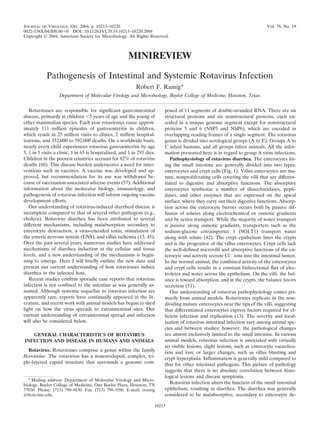
  • 2. 10214 MINIREVIEW J. VIROL. struction (34). In addition to enterocyte destruction, absorp- tion of Na , water, and mucosal disaccharidases are decreased (10, 28), while mucosal cyclic AMP appears not to be altered (16). Malabsorption results in the transit of undigested mono- and disaccharides, carbohydrates, fats, and proteins into the colon. The undigested bolus is osmotically active, and the co- lon is unable to absorb sufficient water, leading to an osmotic diarrhea (27). Another study suggested that the diarrhea was malabsorptive and resulted from epithelial damage caused by villus ischemia (58). A secretory component of the diarrhea was suggested, based on elevated levels of prostaglandin E2 (PGE2) in the infected gut and the stimulation of secretion by PGE2 (79). The fact that gut lesions often do not correlate with the presence of diarrhea stimulated the search for other mech- anisms of diarrhea induction. The viral nonstructural protein NSP4, a secreted fragment of NSP4, or certain NSP4 peptides were found to have toxin-like activity and to induce diarrhea when inoculated into mice (3, 23, 77). The NSP4 enterotoxin activity provides a way to mediate diarrheagenic changes in the absence of significant damage or to mediate changes at unin- fected sites. Recently, it was shown that several drugs that block the action of the ENS attenuate rotavirus-induced secre- tion in the intestine, suggesting a role for the ENS in rotavirus diarrhea (44, 45, 46). It was estimated that 67% of the fluid and electrolyte secretion in rotavirus diarrhea in experiments with mice was due to activation of the ENS (46). Thus, it is clear that rotavirus diarrhea is multifactoral, resulting from the direct effects of virus infection and the indirect effects of in- fection and the host response. Some rotavirus infections are asymptomatic (10), which sug- gests that both viral and host factors can affect disease severity (13). Among the viral factors are the following. (i) Some alleles of VP4 may be associated with asymptomatic disease (24). (ii) Virus strains can be attenuated, particularly by passage in cell culture. Attenuation generally results in a restricted ability to replicate and cause disease in the host (32). (iii) Virus strains plasmic reticulum (blue), increasing [Ca2 ]i. (ii) A cell is secondarily infected after virus release from the initial cell. NSP4 produced by the infection disrupts the tight junctions, allowing paracellular flow of water and electrolytes (green arrow). (iii) NSP4 binds to a specific receptor on a cell and triggers a signaling cascade through PLC and IP3 that results in release of Ca2 and an increase in [Ca2 ]i. Intracellular expression of NSP4 does not stimulate PLC. The increase in [Ca2 ]i acts to disrupt the microvillar cytoskeleton. (iv) The brown cell repre- sents a crypt cell. It can be acted on directly by NSP4, or NSP4 can stimulate the ENS, which in turn signals an increase in [Ca2 ]i that induces Cl secretion. Panel B shows the normal architecture of the small intestine, with the circulatory system removed for clarity. This panel emphasizes the ENS and its ganglia in the different submucosal levels. (Panel B is adapted from reference 25, with the permission of M. D. Gershon and the publisher.) Panel C shows a reflex arc in the ENS that can receive signals from the villus epithelium and activate the crypt epithelium. (Adapted from reference 44, with the permission of FIG. 1. Model of rotavirus-induced diarrhea. Panel A depicts L. Svensson and the publisher.) Inset 1 shows a whole mount of an events in the infected epithelium. For clarity, not all events are shown adult mouse small intestinal villus, stained with antibody to protein in each cell. The following processes are shown in order from left to gene product 9.5 to reveal the rich innervation (yellow stain). (Inset 1 right across the four cells. (i) The initial cell is infected by luminal is used by courtesy of R. O. Heuckeroth and with permission.) Inset 2 virus, with virus entry and uncoating, formation of a viroplasm (Vi), shows that infected villus enterocytes may stimulate the ENS by the and the release of virus and viral proteins. NSP4 (red triangles) may be basolateral release of NSP4 or other effector molecules. (Inset 2 is released via a nonclassical secretory pathway. Intracellular NSP4 also adapted from reference 45 with permission of L. Svensson and the induces release of Ca2 from the internal stores, primarily the endo- publisher.)
  • 3. VOL. 78, 2004 MINIREVIEW 10215 seem to be adapted for growth in particular host species (host triggers a phospholipase C–inositol 1,3,5-triphosphate (PLC- range) (5). Several host factors have also been shown to affect IP3) cascade that culminates in the release of Ca2 from the the severity of rotavirus disease, including the following. (i) endoplasmic reticulum, increasing [Ca2 ]i. If NSP4 acts on Malnutrition is documented to increase the severity of rotavi- enterocytes, one of the results is the disruption of tight junc- rus diarrhea (71), where it delays small intestinal recovery (78) tions, resulting in paracellular permeability. If NSP4 acts on and modifies intestinal inflammatory responses (79). (ii) Ro- crypt cells, the resulting increase in [Ca2 ]i leads to secretion tavirus symptomatic infections are generally age restricted in the crypt, mediated by activation of a Cl transporter, re- (13). The age dependence appears to be unrelated to receptor sulting in an increased secretory component of the diarrhea. expression, as both the viral and the NSP4 receptors are ex- Secreted NSP4, or other effector molecules released from in- pressed in adult animals. However, signaling downstream of fected cells, may also stimulate the ENS (Fig. 1C). Indeed, the NSP4 receptor does appear to be age dependent (53), but experiments with agents that block function of the ENS age restriction may be related to immunity, as neutralizing showed that rotavirus infection induced secretion via stimula- antibodies increase with age and virus exposure. (iii) Rotavirus tion of the ENS. This information begins to shed light on the disease may be related to age-dependent protease expression, mechanism(s) by which relatively few infected cells, causing as viral infectivity requires protease cleavage of VP4 and new- little visible damage to the mucosa, can elicit a diarrheal re- borns have low levels of protease in the gut (29). (iv) The sponse. expression of intestinal mucins and the rate of epithelial cell replacement and fluid absorption are both age dependent and THE MOLECULAR BASIS OF DIARRHEA INDUCTION have been shown to affect rotavirus infection and disease in the host (51, 76). Rotavirus diarrhea is multifactoral, has malabsorption and secretion components, and may have other components sug- THE CURRENT MODEL OF DIARRHEA INDUCTION gested to be related to villus ischemia and intestinal motility. BY ROTAVIRUS Here I present the relevant data on the induction of each of these components (references are selective and for illustrative Many of the salient events in rotavirus induction of diarrhea purposes). are shown in Fig. 1. Most of the data used to construct this Malabsorption. A malabsorptive component of rotavirus di- figure were derived from model animal and cell culture sys- arrhea appears to be related to the primary infection with the tems, although some items were demonstrated in humans as virus. Infection of villus enterocytes leads to a cascade of well. Numerous recent reviews have dealt with specific aspects events involving Ca2 (Fig. 1A). This disruption of Ca2 ho- of diarrhea induction (21–23, 43–45, 50, 52, 67). From these meostasis appears to be mediated by synthesis of viral proteins reviews and the discussion above, it is clear that a multitude of (17, 49). Increased Ca2 permeability at both the plasma mem- viral and host factors have the potential to influence rotavirus brane and the endoplasmic reticulum leads to an increase in infection and disease production. Below, I have assembled [Ca2 ]i, triggering a chain of events that leads to cell lysis (61). what we currently understand about the interplay of these The fact that NSP4 expressed in cells also leads to increases in factors in the induction of rotavirus diarrhea. [Ca2 ]i implicates it as the mediator of virus-induced [Ca2 ]i The process leading to diarrhea is initiated when rotavirus dysregulation (74). A fragment of NSP4 (amino acids 112 to binds to and infects enterocytes in the small intestine (Fig. 1A). 175) is secreted via a nonclassical pathway early after infection Binding is mediated by sequential interaction with a series of (77), and this fragment added exogenously to cells also causes sialic acid-containing and nonsialylated receptor molecules. an increase in [Ca2 ]i (73). The increase in [Ca2 ]i follows The virus is internalized by an unknown mechanism, and the NSP4 binding to a specific apical receptor (22) that triggers a outer capsid is lost, activating the virion-associated transcrip- PLC-IP3 cascade resulting in release of Ca2 from intracellular tase and viral macromolecular syntheses. Viral proteins and stores (19). In contrast, the increase in [Ca2 ]i induced by RNAs concentrate in cytoplasmic structures called viroplasms, intracellular NSP4 is independent of PLC stimulation (73). where RNA replication and packaging take place. Intracellular The NSP4-mediated effects may amplify the diarrheagenic ef- events, probably involving NSP4, cause release of Ca2 from fect of infection in the absence of significant visible tissue the endoplasmic reticulum. The increase in intracellular Ca2 damage. However, the ability of inactivated rotavirus particles concentration ([Ca2 ]i) triggers a number of cellular processes, to induce diarrhea (68) suggests that viral structural proteins including disruption of the microvillar cytoskeletal network, may also play a role in the dysregulation leading to diarrhea. lowered expression of disaccharidases and other enzymes at Rotavirus infection has other effects on enterocytes that may the apical surface, general inhibition of the Na -solute co- contribute to malabsorption (Fig. 1A). Infection leads to an transport systems, and necrosis. NSP4 appears to be released increase in [Na ]i and a decrease in [K ]i, which appear to be specifically by a Ca2 -dependent, nonclassical secretion path- related to increased plasma membrane permeability and not way prior to cell lysis. These events lead to a malabsorption inhibition of the Na /K pump (17). Changes in intracellular component of the diarrhea through reduction in absorptive levels of Na and K could impair electroneutral NaCl ab- capacity of the epithelium, reduced activity of Na -solute co- sorption and Na -linked nutrient absorption, resulting in a loss transporters, and reduction of digestive enzyme expression on of fluid (50). [Na ]i dysregulation may be related to a general the epithelial surface. inhibition of the Na -solute cotransport systems (30). NSP4 The release of NSP4 from infected cells allows paracrine may also be involved, as the NSP4114-135 peptide is a specific effects to occur on uninfected cells (Fig. 1A). NSP4 binds to and noncompetitive inhibitor of SGLT1 (31). Infection also these cells, using specific, unidentified receptor(s) (22), and reduces the expression of digestive enzymes at the apical sur-
  • 4. 10216 MINIREVIEW J. VIROL. face of infected enterocytes. For example, the activities of agents that block ENS stimulation (lidocaine, tetrodotoxin, alkaline phosphatase, lactase, sucrase, and maltase are reduced and mecamylamide) significantly lowered the transmembrane (6, 12, 16). The expression of sucrase and isomaltase in cul- potential difference in a virus dose-dependent manner in tured human intestinal epithelium was also reduced, probably Ussing chamber or organ bath experiments using rotavirus- as a result of perturbation of protein targeting and the mi- infected intestinal tissues. In organ bath experiments, blocking crovillar cytoskeleton (33). Rotavirus infection alters the struc- the ENS could change net secretion to net absorption (45). In ture of polarized enterocytes in a number of ways. The increase live infected animals, repeated administration of lidocaine sig- in [Ca2 ]i induced by rotavirus infection affects the Ca2 - nificantly prevented fluid losses. Thus, it is clear that the ENS sensitive proteins F-actin, villin, and tubulin, damaging the is activated during rotavirus infection, and this activation could microvillar cytoskeleton, whereas rearrangements of other cy- explain how relatively few infected cells at the villus tips could toskeletal proteins (cytokeratin-18) are independent of stimulate crypt cells to secrete electrolytes and water (45). changes in [Ca2 ]i (6, 7). Both rotavirus infection and NSP4 While it is unknown if NSP4 directly stimulates the ENS, the promote functional changes in tight junctions between entero- ENS is known to respond to a number of molecules released cytes that maintain the epithelial barrier (18, 72). The drop in from enterocytes. Cholera toxin induces the release of 5-hy- transepithelial resistance induced by either the virus or NSP4 droxytryptamine (5-HT) from enterochromaffin cells in the suggests that infection can cause paracellular leakage. Rotavi- villus epithelium, and 5-HT is a stimulator of the ENS (44). It rus also induces intestinal epithelial cells to secrete CXC and is possible that secreted NSP4 binds to enterochromaffin cells, CC chemokines, suggesting that enterocyte chemokine secre- inducing a release of 5-HT and stimulation of the ENS (44) tion plays a role in initiating the immune response to infection (Fig. 1C, inset 2). Likewise, the secretion of chemokines and (9, 66, 69). Interleukin-8 (IL-8), GRO- , RANTES, interferon prostaglandins by infected enterocytes may serve to stimulate (IFN)-stimulated protein 10, and granulocyte-macrophage col- the ENS. Currently, the ENS and NSP4 appear to have the ony-stimulating factor (GM-CSF) are stimulated, whereas major roles in the secretory response to infection. other chemokines (tumor necrosis factor alpha, IL-1 , IFN- , Villus ischemia. Although damage to the intestinal epithe- IFN- , MIP- , MCP-1, and IL-6) are unchanged. Induction of lium is minimal in rotavirus-infected mice, villus ischemia was IL-8 and RANTES is noteworthy because these are the most observed in some studies (58, 70). It was proposed that diar- potent chemoattractants for intestinal intraepithelial lympho- rhea could result from virus-induced release of an unknown cytes (20). It is unclear if virus replication is required for vasoactive agent from infected epithelium, causing a local vil- induction of chemokine secretion (9, 66). In addition, the lev- lus ischemia and subsequent functional damage to enterocytes els of PGE2 are increased in infected intestine (79). The che- (59). However, villus ischemia has not been observed in other mokines may activate the immune response rather than di- animal models, so the significance of this observation remains rectly contribute to diarrhea. Thus, rotavirus infection causes a unknown. number of changes in the villus epithelium that contribute to Intestinal motility. In some diarrheal infections, intestinal malabsorption. motility is significantly increased. The intestinal transit time is Secretion. The secretory component of rotavirus diarrhea decreased in rotavirus infection, implying increased motility appears to be secondary to virus-induced functional changes at (50). The ENS generally controls motility, but the molecular the villus epithelium. The central players in secretion appear to stimulator of motility is not known. It could be any of the ENS be NSP4 and the ENS. The precise role and targets of secreted stimulators discussed above. NSP4 are unknown. NSP4 may simply amplify the effects of To summarize, rotavirus diarrhea is clearly a multicompo- infection in the enterocyte epithelium. However, NSP4 may nent disease. Good evidence exists for malabsorptive and se- also act at the crypt epithelium (Fig. 1A), where it would cretory components. The mediators of these disease compo- induce increases in crypt cell [Ca2 ]i, activate Cl secretion, nents range from primary cellular damage to a secreted viral and lead to an outflow of water. This Cl secretion is known to enterotoxic peptide and a virus-induced interaction with the be unrelated to the cAMP-dependent Cl channel of crypt ENS. cells because CFTR-knockout mice are susceptible to rotavirus or NSP4-induced diarrhea (2, 53). The identity of the Cl SYSTEMIC INFECTION WITH ROTAVIRUS channel involved in rotavirus diarrhea in CFTR-knockout mice remains unknown. It is proposed that NSP4 itself may form a L. M. Kraft performed some of the early studies of rotavirus channel or that NSP4 activates a dormant Ca2 -activated an- infection, using epidemic diarrhea of infant mice virus (EDIM) ion channel (53). Interestingly, these studies also showed that before it was recognized as a rotavirus (1). An important but the age dependence of rotavirus diarrhea is not due to age- under-appreciated aspect of this work is that rotavirus spread dependent expression of a NSP4 receptor or age-dependent throughout the bodies of the infected mice following oral in- Ca2 mobilization but rather to the age dependence of Cl fection (39, 40, 41). By 72 h postinfection, infectious EDIM permeability (53). Another possible target of secreted NSP4 is was found in the lungs, liver, spleen, kidney, bladder, brain, the ENS, which is also a target in classic cholera toxin-induced and blood. Since most of the organs where EDIM was detected diarrhea (45). Indeed, the ENS is rich immediately under the are highly vascular, it was generally assumed that virus in the villus epithelium, and it is situated to receive stimuli from the tissues reflected the presence of blood. rotavirus-damaged epithelium (Fig. 1C, inset 1). Although Subsequently, a large number of clinical case reports sug- NSP4 stimulation of the ENS has not been shown experimen- gested that rotaviruses could be found at extraintestinal sites tally, it has been shown that the ENS is involved in rotavirus following infection. It was not clear if the virus was replicating diarrhea (46). The application of a number of pharmacologic and infectious at these sites or passively present in the blood or
  • 5. VOL. 78, 2004 MINIREVIEW 10217 if it resulted from contamination during sample collection. replicate to spread, but it need not replicate to high titer to Examples include the finding of virus in the liver following fatal spread to the liver. A follow-up study showed that virus es- disease (8), the finding of elevated liver enzymes associated caped the intestine via a lymphatic route, appearing sequen- with virus infection (38), and the demonstration of viral repli- tially in the Peyer’s patch, the mesenteric lymph node, and, cation in the liver and kidneys of an immunodeficient child finally, the peripheral tissues (55). These studies also showed (26). Rotavirus involvement in biliary atresia was suggested, that genome segment 6, encoding VP6, was a secondary de- although the best data involve group C rotaviruses (65). Neu- terminant of spread. The combination of segment 6 from a rological involvement was suggested by several reports of chil- spreading virus with segment 7 from a nonspreading virus dren with concomitant convulsions and rotavirus diarrhea (for allowed escape from the intestine but only to the mesenteric examples, see references 35 and 56). Whether putative central lymph node and no farther (55). The association of a nonstruc- nervous system (CNS) infection results from contamination is tural protein, NSP3, with escape of virus from the intestine not known, but one study strongly suggested infection of the suggested that virus transits in infected cells. Indeed, prelimi- CNS (48). In recent studies, rotavirus antigens were detected nary experiments indicate that virus in the mesenteric lymph in myocardium from patients who died unexpectedly (11), and node is internalized by some cells of the lymphocytic or myloid in vitro studies demonstrated the ability of rotavirus to repli- lineage (E. C. Mossel and R. F. Ramig, unpublished data). cate in primary islet cells, an observation that correlates with NSP3 is described as a protein that functions in the regulation the temporal association of infection with development of pan- of protein synthesis by binding to a specific sequence at the 3 creatic islet autoantibodies (14). These case reports strongly termini of the nonpolyadenylated viral mRNAs and interacts indicate that rotavirus infection may have rare systemic sequel- with the translation initiation complex to favor viral protein lae, although one must bear in mind that infectious viruses synthesis in the infected cell (62). How NSP3 functions as the were not isolated and replication was shown only once (26). primary determinant of spread to the liver is not understood. In animal models, rotaviruses have also been documented to Potential role for viremia in rotavirus spread and patho- spread beyond the intestine after oral infection. In the mouse genesis. The numerous clinical reports of rotavirus at systemic model, sites of spread include the lamina propria, Peyer’s sites and work with mice that showed systemic spread patches, mesenteric lymph nodes, lung, liver, kidney, and bile prompted a systematic search for evidence of widespread ro- duct (13). Group A rotaviruses have been shown to induce tavirus viremia in children and animal model systems (4). In a biliary atresia in mice, but this system requires intraperitoneal, retrospective study, 22 of 33 (66%) serum samples from rota- not oral, inoculation of virus (15). The most detailed studies of virus-infected children were positive for rotavirus antigen by extraintestinal spread in the mouse examined spread to the enzyme immunoassay (EIA), whereas 0 of 35 rotavirus-nega- liver following oral inoculation. tive children had antigens in their sera. In a subset of six The liver as a site of systemic infection. Spread of virus in children with paired acute and convalescent-phase sera, viral orally infected mice was shown in persistently infected SCID antigen was present in all acute-phase sera and none of the mice where a diffuse hepatitis was noted. Infectious virus was convalescent-phase sera, indicating that the antigenemia is isolated from the liver, and surviving mice developed chronic transient (4). Only three of six serum samples from rotavirus- liver disease (64, 75). Infectious virus was also found in the infected children were positive by PCR, the lower positivity livers of normal mice, although the hepatitis was less severe probably reflecting the lower sensitivity of the PCR assay used and resolved spontaneously (75). Spread of virus to the liver (4). In another study, three children who died of rotavirus- and development of hepatitis were described as virus strain associated disease were examined retrospectively. Two of the dependent. These results were supported by studies of cultured three children had rotavirus RNA in extraintestinal tissues human hepatoma (HepG2) cells, where some viruses were (spleen, heart, lung, kidney, testes, bladder, adrenal gland, and capable of complete infectious cycles and others were not (37, pancreas), as determined by reverse transcription-PCR. Con- 63). The strain-specific ability to infect HepG2 cells segregated firmation of positive PCR products by hybridization with se- with viral genome segment 4, encoding the outer capsid spike rotype-specific probes indicated that the two children with protein VP4 (36, 63). Subsequent studies indicated that viruses extraintestinal rotavirus were infected with different G sero- unable to replicate in HepG2 cells entered less efficiently, but types (47). These findings show that viral antigen and/or virus the absolute block to infection occurred at a late step in the particles enter the circulation. intracellular replication cycle (S. Jafar and R. F. Ramig, un- Prospective studies with animal models confirmed the anti- published data). genemia seen in humans and also demonstrated viremia. Ro- Recent studies utilized the strain difference in extraintestinal tavirus antigen could be detected by EIA in 100% of the sera spread to the liver as the basis for a genetic approach to from infant and adult mice, rats, rabbits, and calves that were identifying viral determinants of the spread phenotype. A col- experimentally infected (4). Three of three infected infant lection of reassortants made from virus strains RRV (spread mouse serum samples yielded infectious virus when assayed by competent) and SA11-Cl4 (spread incompetent) were inocu- oral inoculation of naive mice with sera, and 9 of 11 sera from lated orally into suckling mice, and the presence of infectious adult mice yielded infectious virus in the same assay (4). Stud- virus in the liver was used as a proxy for extraintestinal spread. ies were subsequently carried out with infant rats infected with A statistical analysis of the data indicated that genome seg- the heterologous virus strains rhesus RRV and human ment 7, encoding the nonstructural protein NSP3, was signif- HAL1166 (S. E. Crawford, D. G. Patel, E. Cheng, Z. Berkova, icantly linked to virus spread to the liver (54). However, rep- J. M. Hyser, A. A. McKinstry, and M. K. Estes, Abstr. 8th lication of virus to high titer in the intestine was not International Symposium on Double-Stranded RNA Viruses, significantly linked to the ability to spread. Thus, a virus must abstr. W6.4, 2003). In these studies, the sacrificed animals were
  • 6. 10218 MINIREVIEW J. VIROL. TABLE 1. Rotavirus antigenemia and viremia in various hosts about 25% of mice experience liver infection after oral infec- % Infected animals with: tion with the same virus strain (54, 55). Host Serum Serum antigenemia infectivity CLOSING COMMENTS Childrena 66 NTd Rotaviruses naturally infect the enteric tract and cause di- Mice (infant)a 100 100 arrheal disease in children and young animals. The pathophys- Mice (adult)a 100 NTd iology of rotavirus diarrhea is clearly multifactoral. There is a Rat (infant)a,b 100 100 Rabbit (adult)a 100 NTd malabsorptive component of the diarrhea that seems related to Bovine (adult)a 100 NTd primary damage to intestinal epithelium by virus infection and Pig (infant)c 100 100 the action of a secreted viral enterotoxin (NSP4). The effects a The children had natural infections, the mice were challenged with homol- observed on the epithelium can be traced primarily to dysregu- ogous or heterologous virus, the rats were challenged with heterologous virus, lation of Ca2 in the epithelium. A secretory component of the rabbits were challenged with homologous virus, and the bovines were natu- rotavirus diarrhea appears to result from stimulation of the rally infected. b Identical results were obtained with rhesus or human challenge virus. ENS. The mode of activation of the ENS is not clear, but it c Challenged with virulent human virus (Saif et al., 8th International Sympo- may be through secreted NSP4 or the chemokines and other sium on Double-Stranded RNA Viruses). d NT, not tested. factors released from infected epithelial cells. The secretory component of the diarrhea appears to result from Ca2 dys- regulation of the secretory crypt cells. We have learned a great perfused with buffer to wash blood out of the tissues. Rotavirus deal of the molecular biology of rotavirus diarrheal disease antigen was detected in small intestine, stomach, liver, kidney, components, but a great deal of work remains to be done and spleen in some animals after perfusion, suggesting that the before rotavirus diarrhea induction is completely understood. virus present was specifically associated with those tissues and In the past several years, interest in rotavirus spread to not passively present in the circulation. Infectious virus could extraintestinal tissues has been renewed. It appears that some be detected in 100% of sera from infected rat pups by fluores- of this spread depends on viral factors, but other components cent focus assay on MA104 cells, at titers up to 103 focus- of the spread may be related to viremic hosts. It remains forming units/ml (Crawford et al., 8th International Symposium unclear if viremia is, in any way, related to diarrheal disease. It on Double-Stranded RNA Viruses). Finally, in a study with is important to determine if systemic infection with rotavirus is pigs, no viremia was detected in gnotobiotic piglets infected responsible for, or plays a role in, clinical syndromes not cur- with attenuated human rotavirus Wa, but viremia was detected rently associated with rotavirus. in 100% of gnotobiotic piglets infected with virulent human The studies reviewed here indicate that we have made sig- strain Wa. Inoculating additional gnotobiotic piglets with the nificant progress in understanding the molecular basis for ro- viremic sera and observing diarrhea confirmed the presence of tavirus intestinal pathogenesis and spread to peripheral sites. infectious virus in the serum (L. J. Saif, M. S. P. Azevedo, L. However, there remain a large number of questions to be Yuan, K. I. Nguyen, S. M. Pouly, and M. Gochnauer, Abstr. 8th answered. For example, we need to know if NSP4 is the key International Symposium on Double-Stranded RNA Viruses, viral protein or if other viral structural and/or nonstructural abstr. W6.3, 2003). It is interesting that viremia was not ob- proteins play a role in induction of diarrhea. How is a NSP4 served routinely in the studies of rotavirus intestinal escape to fragment specifically secreted from infected cells early in in- the liver (55). In these studies, infectious virus was detected in fection? How is the ENS activated by rotavirus infection, and the blood of only 2 of 60 (3%) animals infected with strain what are the roles of NSP4, chemokines, or other stimulatory RRV, the strain that induced antigenemia in 100% of mice (4) molecules released by infected cells? Do rotavirus strains with and viremia in 100% of rats (Crawford et al., 8th International NSP4 polymorphisms differ in activation of the ENS? What is Symposium on Double-Stranded RNA Viruses). The reason the nature of the age-dependent, Ca2 -regulated Cl channel for this difference is unclear, although it may indicate that virus activated by infection? Does the host innate or adaptive im- moves from the intestine to the liver by a mechanism that does mune response play any role in the development of intestinal not involve viremia. disease or systemic spread? How does a viral nonstructural The results presented here indicate that antigenemia is a protein known to regulate translation play a central role in the nearly universal event in children and animal models (Table 1). spread of virus from the intestine to the liver? How does In animals, antigenemia was accompanied by a transient vire- rotavirus gain access to the circulation, and does it leave cir- mia, as demonstrated by the presence of infectious virus in the culation to cause clinically significant infections at systemic sera of virtually all animals tested. While viremia is not yet sites? Finally, it will be important to determine if viremia proven in children for lack of demonstration of infectious virus and/or systemic spread contribute to pathogenesis in the aver- in the serum, it seems likely that viremia also occurs in chil- age rotavirus infection where extraintestinal sequellae are not dren. This viremia may provide a mechanism for seeding pe- seen. Answers to these and other questions are exciting to ripheral tissues with virus during rotavirus infection and may anticipate and will increase our understanding of this ubiqui- account for the reports of systemic sequellae associated with tous disease of childhood. rotavirus infection. However, these data also suggest that sys- temic sequellae to viremia are rare, since, for example, 100% ACKNOWLEDGMENTS of mice and rats are viremic (4; Crawford et al., 8th Interna- I thank Robert O. Heuckeroth for allowing use of his unpublished tional Symposium on Double-Stranded RNA Viruses) but only work (Fig. 1C, inset 1) and M. D. Gershon (Fig. 1B) and L. Svensson
  • 7. VOL. 78, 2004 MINIREVIEW 10219 (Fig. 1C and inset 2) for use of their published figures. I also thank my 24. Flores, J., K. Midthun, Y. Hoshino, K. Green, M. Gorziglia, A. Z. Kapikian, colleagues at Baylor College of Medicine, S. Crawford, S. Blutt, M. and R. M. Chanock. 1986. Conservation of the fourth gene among rotavi- Conner, M. Estes, E. Mossel, and B. V. V. Prasad, for helpful discus- ruses recovered from asymptomatic newborn infants and its possible role in sions and for critically reviewing the manuscript. attenuation. J. Virol. 60:972–979. 25. Gershon, M. D. 1999. The enteric nervous system: a second brain. Hosp. The work from the author’s laboratory was supported by National Pract. 34:31–39. Institutes of Health grants RO1-AI16687 and T32-AI07471. 26. Gilger, M. A., D. O. Matson, M. E. Conner, H. M. Rosenblatt, M. J. Fine- gold, and M. K. Estes. 1992. Extraintestinal rotavirus infections in children REFERENCES with immunodeficiency. J. Pediatr. 120:912–917. 1. Adams, W. R., and L. M. Kraft. 1963. Epizootic diarrheas of infant mice: 27. Graham, D. Y. and M. K. Estes. 1988. Viral infections of the intestine, p. identification of the etiologic agent. Science 141:359–360. 566–578. In G. Gitnick (ed.), Gastroenterology. Medical Examination Pub- 2. Angel, J., B. Tang, N. Feng, H. B. Greenberg, and D. Bass. 1998. Studies of lishing Company, New Hyde Park, N.Y. the role for NSP4 in the pathogenesis of homologous murine rotavirus 28. Graham, D. Y., J. W. Sackman, and M. K. Estes. 1984. Pathogenesis of diarrhea. J. Infect. Dis. 177:455–458. rotavirus-induced diarrhea: preliminary studies in miniature swine piglet. 3. Ball, J. M., P. Tian, C. Q-Y. Zeng, A. P. Morris, and M. K. Estes. 1996. Dig. Dis. Sci. 29:1028–1035. Age-dependent diarrhea induced by a rotavirus nonstructural glycoprotein. 29. Greenberg, H. B., H. F. Clark, and P. A. Offit. 1994. Rotavirus pathology and Science 272:101–104. pathophysiology. Curr. Top. Microbiol. Immunol. 185:255–283. 4. Blutt, S. E., C. D. Kirkwood, V. Parreno, K. L. Warfield, M. Ciarlet, M. K. 30. Halaihel, N., V. Lievin, F. Alvarado, and M. Vasseur. 2000. Rotavirus infec- Estes, K. Bok, R. F. Bishop, and M. E. Conner. 2003. Rotavirus antigenae- tion impairs intestinal brush-border membrane Na -solute cotransport ac- mia and viraemia: a common event? Lancet 362:1445–1449. tivities in young rabbits. Am. J. Physiol. Gastrointest. Liver Physiol. 279: 5. Broome, R. L., P. T. Vo, R. L. Ward, H. F. Clark, and H. B. Greenberg. 1993. G587–G596. Murine rotavirus genes encoding outer capsid proteins VP4 and VP7 are not 31. Halaihel, N., V. Lievin, J. M. Ball, M. K. Estes, F. Alvarado, and M. Vasseur. major determinants of host range restriction and virulence. J. Virol. 67:2448– 2000. Direct inhibitory effect of rotavirus NSP4(114–135) peptide on the 2455. Na -D-glucose symporter of rabbit intestinal brush border membrane. J. Vi- 6. Brunet, J. P., J. Cotte-Laffitte, C. Linxe, A. M. Quero, M. Geniteau-Leg- rol. 74:9464–9470. endre, and A. L. Servin. 2000. Rotavirus infection induces an increase in 32. Hall, G. A., J. C. Bridger, K. R. Parsons, and R. Cook. 1993. Variation in intracellular calcium concentration in human intestinal epithelial cells: role rotavirurus virulence: a comparison of pathogenesis in calves between two in microvillar actin alteration. J. Virol. 74:2323–2332. rotaviruses of different virulence. Vet. Pathol. 30:223–233. 7. Brunet, J. P., N. Jourdan, J. Cotte-Laffitte, C. Linxe, M. Geniteau-Legendre, 33. Jourdan, N., J. P. Brunet, C. Sapin, A. Blais, J. Cotte-Laffitte, F. Forestier, A. L. Servin, and A. M. Quero. 2000. Rotavirus infection induces cytoskel- A. M. Quero, G. Trugnan, and A. L. Servin. 1998. Rotavirus infection eton disorganization in human intestinal epithelial cells: implication of an reduces sucrase-isomaltase expression in human intestinal epithelial cells by increase in intracellular calcium concentration. J. Virol. 74:10801–10806. perturbing protein targeting and organization of microvillar cytoskeleton. 8. Carlson, J. A., P. J. Middleton, M. T. Szymanski, J. Huber, and M. Petric. J. Virol. 72:7228–7236. 1978. Fatal rotavirus gastroenteritis: an analysis of 21 cases. Am. J. Dis. 34. Kapikian, A. Z., Y. Hoshino, and R. M. Chanock. 2001. Rotaviruses, p. Child. 132:477–479. 1787–1833. In D. M. Knipe, P. M. Howley, D. E. Griffin, R. A. Lamb, M. A. 9. Casola, A., M. K. Estes, S. E. Crawford, P. L. Ogra, P. B. Ernst, R. P. Martin, B. Roizman, and S. E. Straus, Fields virology, 4th ed. Lippincott Garfalo, and S. E. Crowe. 1998. Rotavirus infection of cultured intestinal Williams & Wilkins, Philadelphia, Pa. epithelial cells induces secretion of CXC and CC chemokines. Gastroenter- 35. Keidan, I., I. Shif, G. Keren, and J. H. Passwell. 1992. Rotavirus encepha- ology 114:947–955. lopathy: evidence of central nervous system involvement during rotavirus 10. Chrystie, I. L., B. M. Totterdell, and J. E. Banatvala. 1978. Asymptomatic infection. Pediatr. Infect. Dis. J. 11:773–775. endemic rotavirus infections of the newborn. Lancet i:1176–1178. 36. Kitamoto, N., N. M. Mattion, and M. K. Estes. 1993. Alterations in the 11. Cioc, A. M., and G. J. Nuovo. 2002. Histologic and in situ viral findings in the sequence of the gene 4 from a human rotavirus after multiple passages in myocardium in cases of sudden unexpected death. Mod. Pathol. 9:914–922. HepG2 cells. Arch. Virol. 130:179–185. 12. Collins, J., W. G. Starkey, T. S. Wallis, G. J. Clarke, K. J. Worton, A. J. 37. Kitamoto, N., R. F. Ramig, D. O. Matson, and M. K. Estes. 1991. Compar- Spencer, S. J. Haddon, M. P. Osborne, C. D. Candy, and J. Stephen. 1988. ative growth of different rotavirus strains in differentiated cells (MA104, Intestinal enzyme profiles in normal and rotavirus-infected mice. J. Pediatr. HepG2, and CaCo-2). Virology 184:729–737. Gastroenterol. Nutr. 7:264–272. 38. Kovacs, A., L. Chan, C. Hotrakitya, G. Overturf, and B. Portnoy. 1986. 13. Conner, M. E., and R. F. Ramig. 1997. Viral enteric diseases, p. 713–743. In Serum transaminase elevations in infants with rotavirus gastroenteritis. J. Pe- N. Nathanson (ed.), Viral pathogenesis. Lippincott-Raven Publishers, Phil- diatr. Gastroenterol. Nutr. 5:873–877. adelphia, Pa. 39. Kraft, L. M. 1958. Observations on the control and natural history of epi- 14. Coulson, B. S., P. D. Witterick, Y. Tan, M. J. Hewish, J. N. Mountford, L. C. demic diarrhea of infant mice (EDIM). Yale J. Biol. Med. 31:121–137. Harrison, and M. C. Honeyman. 2002. Growth of rotaviruses in primary 40. Kraft, L. M. 1962. Two viruses causing diarrhea in infant mice, p.115–127. In pancreatic cells. J. Virol. 76:9537–9544. R. J. C. Harris (ed.), The problems of laboratory animal disease. Academic 15. Czech-Schmidt, G., W. Verhagen, P. Szavay, J. Leonhardt, and C. Petersen. Press, New York, N.Y. 2001. Immunological gap in the infectious animal model for biliary atresia. 41. Kraft, L. M. 1982. Viral diseases of the digestive system, p. 159–191. In H. L. J. Surg. Res. 101:62–67. Foster, J. G. Fox and D. J. Small (ed.), The mouse in biomedical research, 16. Davidson, G. P., D. G. Gall, M. Petric, D. G. Butler, and J. R. Hamilton. vol. 2. Academic Press, New York, N.Y. 1977. Human rotavirus enteritis induced in conventional piglets: intestinal 42. Loo, D. D. F., E. M. Wright, and T. Zeuthen. 2002. Water pumps. J. Physiol. structure and transport. J. Clin. Investig. 60:1402–1409. 542:53–60. 17. del Castillo, J. R., J. E. Ludert, A. Sanchez, M. C. Ruiz, F. Michelangeli, and 43. Lopez, S., and C. F. Arias. 2003. Attachment and post-attachment receptors F. Liprandi. 1991. Rotavirus infection alters Na and K homeostasis in for rotavirus, p. 143–163. In U. Desselberger and J. Gray (ed.), Viral gas- MA104 cells. J. Gen. Virol. 72:541–547. troenteritis. Elsevier Science BV, Amsterdam, The Netherlands. 18. Dickman, K. G., S. J. Mempson, J. Anderson, S. Lippe, L. Shao, and R. D. 44. Lundgren, O., and L. Svensson. 2001. Pathogenesis of rotavirus diarrhea. Shaw. 2000. Rotavirus alters paracellular permeability and energy metabo- Microbes Infect. 3:1145–1156. lism in Caco-2 cells. Am. J. Physiol. 279:G757–G766. 45. Lundgren, O., and L. Svensson. 2003. The enteric nervous system and in- 19. Dong, Y., C. Q. Zeng, J. M. Ball, M. K. Este, and A. P. Morris. 1997. The fectious diarrhea, p. 51–68. In U. Desselberger and J. Gray (ed.), Viral rotavirus enterotoxin NSP4 mobilizes intracellular calcium in human intes- gastroenteritis. Elsevier Science BV, Amsterdam, The Netherlands. tinal cells by stimulating phospholipase C-mediated inositol 1,4,5-trisphos- 46. Lundgren, O., A. Timar-Peregrin, K. Persson, S. Kordasti, I. Uhnoo, and L. phate production. Proc. Natl. Acad. Sci. USA 94:3960–3965. Svensson. 2000. Role of the enteric nervous system in the fluid and electro- 20. Ebert, E. 1995. Human intestinal intraepithelial lymphocyres have potent lyte secretion of rotavirus diarrhea. Science 287:491–495. chemotactic activity. Gastroenterology 109:1154–1159. 47. Lynch, M., W.-J. Sheih, K. Tatti, J. R. Gentsch, T. Ferebee-Harris, B. Jiang, 21. Estes, M. K., G. Kang, C. Q. Zeng, S. E. Crawford, and M. Ciarlet. 2001. J. Guarner, J. S. Bresee, M. Greenwald, S. Cullen, H. D. Davies, C. Trevens, Pathogenesis of rotavirus gastroenteritis, p. 82–96. In D. J. Chadwick and S. R. Zaki, and R. I. Glass. 2003. The pathology of rotavirus-associated J. A. Goode, (ed.), Gastroenteritis viruses. Novartis Foundation Symposium deaths using new molecular diagnostics. Clin. Infect. Dis. 37:1327–1333. 238. John Wiley & Sons, Inc., New York, N.Y. 48. Lynch, M., B. Lee, P. Azimi, J. Gentsch, C. Glaser, S. Gilliam, H. G-H. 22. Estes, M. K. 2003. The rotavirus NSP4 enterotoxin: current status and Chang, R. Ward, and R. I. Glass. 2001. Rotavirus and central nervous system challenges, p. 207–224. In U. Desselberger and J. Gray (ed.), Viral gastro- symptoms: cause or contaminant? Case reports and review. Clin. Infect. Dis. enteritis. Elsevier Science BV, Amsterdam, The Netherlands. 33:932–938. 23. Estes, M. K. and A. P. Morris. 1999. A viral enterotoxin: a new mechanism 49. Michelangeli, F., M. C. Ruiz, J. R. del Castillo, J. E. Ludert, and F. Liprandi. of virus induced pathogenesis, p. 73–82. In P. S. Paul and D. H. Francis (ed.), 1991. Effect of rotavirus infection on intracellular calcium homeostasis in Mechanisms in the pathogenesis of enteric diseases 2. Kluwer Academic/ cultured cells. Virology 181:520–527. Plenum Publishers, New York, N.Y. 50. Michelangeli, F., and M. C. Ruiz. 2003. Physiology and pathophysiology of
  • 8. 10220 MINIREVIEW J. VIROL. the gut in relation to viral diarrhea, p. 23–50. In U. Desselberger and J. Gray 65. Riepenhoff-Talty, M., V. Gouvea, M. J. Evans, L. Svensson, E. Hoffenberg, (ed.), Viral gastroenteritis. Elsevier Science BV, Amsterdam, The Nether- R. J. Sokol, I. Uhnoo, S. J. Greenberg, K. Schakel, G. Zhaori, J. Fitzgerald, lands. S. Chong, M. El-Yousef, A. Nemeth, M. Brown, D. Piccoli, J. Hyans, D. 51. Moon, H. W. 1994. Pathophysiology of viral diarrhea, p. 27–52. In A. Z. Ruffin, and T. Rossi. 1996. Detection of group C rotavirus in infants with Kapikian (ed.), Viral infections of the gastrointestinal tract. Marcel Dekker, extrahepatic biliary atresia. J. Infect. Dis. 174:8–15. Inc., New York. 66. Rollo, E. E., K. P. Kumar, N. C. Reich, J. Cohen, J. Angel, H. B. Greenberg, 52. Morris, A. P., and M. K. Estes. 2001. Microbes and microbial toxins: para- R. Sheth, J. Anderson, B. Oh, S. J. Hempson, E. R. Mackow, and R. D. Shaw. digms for microbial-mucosal interactions. VIII. Pathological consequences 1999. The epithelial cell response to rotavirus infection. J. Immunol. 163: of rotavirus infection and its enterotoxin. Am. J. Physiol. Gastrointest. Liver 4442–4452. Physiol. 281:G303–G310. 67. Ruiz, M., J. Cohen, and F. Michelangeli. 2000. Role of Ca2 in the replica- 53. Morris, A. P., J. K. Scott, J. M. Ball, C. Q. Zeng, W. K. O’Neal, and M. K. tion and pathogenesis of rotavirus and other viral infections. Cell Calcium Estes. 1999. NSP4 elicits age-dependent diarrhea and Ca2 mediated I 28:137–149. influx into intestinal crypts of CF mice. Am. J. Physiol. 277:G431–G444. 68. Shaw, R. D., S. J. Hempson, and E. R. Mackow. 1995. Rotavirus diarrhea is 54. Mossel, E. C., and R. F. Ramig. 2002. Rotavirus genome segment 7 (NSP3) caused by nonreplicating viral particles. J. Virol. 69:5946–5950. is a determinant of extraintestinal spread in the neonatal mouse. J. Virol. 69. Sheth, R., J. Anderson, T. Sato, B. Oh, S. J. Hempson, E. Rollo, E. R. 76:6502–6509. Mackow, and R. D. Shaw. 1996. Rotavirus stimulates IL-8 secretion from 55. Mossel, E. C., and R. F. Ramig. 2003. A lymphatic mechanism of rotavirus cultured epithelial cells. Virology 221:251–259. extraintestinal spread in the neonatal mouse. J. Virol. 77:12352–12356. 70. Starkey, W. G., J. Collins, T. S. Wallis, G. J. Clarke, A. J. Spencer, S. J. 56. Nishimura, S., H. Ushijima, and H. Shiraishi. 1993. Detection of rotavirus Haddon, M. P. Osborne, D. C. Candy, and J. Stephen. 1986. Kinetics, tissue in cerebrospinal fluid and blood of patients with convulsions and gastroen- specificity and pathological changes in murine rotavirus infection of mice. teritis by means of the reverse transcriptions polymerase chain reaction. J. Gen. Virol. 67:2625–2634. Brain Dev. 15:457–459. 71. Steel, R. B., and A. Torres-Medina. 1984. Effects of environmental and 57. Offit, P. A., H. F. Clark, and R. L. Ward. 2003. Current state of development dietary factors on human rotavirus infection in gnotobiotic piglets. Infect. of human rotavirus vaccines, p. 345–356. In U. Desselberger and J. Gray Immun. 43:906–911. (ed.), Viral gastroenteritis. Elsevier Science BV, Amsterdam, The Nether- 72. Tafazoli, F., C. Q. Zeng, M. K. Estes, K.-E. Magnusson, and L. Svensson. lands. 2001. NSP4 enterotoxin of rotavirus induces paracellular leakage in polar- 58. Osborne, M. P., S. J. Haddon, A. J. Spencer, J. Collings, W. G. Starkey, T. S. ized epithelial cells. J. Virol. 75:1540–1546. Wallis, G. J. Clarke, K. J. Worton, D. C. Candy, and J. Stephen. 1988. An 73. Tian, P., M. K. Estes, Y. Hu, J. M. Ball, C. Q-Y. Zeng, and W. P. Schilling. electron microscopic investigation of time-related changes in the intestine of 1995. The rotavirus nonstructural glycoprotein NSP4 mobilizes Ca2 from neonatal mice infected with murine rotavirus. J. Pediat. Gastroenterol. Nutr. the endoplasmic reticulum. J. Virol. 69:5763–5772. 7:236–248. 74. Tian, P., Y. Hu, W. P. Schilling, D. A. Lindsay, J. Eiden, and M. K. Estes. 59. Osborne, M. P., S. J. Haddon, K. J. Worton, A. J. Spencer, W. G. Starkey, D. 1994. The nonstructural glycoprotein of rotavirus affects intracellular cal- Thornber, and J. Stephen. 1991. Rotavirus-induced changes in the micro- cium levels. J. Virol. 68:251–257. circulation of intestinal villi of neonatal mice in relation to the induction and 75. Uhnoo, I., M. Riepenhoff-Talty, T. Dharakul, P. Chegas, J. E. Fisher, H. B. persistence of diarrhea. J. Pediatr. Gastroenterol. Nutr. 12:111–120. Greenberg, and P. L. Ogra. 1990. Extramucosal spread and development of 60. Parashar, U. M., E. G. Hummelman, J. S. Bresee, M. A. Miller, and R. I. hepatitis in immunodeficient and normal mice infected with rhesus rotavirus. Glass. 2003. Global illness and deaths caused by rotavirus disease in chil- J. Virol. 64:361–368. dren. Emerg. Infect. Dis. 9:565–572. 76. Yolken, R. H., L. A. Peterson, S. L. Vondrfecht, E. T. Fouts, K. Midthun, and 61. Perez, J. F., M. E. Chemello, F. Liprandi, M. C. Ruiz, and F. Michelangeli. D. S. Newburg. 1992. Human milk mucin inhibits rotavirus replication and 1998. Oncosis in MA104 cells is induced by rotavirus infection through an prevents experimental gastroenteritis. J. Clin. Investig. 90:1984–1991. increase in intracellular Ca2 concentration. Virology 252:17–27. 77. Zhang, M., C. Q.-Y. Zeng, A. P. Morris, and M. K. Estes. 2000. A functional 62. Poncet, D. 2003. Translation of rotavirus mRNAs in the infected cell, p. NSP4 enterotoxin peptide secreted from rotavirus-infected cells. J. Virol. 185–205. In U. Desselberger and J. Gray (ed.), Viral gastroenteritis. Elsevier 74:11663–11670. Science BV, Amsterdam, The Netherlands. 78. Zijlstra, R. T., S. M. Donovan, J. Odle, H. B. Gelberg, B. W. Petschow, and 63. Ramig, R. F., and K. L. Galle. 1990. Rotavirus genome segment 4 determines H. R. Gaskins. 1997. Protein-energy malnutrition delays small-intestinal viral replication phenotype in cultured liver cells (HepG2). J. Virol. 64:1044– recovery in neonatal pigs infected with rotavirus. J. Nutr. 127:1118–1127. 1049. 79. Zijlstra, R. T., B. A. McCracken, J. Odle, S. M. Donovan, H. B. Gelberg, 64. Riepenhoff-Talty, M., T. Dharakul, E. Kowalski, S. Michalak, and P. L. B. W. Petschow, F. A. Zuckermann, and H. R. Gaskins. 1999. Malnutrition Ogra. 1987. Persistent rotavirus infection in mice with severe combined modified pig small intestinal inflammatory responses to rotavirus. J. Nutr. immunodeficiency. J. Virol. 61:3345–3348. 129:838–843.