1. Literature available on the coronary arteries (CAs)
of the pig (Sus scrofa) is meagre. The pattern of the CAs
in the pig was similar to that of the humans and the right
coronary artery (RCA) was the dominant artery1-3
.
Anatomy & distribution of coronary arteries in pig in comparison
with man
Daisy Sahni, G.D. Kaur, Harjeet & Indar Jit*
Department of Anatomy, Postgraduate Institute of Medical Education & Research, Chandigarh, India
Received April 23, 2007
Background & objectives: The suitability of pig as an animal model for research in coronary artery
disease is well established. As coronary arteries (CAs) of the pig are reportedly closely resemble
those of man. We investigated the CAs of the pig (Sus scrofa) and study differences between the
two, if any.
Methods: The origin and pattern of the coronary arteries were studied in the hearts of 30 fully
grown pigs obtained from a slaughter house in Chandigarh (India). The openings of the CAs were
identified at the commencement of the ascending aorta. The arteries were washed with acetone by
introducing appropriate sized cannulae in their ostia. A 20 per cent solution of cellulose acetate
butyrate (CAB), a plastic material, dissolved in acetone was injected in the CAs. The hearts were
fixed in 10 per cent formalin solution for three to four days after which the CAs and their branches
were dissected.
Results: Both coronary arteries arose from the aortic sinuses below the supravalvular ridge in all
the cases. Sinuatrial nodal artery (SAN) arose from the RCA in 70 per cent and from the circumflex
artery (CX) in 30 per cent of instances. There was RCA dominance in all hearts of the pig. The
atrioventricular nodal artery (AVN) and the posterior interventricular artery (PIV) were branches
of RCA. The coronary arterial circulation in the pig was found to be similar to that in human.
Interpretation & conclusions: By and large the coronary arterial pattern of the pig was similar in
that of the humans. We can conclude that the heart of a pig can be used for experiments but differences
have to be kept in mind.
Key words Comparison - coronary arteries - heart - man - pig
Comparison between the distribution patterns of CAs in
pig and man has not been studied. In India, no
investigation seems to have been conducted on CAs of
the pig. We carried out this study to assess the coronary
Indian J Med Res 127, June 2008, pp 564-570
564
* Late Emeritus Professor, Department of Anatomy, Postgraduate Institute of Medical Education & Research, Chandigarh 160 012, India
2. arterial pattern in the pig (Sus scrofa), and an attempt
was made to compare it with those of our findings in the
human heart4-8
to elucidate the differences, if any, in the
pattern of CAs in the two species.
Material & Methods
The study was conducted in the Department of
Anatomy, Postgraduate Institute of Medical Education
& Research, Chandigarh, on 30 hearts of domesticated
pigs. The hearts were obtained from a slaughter house
in Chandigarh, India. The hearts were intact without
any injury to blood vessels. The animals were fully
grown but their weight, age and sex were not known.
Each heart was washed under running tap water as in
the case of human heart4
. After a gross inspection of
each heart, the openings of the CAs were identified at
the commencement of the ascending aorta. The arteries
were washed with acetone by introducing appropriate
sized cannulae in their ostia. A 20 per cent solution of
cellulose acetate butyrate (CAB), a plastic material
dissolved in acetone was injected in the CAs4
. The hearts
were fixed in 10 per cent formalin solution for three to
four days. Fat and connective tissues surrounding the
CAs and their branches were carefully dissected without
injuring the blood vessels. The interventricular septum
(IVS) was exposed as in human hearts6
, the septal
arteries (perforators) running subendocardially were
dissected. The study protocol was approved by the
Ethics committee of the Institute.
Results
Right coronary artery (RCA)
The RCA arose from the middle of the anterior aortic
sinus below the supravalvular ridge in all specimens.
After origin, it extended to the right behind the pulmonary
trunk and thereafter between the pulmonary trunk and
the right atrium and then in the right anterior
atrioventricular sulcus to reach the acute margin of the
heart, where it turned around to lie in the posterior
atrioventricular sulcus (Fig. 1). The portion of the artery
which was lying in the anterior atrioventricular sulcus
was called the first segment while the part lying in the
posterior atrioventricular sulcus was named the second
segment. About 5 mm proximal to the crux, the artery
turned downwards and to the left describing a curve (Fig.
2, see arrow), the convexity of which was directed
upwards and to the left. This curve was present in all the
hearts. Beyond the curve the artery extended in the
posterior interventricular sulcus, where it constituted the
third segment. Extending in that for three-fourths of the
distance between the crux and the apex, it divided into
two branches the right and the left. The right branch
extended on the posterior surface of the right ventricle
while the left branch either continued in the posterior
interventricular sulcus or on the posterior surface of the
left ventricle (Fig. 2). There was no visible surface
anastomosis between the terminal branches of this artery
and the ventricular branches of the left coronary artery
(LCA).
Branches from the first segment
Sinuatrial nodal artery (SAN): In 21 (70%) hearts the
SAN artery arose as the first branch from its right side
at a mean distance of 20 mm distal to the origin of the
RCA (Fig.1). It extended upwards and to the left on the
surface of the right atrium to reach the S.A. node
between the right atrial appendage (RAA) and the
superior vena cava (SVC).
Atrial branches: One to three atrial branches arose from
the right side of the RCA and extended upwards and to
the right on the anterior surface of the right atrium. They
were very small branches. There was no visible
anastomosis between them.
Right conus artery (RC): It was the first but a small
branch which arose from the left side of the RCA and
extended to the left on the anterior surface of the right
ventricle. It did not show any anastomosis with a similar
left coronary branch. Hence, there was no anastomotic
circle around the pulmonary trunk (‘annulus of
Vieussens’).There was no third coronary artery arising
directly from the aorta.
Ventricular branches (VB) : Three to four ventricular
branches arose from the left side of the RCA and passed
to the anterior surface of the right ventricle; they
descended obliquely downwards and to the left towards
the incisura (Fig. 3). There were no gross anastomosis
seen between them and the branches of the anterior
interventricular artery (AIV).
Acute marginal branch (AM): On reaching the acute
margin of the heart, the RCA gave a small marginal
branch which extended to the left towards the apex
(Fig. 3). It did not show any anastomosis with the
branches of the AIV.
No specimen showed the middle or a posterior
border branches.
Branches from the second segment
Atrial branches: Usually, there were two to three atrial
branches which extended upwards from the second
segment to the posterior surface of the right atrium.
SAHNI et al: CORONARY ARTERIES IN PIG & MEN 565
3. 566 INDIAN J MED RES, JUNE 2008
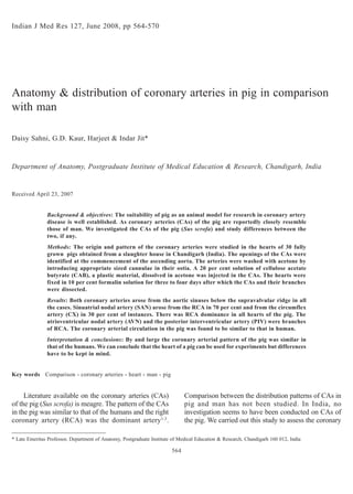
Fig. 1. Anterosuperior view of pig’s heart showing the origin of the two coronary arteries, the right coronary artery (RCA) lies in the right anterior
atrioventricular sulcus and gives a few branches (VB) to the anterior surface of right ventricle (RV). The left coronary artery (LCA) is seen to divide
into anterior interventricular artery (AIV) and circumflex artery (CX). The RCA gives the sinuatrial nodal artery (SAN) which is seen to extend
upwards and to the left on the right atrium to reach the sinuatrial node. Fig. 2. Posterior view of the heart showing the second and third segments
including the intervening curve (see arrow) of right coronary artery (RCA). On the left side, second segment of circumflex artery (CX) and its
termination into ventricular (VB) and sulcal branches (SB) are also visible. Fig.3. Right anterior oblique view of the heart, acute marginal artery
(AM) is seen to arise from the right coronary artery (RCA), it extends towards the apex on the acute margin of the heart. Fig. 4. Left anterior view of
the heart showing the right coronary artery (RCA) and its right conus (RC), and ventricular branch (VB). The left coronary artery (LCA) and its two
branches, anterior interventricular (AIV) and circumflex arteries (CX), a long diagonal branch (DB) from the left side of AIV and left conus (LC) and
a long ventricular branch (VB) arising from its right side is also seen.
4. Ventricular branches (Fig. 2): This segment also gave
three to four short ventricular branches (VB) which
extended downwards and to the right on the posterior
surface of the right ventricle.
A.V. nodal artery (AVN): An AVN artery arose separately
or jointly with the left atrioventricular sulcal branch
(SB) from the convexity of RCA near the crux of the
heart, the AVN artery could be traced to the AV node.
The sulcal branch passed to the left in the posterior
atrioventricular sulcus and might anastomose with the
sulcal branch of the circumflex artery (CX).
Branches from the third segment
Posterior interventricular artery - Ventricular branches:
Three or four obliquely directed ventricular branches
arose from the right side of the third segment of RCA
and extended downwards and to the right and passed
on the posterior surface of the right ventricle (Fig .2).
Small septal perforators arose from the deeper surface
of posterior interventricularartery.
Left coronary artery (LCA)
In all instances, LCA arose from the left posterior
aortic sinus and extended to the left behind the pulmonary
trunk for a mean distance of 5 mm and divided into
anterior interventricular and CX arteries (Fig. 4).
Anterior interventricular artery (AIV)
It passed in the anterior interventricular sulcus
usually for three or fourths of its length when it divided
into two terminal branches (Fig. 5). In four hearts, the
AIV terminated as three to four branches near the
incisura on the acute margin. There seemed to be
anastomosis between the right terminal branches of the
AIV and ventricular branches of the first segment of
RCA including the acute marginal artery.
Branches from AIV (Figs 4 & 5): A left conus (LC)
branch and three to four short ventricular branches arose
from the right side of the AIV. The conus artery was the
first branch. It was a short artery; it did not show any
anastomosis with the conus artery of the RCA. Three
to four diagonally directed branches also arose from
the left side of AIV and continued on the anterior surface
of the left ventricle. One of them usually the upper could
be long and considered to be the diagonal artery (DB).
Septal perforators also arose from its deeper aspect.
Circumflex artery (CX)
After origin, it continued in the left anterior
atrioventricular sulcus to reach the obtuse margin of
the heart (Fig. 6), crossing the same it passed in the left
posterior atrioventricular sulcus usually for half the
distance between the obtuse margin and the crux, where
it divided into a sulcal branch and a ventricular branch
(Fig. 2). The former usually anastomosed with the sulcal
branch of RCA while the later extended downwards on
the posterior surface of the left ventricle. The part of
the artery lying in the anterior atrioventricular sulcus
was called the first segment while the portion lying in
the posterior part of the atrioventricular sulcus was
named the second segment.
Branches from the first segment of circumflex artery
S.A. nodal (SAN) artery: The SAN artery was found to
arise from the upper border of proximal part of the CX
in 9 of the 30 hearts (30%). After origin, it ran upwards
and to the right on the anterior surface of the left atrium
behind the aorta and pulmonary trunk to reach the S.A.
node (Fig. 6).
Atrial arteries: There were one to two atrial branches
which arose from its upper border. They ran upwards
on the anterior surface of the left atrium to supply the
same.
Ventricular branches: One to two ventricular branches
arose from its lower border; they passed downwards
on the anterior surface of the left ventricle.
Diagonal branch : In 50 per cent hearts a long diagonal
branch extended on the anterior surface of the left
ventricle towards the apex of the heart.
Obtuse marginal branch (OM): The obtuse marginal
branch was seen in 90 per cent hearts. It extended along
the obtuse margin of the left ventricle (Fig. 5)
Branches from the second segment of circumflex
artery (Fig. 2)
One to two atrial branches arose from its upper
border and passed upwards on the posterior surface of
the left atrium. Two to four ventricular branches arose
from the lower border of the CX which extended on
the posterior surface of the left ventricle. In four
instances even six ventricular branches were also seen.
There was RCA dominance in all the specimens
(100%).
Blood supply to the interventricular septum
As in human heart septal branches which may be
called as perforators arose from the anterior and
posterior interventricular arteries. Anterior
SAHNI et al: CORONARY ARTERIES IN PIG & MEN 567
5. Fig. 5. Anterior view of the heart showing the anterior interventricular artery (AIV) extending in the anterior interventricular sulcus and terminating as
two branches proximal to the apex of the heart. A long diagonal branch (DB) arising from the AIV and obtuse marginal (OM) from the proximal part
of circumflex artery (CX) extending on the left ventricle (LV).Ventricle branches (VB) arising from the RCA and extending on the anterior wall of the
right ventricle (RV) are visible also seen. Fig. 6. Left anterosuperior view of the heart showing the left coronary artery (LCA) dividing into the anterior
interventricular artery (AIV) and circumflex artery (CX).The sinuatrial nodal artery (SAN) is seen to arise from the circumflex artery (CX), which
extends to the right behind the aorta to reach the sinuatrial node. Fig. 7. A dissection of the heart showing the right convex surface of the interventricular
septum on which a long septal artery (1) arising as a first branch from the anterior interventricular artery (AIV) is extending towards the right and
downwards dividing into number of branches. Two to Six perforators (2-6) arising from the deeper aspect of the anterior interventricular artery are also
seen. Fig.8. A dissection of the inferior surface of the heart showing the posterior one third of the interventricular septum; nine small perforators (1-9)
are seen to arise from the deeper aspect of posterior interventricular artery (PIV) which extends upto the apex of the heart from the RCA.
568 INDIAN J MED RES, JUNE 2008
6. interventricular artery was a branch of LCA while the
posterior was that of RCA in all instances.
Perforators from the anterior interventricular artery
(AIV)
The origin of the first anterior perforator was very
constant; it arose from the deeper surface of the AIV, a
branch of the LCA, at approximately 15 mm from the
origin of the later. It extended across the posterior wall
of the infundibulum of the right ventricle. In the
interventricular septum it divided into number of
branches (Fig. 7). There were five additional small
perforators which supplied the septum and anastomosed
with the perforators from PIV in all the hearts.
Perforators from the posterior interventricular artery
(PIV)
Nine or ten small perforators arose from the deeper
surface of PIV. They extended anteriorly in the septum
as perforators and anastomosed with the perforators
from AIV (Fig. 8).
Discussion
The prospect of using pig organs for human
xenotransplantation is becoming increasingly likely due
to advances in the transgenic technology. Futhermore,
pigs share important characteristics with the anatomy
of human cardiovascular system, making them useful
models for the study of human diseases. Comparative
morphological studies on the coronary arteries of the
left ventricular free wall were carried out on human,
dog, and monkey hearts by using postmortem coronary
arteriography, soft X-ray photograms, and the clearing
method. The results showed that the types of coronary
arteries (types I, II, and III) and connecting portion of
anastomotic vessels in the pig and monkey hearts closely
resembled those in man9
. With this in mind we discuss
the anatomy and distribution of coronary arteries of the
porcine heart and compared the same to that of our
earlier study in man4-6
. The study showed that the
distribution and branching of the CAs in the pig heart
appeared about the same and similar to earlier general
descriptions given in the literature except some
variations observed in the present study.
Trifurcation of LCA: The division of the LCA into three
branches-AIV, CX and an intermedium branch had been
observed previously in 20 per cent cases10
, such a
variation has neither been seen in the present study nor
has been mentioned by any other author.
Dominance: Variation in the total cardiac supply mainly
affects the diaphragmatic surface of the heart as the
posterior surfaces of the both ventricles consist of the
relative ‘dominance’ of blood supply of a branch from
RCA or LCA. Although the term is misleading as the
LCA supplies a greater volume of heart tissue, yet the
most acceptable definition of ‘dominance’ is designated
as the coronary artery which extends in the posterior
interventricular sulcus.In ‘right dominance’ the PIV is
derived from the RCA, in ‘left dominance’ it is from
the LCA and in ‘balanced’ circulation branches of both
arteries in or near the groove. In the present study,
consisting of 30 specimens, the RCA was always seen
to extend as the PIV and extended along the entire
posterior length of interventricular sulcus.This is in
agreement with the observations of earlier authors1,2
but
in contradiction to Weaver et al10
who found dominance
of RCA in 78 per cent and LCA in 5 per cent and
balanced circulation in 17 per cent swine hearts.
Intercoronary anastomosis: Blumgart et al1
studied the
coronary arteries of 44 pigs and did not find any visible
intercoronary anastomoses in 43 hearts. However, in
one heart inter-communicating twigs were seen between
left anterior descending and branches of RCA at the
apex. In the present study, surface anastomosis was
hardly observed which is in confirmation with the
observations of earlier authors.
Comparison between the pattern of distribution of the
coronary arteries of the pig and man: On comparing
the observations of the pattern of distribution of CAs
in the pig to that of man4-8
, it was found that the pattern
was almost similar to the later except some gross
percentage frequency differences. In all instances in
pig’s heart both CAs arose from the sinus below the
supravalvular ridge, however, in the human hearts the
RCA arose below the supravalvular ridge in 85 per cent
of males and 87 per cent of females; the LCA arose
from the sinus in 67.8 per cent of males and 71.3 per
cent of female5
. In the pig the SAN arose from the RCA
in 70 per cent instances while the incidence in human
was 74.7 per cent in male and 68 per cent female hearts4
.
The acute marginal artery arising from RCA was present
in 60 per cent hearts of the pig; 87 per cent in male and
90 per cent in female hearts in the humans. It has been
emphasized that the coronary arterial circulation of the
pig is remarkably similar to man1-3,10,11
.
Ourobservations,havedemonstratedsomesignificant
differences in the coronary circulation of pig and human
hearts; recognition of these differences is important for
the surgeons. The main differences in the pattern of CAs
of the pig from that of man was that the third coronary
artery and the border branches were not seen in any of the
SAHNI et al: CORONARY ARTERIES IN PIG & MEN 569
7. specimen in the former. The posterior interventricular
artery was always a branch of RCA in the hearts of pig
hence there was 100 per cent RCA dominance which was
not so in human hearts. Besides this, AIV terminated
proximal to apex in the pig hearts while in human hearts it
usually crossed the apex. The origin and course of both
the anterior and posterior septal arteries were similar to
the analogous arteries in the human heart except that
posterior perforators were always from the RCA in pig’s
heart. In about 85 per cent of human hearts the RCA or its
conusbranchgaveaninterventricularseptalbranch,which
establishedanastomoseswiththeseptalperforatorsofAIV;
this artery was not seen in the hearts of pig.
In conclusion, as far as the coronary blood supply
is concerned the heart of the pig was similar to that of
man with small differences. Therefore, pig’s heart could
be used for conducting experiments on coronary arteries.
Acknowledgment
Authors thank the Director of the Postgraduate Institute of
Medical Education and Research, Chandigarh, for financial support.
References
1. Blumgart HL, Zoll PM, Freedberg AS, Gilligan DR. The
experimental production of intercoronary arterial anastomoses
and their functional significance. Circ J 1950; 10 :10-27.
Reprint requests: Dr Daisy Sahni, Additional Professor, Department of Anatomy, Postgraduate Institute of Medical Education
& Research, Chandigarh 160 012, India
e-mail: daisy_sahni@rediffmail.com
2. Lumb G, Singletary HP. Blood supply to the antrioventricular
node and bundle of His: A comparative study in pig, dog, and
man. Am J Pathol 1962; 41 : 65-71.
3. Schwarze E, Schröder L. Kompendium der Veterinar
Anatomie, Band III Jena, VEB Gustav Fisher Verlag 1964.
4. Sahni D, Jit I. Sinuatrial nodal artery in the north-west Indians.
Indian Heart J 1988; 40 : 29-36.
5. Sahni D, Jit I. Origin and size of the coronary arteries in the
north-west Indians. Indian Heart J 1989; 41 : 221-8.
6. Sahni D, Jit I. Blood supply of the human interventricular
septum in north-west Indians. Indian Heart J 1990; 42 : 161-
9.
7. Sahni D, Jit I. Size of the valves and thicknesses of anterior
walls of the ventricles of the adult Indian hearts. Indian Heart
J 1991; 43 : 361-5.
8. Sahni D, Jit I. Incidence of myocardial bridges in north-west
Indians. Indian Heart J 1991; 43 : 431-6.
9. Kato T, Yasue T, Shoji Y, Shimabukuro S, ItoY, Goto S, et al.
Angiographic difference in coronary artery of man, dog, pig,
and monkey. Acta Pathol Jpn 1987; 37 : 361-73.
10. Weaver ME, Pantely GA, Bristow JD, Ladely HD. A
quantitative study of the anatomy distribution of coronary
arteries in swine in comparison with other animals and man.
Cardiovasc Res 1986; 20 : 907-17
11. Christensen GC, Campeti FL. Anatomic and functional studies
of the coronary circulation in the dog and pig. Am J Vet Res
1959; 20 : 18-26.
570 INDIAN J MED RES, JUNE 2008