Pulmonary Embolism Diagnosis and Treatment
•Transferir como PPTX, PDF•
8 gostaram•3,777 visualizações


Recomendados
Mais conteúdo relacionado
Mais procurados
Mais procurados (20)
Destaque
Destaque (9)
Semelhante a Pulmonary Embolism Diagnosis and Treatment
Semelhante a Pulmonary Embolism Diagnosis and Treatment (20)
Último
Último (20)
Pulmonary Embolism Diagnosis and Treatment
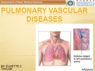
- 2. . • Pulmonary embolism (P.E) is mainly the end result of DVT’s or a general blood clot elsewhere in the body • The blood clot has the potential to break-off and enter the circulatory system and travel • A clot the travels through the circular system to another location is known • as an embolus • The embolus from the DVT usually gets dislodged and gets lodged in the heart • And then to one of the pulmonary arteries. • A P.E clogs the artery that provides blood supply to part of the lung. • Hence O2 and CO2 exchange is blocked • And general blood supply to the lung is decreased • Would as a result cause the lung to infarct.
- 3. . • Life threatening causes of chest pain and shortness of breath “DYSPNOEA • Pain sharp worse fro deep breathe “often called pleuritic pain”. • They may present with HEMOPTYSIS •The patient may have stable vital signs (blood pressure, heart rate, respiratory rate, and oxygen saturation) but frequently presents with an elevated heart rate. •A severe pulmonary embolus can present with shock or cardiac arrest, particularly when a large clot blocks the outflow of blood from the right side of the heart to the lungs (saddle embolus). • Depending on the amount of blood clot (clot burden or clot load), • oxygen saturation can be variably compromised as can the blood pressure and heart rate. In a classic presentation, •The heart rate and respiratory rate are elevated as the body tries to compensate.
- 4. • The risk factors for a P.E are the same as risk factors for D.V.T , these are referred to as Virchow's TRIAD and include: Prolonged immobilization alteration in normal blood flow(stasis) Increased clotting potential of the blood hypercoagulability Any damage to the walls of the veins • Prolonged immobilization Extended travelling (sitting in a car , airplane , train etc.) Hospitality or prolonged bed vest. Increase blood clotting potential Medication , birth control pills ,estrogen.
- 5. INVESTIGATION ATERIAL BLOOD • PaCO2 – Partial pressure of CO2 in the blood ,critical in regulating levels and maintaining body ph • PaCO2 is maintained at 5.3 kPa (40 mmHg) • D- dimer and other circulating markers. • D-dimers is a specific degradation product released into the circulation when cross-linked fibrin undergoes endogenosis. • An elevated D-dimer is limited value, as it occurs in a number of conditions including P.E
- 6. DIFFERENT DEGREES OF PE .
- 7. . • Take note of the chest pain and breathlessness • Physical examination will concentrate Heart and lungs Since the chest pain may be presenting complains of heart attack Pneumonia, pneumothorax ( collapsed lung) And dissection of an aortic aneurysm • The physical exam will also include looking for signs of a D.V.T in an extremely warmth swelling redness , and tenderness. • NB note that the signs associated with deep vein thrombosis may be completely absent even in the PRESENTS of a clot.
- 8. ‘ • Full blood count • Electrolytes • BUN (blood urea nitrogen) • Creatinine blood test • Chest x-ray, and • Electrocardiogram • The chest x-ray is often normal in P.E • The EKG/ECG may be normal, but usually demonstration a rapid heart rate • So called sinus tachycardia (heart > 100 bpm). • If there is significant blockage in a pulmonary artery. • It acts like a dam and it harder for the heart to push blood pas t the obstruction clot or clots. • This can result in the change in the electrical signal passing through the heart by stretching the heart muscle, revealed on a EKG a so called right heart strain. • Since the cost of missing the diagnosis of P.E can be death, the approach to diagnosis is to prove that no P.E exists.
- 9. . Patients who have suffered symptomatic Venous thromboembolism (VTE) carry an increased risk of further events , particularly if persisting risk factor is present. The risk of recurrence is highest at 6- 12mnths after initial event Immediate mortality may be greatest in patients with echocardiography evidence of right ventricular dysfunction or cardiogenic shock A minority progress to overt right ventricular failure .
- 10. ,
- 11. Pulmonary hypertension( PH) is the narrowing of the pulmonary arterioles within the lung. The narrowing of the arteries creates resistance and an increased work load for the heart. The heart becomes enlarged from pumping blood against the resistance. PH is defined as the mean pulmonary artery pressure > 25mmHg @ rest or 30mmHg with exercise
- 12. PH The right ventricle and right atrium are the two chambers on the right side of the heart. High pressures in the lung’s vessels causes these chambers to become enlarged and weak and to not pump as well, resulting in right sided heart failure. Pulmonary Hypertension is a long-term or chronic disease. It affects both sexes, but is more common in women, and occurs between 30- 45 years of age.
- 13. Fatigue Hoarseness Difficulty breathing (dyspnoea) Dizziness Palpitations Fainting spells ( syncope ) Swelling of legs and ankles ( edema) Bluish Lips, skin ( cyanosis ) Chest pain
- 14. PULMONARY HYPERTENSION THERE ARE 2 TYPES OF PULMONARY HYPERTENSION 1. PRIMARY HYPERTENSION : This type occurs with no known underlying disease Affects predominantly young people, more so women between 20 and 30 years The use of certain weight loss drugs ( Redux, Pondimin and fen-phen), Street drugs such as Heroin or Cocaine, and AIDS and cirrhosis of the liver can trigger the disease. Pathological features include hypertrophy of both the media and intima of the vessel wall. Clonal expansion of endothelial cells which take the appearance of plexiform lesions .
- 15. SECONDARY PULMONARY HYPERTENSION This type of pulmonary hypertension is a result of heart and lung disease. Examples of heart and lung diseases that can cause pulmonary hypertension include: < Emphysema or asthma < Blood clots that have traveled to the lung < Collagen vascular diseases such as scleroderma < Rheumatoid arthritis, systemic lupus < Congenital heart diseases such as defects and shunts < Lack of oxygen from obstructions during sleep known as sleep apnea.
- 16. . A complete history and physical exam is done. An electrocardiogram (ECG) may show a strain on the right side of your heart. Blood tests are done to indicate how much oxygen is in your blood, or to test if you have a collagen vascular disease. A chest x-ray may show a large pulmonary artery and right-sided heart. This test may also show diseases of the lung such as emphysema. A lung scan is done to show the blood supply in your lungs A CT or CAT scan is a computerized x-ray that can get a better view of the lungs and your heart. Echocardiogram uses sonar (sound waves) to show the pumping function of your heart and how the valves work. A pulmonary function test is done to measure the volume of air in your lungs. Results are obtained by breathing into a mouth piece while exercising on a treadmill or bicycle. An exercise tolerance test will require you to walk on a treadmill as fast as you can for 6 minutes to evaluate how much exercise you can do before you have symptoms. A right heart catheterization is the most accurate way to diagnose pulmonary hypertension. A small tube or catheter is put into a vein in your neck and then guided into the right side of your heart and pulmonary artery to measure pressures.
- 17. . Treatment for Pulmonary Hypertension may include: Oxygen Medications Lifestyle and dietary changes Surgery
- 18. . ACE inhibitors (Captopril, Enalapril, Lisinopril). This drug dilates the blood vessels to improve the heart function and blood flow. Anticoagulants (Coumadin) " blood thinners" are used to decrease the tendency of the blood to clot so that it flows more freely through the blood vessels. It is very important to have this drug monitored for bleeding complications by having routine blood work done. Diuretics ( Lasix, Aldactone ) are used to rid your body of excess fluid. They work to reduce swelling that is caused by pulmonary hypertension and Right sided heart failure. Digoxin improves the pumping ability of your heart. It is important to have this drug level checked at regular intervals of time. Calcium Channel Blocker ( Cardizem, Procardia, Norvasc) are drugs used at high doses to lower the pulmonary pressure.
- 19. It is incurable but the treatments rendered have delivered significant improvements In exercise performance, symptoms and prognosis. Warfarin also helps as an anticoagulant.
- 20. . •Mitchell, Richard Sheppard; Kumar, Vinay; Robbins, Stanley L.; Abbas, Abul K.; Fausto, Nelson (2007). Robbins basic pathology. Saunders/Elsevier. •Pulmonary hypertension. (online).2010.Available www.adams.com (accessed 08 april 2010). •Pulmonary embolism.(online).2010.Available www.medicinenet.com /pulmonary embolism(Accessed 08 April2010). •ColledgeN.R. et al.2010.21st edition.Davidson,s Principles and Practice of Medicine.Churchill Livingstone