Case Review #4: 51 year old female with adjacent segment degeneration
•
1 gostou•3,154 visualizações

A 51 year old female, presented after a previous 3 level cervical spine fusion. The patient developed a cervical disc herniation adjacent to the previous fusion. Dr. Pashman treated her by extending her cervical fusion.

Recomendados
Recomendados
Mais conteúdo relacionado
Mais procurados
Mais procurados (20)
Semelhante a Case Review #4: 51 year old female with adjacent segment degeneration
Semelhante a Case Review #4: 51 year old female with adjacent segment degeneration (20)
Mais de Robert Pashman
Mais de Robert Pashman (20)
Último
Último (20)
Case Review #4: 51 year old female with adjacent segment degeneration
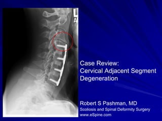
- 1. Case Review: Cervical Adjacent Segment Degeneration Robert S Pashman, MD Scoliosis and Spinal Deformity Surgery www.eSpine.com
- 2. Patient History 51 year old female Status post fusion at C4-5, C5-6 and C6-7 The patient has superadjacent degeneration at C3-4 above the previous fusion. The patient's symptoms have gotten very bad. She has right shoulder pain. There is no problem at C7-T1. The effacement of the spinal cord and fractional kyphosis is noted. She has been treated conservatively with blocks and physical therapy for a long time.
- 4. Indications for Surgery Status post anterior cervical discectomy and fusion, November 2004, at C4-C5, C5-C6, and C6-C7. Superjacent degeneration at C3-C4 with spinal cord compression. Fractional kyphosis. Some motor/sensory deficit. Significant proximal pain radiating to the trap region into her arms and intermittently into her back. Failed conservative therapy.
- 5. Surgical Strategy Radical discectomy, C3-C4, with spinal cord decompression under the microscope. Anterior interbody fusion using Cornerstone 7-mm device, C3-C4, with autogenous bone centrally. Anterior cervical plate fixation with four-hole Atlantis Vision plate, C3-C4. Removal of retained instrumentation and exploration of fusion mass, C4 to C7. Intraoperative somatosensory-evoked potentials. Intraoperative fluoroscopy.
- 7. Pre-Op/Post-op Comparison Normal disc height has been restored Pre-op Post-op
- 8. Pre-Op/Post-op Comparison Pre-op Post-op Patient is doing well. Incision well-healed. X-rays look good. No evidence of problems with the instrumentation. Sub adjacent spine is fused.