Nrgastro.2012.168

This document reviews the regulation of gastrointestinal motility. It discusses how gastrointestinal motility results from coordinated smooth muscle contractions facilitated by electrical and mechanical junctions between smooth muscle cells. Excitation-contraction coupling in smooth muscle cells occurs via calcium entry into cells through ion channels, which triggers phosphorylation of myosin and muscle contraction. Gastrointestinal smooth muscles generate spontaneous contractions driven by pacemaker cells called interstitial cells of Cajal, but enteric motor neurons help coordinate contractions into functional motility patterns. A variety of regulatory factors including neurons, hormones, and paracrine substances further integrate smooth muscle activity to produce normal and abnormal motility behaviors throughout the gastrointestinal tract.
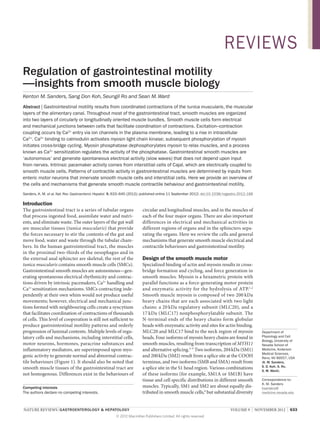
![REVIEWS
Key points
■■ Gastrointestinal motility occurs by the coordinated contractions of the tunica
muscularis, which forms the outer wall of the alimentary canal from the distal
oesophagus to the external anal sphincter
■■ Excitation–contraction coupling results from Ca2+ entry into smooth muscle
cells, Ca2+ release from the sarcoplasmic reticulum, activation of myosin light
chain kinase and phosphorylation of the regulatory light chains of myosin
■■ Contractile force is tuned by Ca2+ sensitization mechanisms that balance rates
of myosin phosphorylation and dephosphorylation
■■ Interstitial cells of Cajal (ICC) provide spontaneous pacemaker activity in
gastrointestinal muscles; ICC and PDGFRα+ cells also contribute to mediation
of inputs from enteric motor neurons
■■ Gastrointestinal motility patterns are highly integrated behaviours requiring
coordination between smooth muscle cells and utilizing regulatory inputs from
interstitial cells, neurons, and endocrine and immune cells
■■ Therapeutic regulation and tissue engineering of gastrointestinal motility is
proving difficult
CNS
Myenteric plexus
Efferent neurons
Mast
cell
Excitation–contraction coupling
Macrophage
Afferent
neurons
Enteric motor
neurons
Eicosanoids Nitric oxide
Histamine
Eicosanoids
PDGFRα+
cell
ICC
SIP
syncytium
Hormones
SMCs
Blood vessel
Integrated motor behaviour
Figure 1 | Regulation of gastrointestinal motility. Major factors involved in
regulation of the tunica muscularis of the gastrointestinal tract. SMCs are shown in
close contact with ICC and PDGFRα+ cells. SMCs form gap junctions with both
classes of interstitial cells, creating an electrical syncytium (SIP syncytium).
Excitatory (orange) and inhibitory (blue) enteric motor neurons, with cell bodies in
the myenteric plexus, innervate ICC, PDGFRα+ and SMCs, so motor responses are
integrated by all three cell types. Information is sent by afferent (sensory) neurons
from the gut to the enteric ganglia, the CNS, and to autonomic ganglia (not shown).
Information is sent by efferent neurons from the CNS to enteric ganglia. Hormones,
reaching the gut via the circulation, and paracrine and immune factors, produced
by mast cells and resident macrophages, also affect motor output. Abbreviations:
ICC, interstitial cells of Cajal; SMC, smooth muscle cell; SIP, syncytium of neuroeffector cells consisting of SMCs, ICC and PDGFRα+ cells.
634 | NOVEMBER 2012 | VOLUME 9
exists in the expression of SMA and SMB isoforms. In
the fundus, for example, SMA is the dominant isoform,
whereas SMB increases with distance from the fundus
to the antrum and represents the dominant isoform in
antral muscles.6 There is also diversity in myosin heavy
chain isoform expression between cells within a given
tissue, but the functional importance of this cellular
hetero eneity is not yet understood.7
g
Contractions are initiated by phosphorylation of
MLC20 by Ca2+/calmodulin-dependent myosin light
chain kinase or Ca 2+-independent kinases, including Rho-kinase, integrin-linked kinase and zipper-
interacting protein kinase (ZIPK; Figure 2). 8–11 An
increasing level of cytoplasmic Ca2+ ([Ca2+]i) is the main
physiological event that activates myosin light chain
kinase. Phosphorylation of MLC20 facilitates myosin
binding to actin, initiating cross-bridge cycling and
force development. MLC20 phosphorylation and contraction are balanced by myosin light chain phosphatase
(MLCP). MLCP is composed of three subunits.12 One
of these subunits (myosin phosphatase target subunit;
MYPT) anchors MLCP to phosphorylated MLC20 and
targets a 37 kDa catalytic subunit (type 1 serine/threonine phosphatase, PP1c), to myosin.13 Phosphorylation
of MYPT (see below) can dramatically affect the balance
between phosphorylated and dephosphorylated MLC20;
therefore, regulation of MLCP can be a powerful means
for modulating the force of contractions.
Studies of several species have provided a general
concept of excitation–contraction coupling in mammalian gastro ntestinal smooth muscles, but these studies
i
have also demonstrated considerable diversity in the
expression of ionic conductances in different species
and in different regions of the gastrointestinal tract.
The ionic channels in human gastrointestinal smooth
muscles responsible for excitation–contraction coupling
and the specific responses of human muscles to neurotransmitters and other regulatory agents have not been
studied in enough depth to clearly describe excitation–
contraction coupling mechanisms in human gastrointestinal smooth muscles. Nonetheless, pertinent data
are described below, including seminal findings from
other smooth muscle types and gastrointestinal muscles
of laboratory animals.
Voltage-dependent calcium channels
Like all excitable cells, the resting potentials of SMCs
in gastrointestinal muscles are negative relative to the
interstitium. Na+ and Ca2+ are not in equilibrium across
the cell membrane, such that a rise in membrane permeability to these ions causes rapid influx. Inward currents
(defined as an influx of positive charges) result in membrane depolarization. Excitation–contraction coupling
is triggered by a rise in [Ca2+]i.8,9 An important feature
of the excitation–contraction coupling mechanism is
linked to the depolarization of SMCs. Depolarization
activates voltage-dependent Ca2+ channels and Ca2+
entry into SMCs, which increases [Ca2+]i.14–16 Smooth
www.nature.com/nrgastro
© 2012 Macmillan Publishers Limited. All rights reserved](data:image/gif;base64,R0lGODlhAQABAIAAAAAAAP///yH5BAEAAAAALAAAAAABAAEAAAIBRAA7)
Recomendados
Mais conteúdo relacionado
Mais procurados
Mais procurados (20)
Destaque
Semelhante a Nrgastro.2012.168
Semelhante a Nrgastro.2012.168 (20)
Mais de Elsa von Licy
Mais de Elsa von Licy (20)
Último
Último (20)
Nrgastro.2012.168
- 1. REVIEWS Regulation of gastrointestinal motility —insights from smooth muscle biology Kenton M. Sanders, Sang Don Koh, Seungil Ro and Sean M. Ward Abstract | Gastrointestinal motility results from coordinated contractions of the tunica muscularis, the muscular layers of the alimentary canal. Throughout most of the gastrointestinal tract, smooth muscles are organized into two layers of circularly or longitudinally oriented muscle bundles. Smooth muscle cells form electrical and mechanical junctions between cells that facilitate coordination of contractions. Excitation–contraction coupling occurs by Ca2+ entry via ion channels in the plasma membrane, leading to a rise in intracellular Ca2+. Ca2+ binding to calmodulin activates myosin light chain kinase; subsequent phosphorylation of myosin initiates cross-bridge cycling. Myosin phosphatase dephosphorylates myosin to relax muscles, and a process known as Ca2+ sensitization regulates the activity of the phosphatase. Gastrointestinal smooth muscles are ‘autonomous’ and generate spontaneous electrical activity (slow waves) that does not depend upon input from nerves. Intrinsic pacemaker activity comes from interstitial cells of Cajal, which are electrically coupled to smooth muscle cells. Patterns of contractile activity in gastrointestinal muscles are determined by inputs from enteric motor neurons that innervate smooth muscle cells and interstitial cells. Here we provide an overview of the cells and mechanisms that generate smooth muscle contractile behaviour and gastrointestinal motility. Sanders, K. M. et al. Nat. Rev. Gastroenterol. Hepatol. 9, 633–645 (2012); published online 11 September 2012; doi:10.1038/nrgastro.2012.168 Introduction The gastrointestinal tract is a series of tubular organs that process ingested food, assimilate water and nutrients, and eliminate waste. The outer layers of the gut wall are muscular tissues (tunica muscularis) that provide the forces necessary to stir the contents of the gut and move food, water and waste through the tubular chambers. In the human gastrointestinal tract, the muscles in the proximal two-thirds of the oesophagus and in the external anal sphincter are skeletal; the rest of the tunica muscularis contains smooth muscle cells (SMCs). Gastrointestinal smooth muscles are autonomous—generating spontaneous electrical rhythmicity and contractions driven by intrinsic pacemakers, Ca2+ handling and Ca2+ sensitization mechanisms. SMCs contracting independently at their own whim would not produce useful movements; however, electrical and mechanical junctions formed with neighbouring cells create a syncytium that facilitates coordination of contractions of thousands of cells. This level of cooperation is still not sufficient to produce gastrointestinal motility patterns and orderly progression of luminal contents. Multiple levels of regulatory cells and mechanisms, including interstitial cells, motor neurons, hormones, paracrine substances and inflammatory mediators, are superimposed upon myogenic activity to generate normal and abnormal contractile behaviours (Figure 1). It should also be noted that smooth muscle tissues of the gastrointestinal tract are not homogenous. Differences exist in the behaviours of Competing interests The authors declare no competing interests. circular and longitudinal muscles, and in the muscles of each of the four major organs. There are also important differences in electrical and mechanical activities in different regions of organs and in the sphincters separating the organs. Here we review the cells and general mechanisms that generate smooth muscle electrical and contractile behaviours and gastrointestinal motility. Design of the smooth muscle motor Specialized binding of actin and myosin results in crossbridge formation and cycling, and force generation in smooth muscles. Myosin is a hexametric protein with parallel functions as a force-generating motor protein and enzymatic activity for the hydrolysis of ATP. 1,2 Smooth muscle myosin is composed of two 200 kDa heavy chains that are each associated with two light chains: a 20 kDa regulatory subunit (MLC20), and a 17 kDa (MLC17) nonphosphorylatable subunit. The N‑terminal ends of the heavy chains form globular heads with enzymatic activity and sites for actin binding. MLC20 and MLC17 bind to the neck region of myosin heads. Four isoforms of myosin heavy chains are found in smooth muscles, resulting from transcription of MYH11 and alternative splicing.3–5 Two isoforms, 204 kDa (SM1) and 200 kDa (SM2) result from a splice site at the COOH terminus, and two isoforms (SMB and SMA) result from a splice site in the S1 head region. Various combinations of these isoforms (for example, SM1A or SM1B) have tissue and cell specific distributions in different smooth muscles. Typically, SM1 and SM2 are about equally distributed in smooth muscle cells,6 but substantial diversity NATURE REVIEWS | GASTROENTEROLOGY & HEPATOLOGY © 2012 Macmillan Publishers Limited. All rights reserved Department of Physiology and Cell Biology, University of Nevada School of Medicine, Anderson Medical Sciences, Reno, NV 89557, USA (K. M. Sanders, S. D. Koh, S. Ro, S. M. Ward). Correspondence to: K. M. Sanders ksanders@ medicine.nevada.edu VOLUME 9 | NOVEMBER 2012 | 633
- 2. REVIEWS Key points ■■ Gastrointestinal motility occurs by the coordinated contractions of the tunica muscularis, which forms the outer wall of the alimentary canal from the distal oesophagus to the external anal sphincter ■■ Excitation–contraction coupling results from Ca2+ entry into smooth muscle cells, Ca2+ release from the sarcoplasmic reticulum, activation of myosin light chain kinase and phosphorylation of the regulatory light chains of myosin ■■ Contractile force is tuned by Ca2+ sensitization mechanisms that balance rates of myosin phosphorylation and dephosphorylation ■■ Interstitial cells of Cajal (ICC) provide spontaneous pacemaker activity in gastrointestinal muscles; ICC and PDGFRα+ cells also contribute to mediation of inputs from enteric motor neurons ■■ Gastrointestinal motility patterns are highly integrated behaviours requiring coordination between smooth muscle cells and utilizing regulatory inputs from interstitial cells, neurons, and endocrine and immune cells ■■ Therapeutic regulation and tissue engineering of gastrointestinal motility is proving difficult CNS Myenteric plexus Efferent neurons Mast cell Excitation–contraction coupling Macrophage Afferent neurons Enteric motor neurons Eicosanoids Nitric oxide Histamine Eicosanoids PDGFRα+ cell ICC SIP syncytium Hormones SMCs Blood vessel Integrated motor behaviour Figure 1 | Regulation of gastrointestinal motility. Major factors involved in regulation of the tunica muscularis of the gastrointestinal tract. SMCs are shown in close contact with ICC and PDGFRα+ cells. SMCs form gap junctions with both classes of interstitial cells, creating an electrical syncytium (SIP syncytium). Excitatory (orange) and inhibitory (blue) enteric motor neurons, with cell bodies in the myenteric plexus, innervate ICC, PDGFRα+ and SMCs, so motor responses are integrated by all three cell types. Information is sent by afferent (sensory) neurons from the gut to the enteric ganglia, the CNS, and to autonomic ganglia (not shown). Information is sent by efferent neurons from the CNS to enteric ganglia. Hormones, reaching the gut via the circulation, and paracrine and immune factors, produced by mast cells and resident macrophages, also affect motor output. Abbreviations: ICC, interstitial cells of Cajal; SMC, smooth muscle cell; SIP, syncytium of neuroeffector cells consisting of SMCs, ICC and PDGFRα+ cells. 634 | NOVEMBER 2012 | VOLUME 9 exists in the expression of SMA and SMB isoforms. In the fundus, for example, SMA is the dominant isoform, whereas SMB increases with distance from the fundus to the antrum and represents the dominant isoform in antral muscles.6 There is also diversity in myosin heavy chain isoform expression between cells within a given tissue, but the functional importance of this cellular hetero eneity is not yet understood.7 g Contractions are initiated by phosphorylation of MLC20 by Ca2+/calmodulin-dependent myosin light chain kinase or Ca 2+-independent kinases, including Rho-kinase, integrin-linked kinase and zipper- interacting protein kinase (ZIPK; Figure 2). 8–11 An increasing level of cytoplasmic Ca2+ ([Ca2+]i) is the main physiological event that activates myosin light chain kinase. Phosphorylation of MLC20 facilitates myosin binding to actin, initiating cross-bridge cycling and force development. MLC20 phosphorylation and contraction are balanced by myosin light chain phosphatase (MLCP). MLCP is composed of three subunits.12 One of these subunits (myosin phosphatase target subunit; MYPT) anchors MLCP to phosphorylated MLC20 and targets a 37 kDa catalytic subunit (type 1 serine/threonine phosphatase, PP1c), to myosin.13 Phosphorylation of MYPT (see below) can dramatically affect the balance between phosphorylated and dephosphorylated MLC20; therefore, regulation of MLCP can be a powerful means for modulating the force of contractions. Studies of several species have provided a general concept of excitation–contraction coupling in mammalian gastro ntestinal smooth muscles, but these studies i have also demonstrated considerable diversity in the expression of ionic conductances in different species and in different regions of the gastrointestinal tract. The ionic channels in human gastrointestinal smooth muscles responsible for excitation–contraction coupling and the specific responses of human muscles to neurotransmitters and other regulatory agents have not been studied in enough depth to clearly describe excitation– contraction coupling mechanisms in human gastrointestinal smooth muscles. Nonetheless, pertinent data are described below, including seminal findings from other smooth muscle types and gastrointestinal muscles of laboratory animals. Voltage-dependent calcium channels Like all excitable cells, the resting potentials of SMCs in gastrointestinal muscles are negative relative to the interstitium. Na+ and Ca2+ are not in equilibrium across the cell membrane, such that a rise in membrane permeability to these ions causes rapid influx. Inward currents (defined as an influx of positive charges) result in membrane depolarization. Excitation–contraction coupling is triggered by a rise in [Ca2+]i.8,9 An important feature of the excitation–contraction coupling mechanism is linked to the depolarization of SMCs. Depolarization activates voltage-dependent Ca2+ channels and Ca2+ entry into SMCs, which increases [Ca2+]i.14–16 Smooth www.nature.com/nrgastro © 2012 Macmillan Publishers Limited. All rights reserved
- 3. REVIEWS G-protein-coupled receptors Potassium channels VDCC NO NSCC Adenylate cyclase Plasma membrane – K+ + Gαs Ca2+ Na+Ca2+ + Ca2+ [Ca2+]i IP3 Gαq/11 PLCβ Sarcoplasmic reticulum NO Gα12/13 GCα GCβ DAG CPI-17 Rho-GEF cGMP cAMP PKC IP3 receptor RhoA PKG PKA – RhoK Ca2+/calmodulin CPI-17 Relaxation MLC20 P T38 Active MLCP + – PP1c M20 MYPT + Myosin light chain kinase ZIPK Telokin P S13 RhoK ZIPK PP1c MLC20 P Contraction T853 P M20 MYPT P T696 Inactive MLCP Figure 2 | Major cellular mechanisms controlling contraction in gastrointestinal smooth muscle cells. Mechanisms leading to enhanced contraction are depicted in red, and pathways linked to decreased contraction are shown in blue. Ca 2+ required for excitation–contraction coupling can enter cells through VDCC or NSCC. The open probability of VDCC is enhanced (circle with + sign) by depolarization caused by opening of NSCC and influx of Na + or Ca2+. Openings of VDCC are decreased by a variety of K+ channels expressed by SMCs; most inhibitory agonists regulate Ca2+ influx by activating K+ channels. Ca2+ entry can also be supplemented by release of Ca2+ from IP3 receptor-operated Ca2+ channels in the sarcoplasmic reticulum membrane. Ca2+ release from sacroplasmic reticulum can also occur through ryanodine receptors (not shown). IP3 is synthesized by PLCβ in response to agonist binding to G-protein-coupled receptors and coupling through Gαq/11. [Ca2+]i binds to calmodulin and activates myosin light chain kinase, which phosphorylates MLC20 to facilitate crossbridge formation. Phosphorylation of MLC20 is balanced by the action of MLCP. Dephosphorylation of MLC20 reduces cross-bridge cycling and leads to muscle relaxation. Factors that lead to inhibition of MLCP increase contraction and, in effect, enhance Ca2+ sensitivity of the contractile apparatus. The opposite is true for factors that activate MLCP. A pathway that increases Ca2+ sensitization (and therefore increases contraction) occurs through binding of G-protein-coupled (Gαq/11 or Gα12/13) receptors and regulation of the GDP–GTP exchange factor (Rho-GEF), RhoA and activation of Rho kinase. Rho kinase and protein kinase C can phosphorylate CPI‑17 (at T38), a protein that when phosphorylated inhibits the catalytic subunit of MLCP (PP1c; circle with negative sign). Rho kinase can also phosphorylate the regulatory subunit of MLCP (MYPT) at T696 and T853. Phosphorylation of MYPT decreases the activity of MLCP, preserving phosphorylation of MLC20. ZIPK also phosphorylates CPI‑17 and MYPT. Cyclic nucleotide-dependent pathways decrease Ca 2+ sensitivity. NO, for example, binds to guanylyl cyclase (composed of GCα and GCβ subunits) and generates cGMP. cGMP activates cGMPdependent protein kinase (PKG), which can phosphorylate RhoA and reduce activation of Rho kinase, thus reducing Ca 2+ sensitization, or it can phosphorylate telokin (S13), which stimulates MLCP (circle with + sign). Binding of receptors coupled through GαS activates adenylate cyclase and production of cAMP. PKA can also phosphorylate telokin and increase MLCP activity. Abbreviations; [Ca2+]i, cytoplasmic Ca2+; DAG, diacylglycerol; IP3, inositol 1,4,5-trisphosphate; MLC20, 20 kDA light chain of myosin; MLCP, myosin light chain phosphatase; NO, nitric oxide; NSCC, nonselective cation channels; PKA, cAMP-dependent protein kinase; PKG, cGMP-dependent protein kinase; PLCβ, phospholipase Cβ; VDCC, voltage-dependent Ca2+ channels; ZIPK, zipper-interacting protein kinase. Permission obtained Wiley © Sanders, K. M. Neurogastroenterol. Motil. 20 (Suppl. 1), 39–53 (2008). muscles express voltage-dependent Ca2+ channels that are characterized by α subunits, encoded by CACNA1C.17 The resulting channels are ‘L-type Ca2+ channels’ that are blocked by dihydropyridines (although these agents fail to fully block contractile response in some species, including humans [unpublished data, Ward and Sanders]). Additional voltage-dependent Ca2+ channels (for example, T‑type channels) contribute to Ca2+ entry in gastrointestinal smooth muscles.18 Nonselective cation channels Ca2+ entry into SMCs also occurs via nonselective cation channels.19 These channels are activated by second messengers, which are mobilized by agonist stimulation or by stretch. Nonselective cation channels can contribute directly to Ca2+ entry, or indirectly through Na+ entry and depolarization. Transient receptor potential (TRP) channels are prominent nonselective cation channels in gastrointestinal smooth muscles. For example, TRPC4 NATURE REVIEWS | GASTROENTEROLOGY HEPATOLOGY © 2012 Macmillan Publishers Limited. All rights reserved VOLUME 9 | NOVEMBER 2012 | 635
- 4. REVIEWS and TRPC6 encode the nonselective cation channels activated by muscarinic stimulation (as shown by gene deactivation).20 TRP channels are voltage-independent and activated either by intracellular Ca2+ or by G‑proteincoupled mechanisms.21 Other nonselective cation channels—activated by other agonists, such as neurokinins and hormones—have slightly different properties.22 For example, a well-known property of nonselective cation channels activated by muscarinic stimulation is potentiation by intracellular Ca2+.23 This property is not apparent in channels activated by neurokinins. The effects of neurokinins in intact muscles are mediated by NK1 receptors (primarily expressed by interstitial cells of Cajal; ICC), and by NK2 receptors (primarily expressed by SMCs).24–26 Regulation of excitability Depolarization of SMCs generates action potentials, owing to the activation of voltage-dependent Ca2+ channels. Ca2+ action potentials substantially increase [Ca2+]i levels. Direct regulation of voltage-dependent Ca2+ channels is not typical in gastrointestinal muscles. Other conductances causing depolarization of gastrointestinal smooth muscles (that is, nonselective cation channels in SMCs or Ca2+-activated Cl– channels in ICC) or hyperpolarization (K+ conductances such as ATP-dependent K+ channels, 2‑pore K+ channels, delayed rectifiers and Ca2+-activated K+ channels in SMCs and small conductance Ca 2+-activated K + channels in platelet-derived growth factor α [PDGFRα]+ cells) are targeted by excitatory or inhibitory agonists (see below). Inward currents tend to depolarize SMCs and increase the openings of voltage-dependent Ca2+ channels. Outward currents tend to hyperpolarize or stabilize the membrane potentials of SMCs and decrease the openings of voltage-dependent Ca2+ channels.27 The role of the sarcoplasmic reticulum SMCs store Ca2+ in intracellular organelles, the most prominent of which, in relation to excitation–contraction coupling, is the sarcoplasmic reticulum. 28,29 The concentration of Ca 2+ in stores far exceeds [Ca 2+] i. Thus, opening Ca2+ permeable ion channels in sarcoplasmic reticulum membranes releases Ca 2+ into the cytoplasm. Inositol 1,4,5-trisphosphate receptors (IP3R; encoded by ITPR1) and ryanodine receptors (encoded by RYR2 and RYR3) provide Ca 2+ permeable sarcoplasmic reticulum channels. Ryanodine receptors are activated by caffeine and by increased [Ca2+]i;30 thus, a small amount of Ca2+ entering the cells can be amplified by Ca2+ release from the sarcoplasmic reticulum.31 IP3R are also sensitive to [Ca2+]i but IP3 is the primary regulator of the openings of these channels. IP3 is formed by the action of phospholipase Cβ (PLCβ) on phosphatidylinositol 4,5 bisphosphate (PIP2) during the response of G-protein-coupled receptors to a variety of agonists. For example, gastrointestinal SMCs express two forms of muscarinic receptors, M2 and M3,32,33 and M3 receptors are coupled through Gq/11 to activation of PLCβ (Figure 2). 636 | NOVEMBER 2012 | VOLUME 9 The sensitivity of sarcoplasmic reticulum channels to second messenger substances, such as Ca2+ and IP3, can lead to interactions between Ca2+ transients (termed sparks from ryanodine receptors and puffs from IP3R) and can induce oscillatory release of Ca 2+ transients from the sarcoplasmic reticulum or even Ca2+ waves that can travel along SMCs. Alignment of the sarcoplasmic reticulum close to the plasma membrane allows Ca 2+ transients to reach quite high concentrations (1 μM) in microdomains between the sarcoplasmic reticulum and plasma membrane.34,35 Ca2+ sparks and puffs regulate Ca2+ sensitive proteins in the plasma membrane, such as ion channels.36–38 In gastrointestinal SMCs, small and large conductance Ca2+-activated K+ (SK and BK) channels and nonselective cation channels are regulated by release of Ca2+ from the sarcoplasmic reticulum.39–43 In some smooth muscles, Ca2+ sparks and puffs can activate Ca2+-activated Cl– channels,41,44 but these channels are not common in gastrointestinal SMCs. Ca 2+ transients, by their ability to activate ion channels, contribute to the regulation of excitability of smooth muscles. Interactions between Ca2+ sparks and puffs can induce regenerative Ca2+ waves 45 These waves seem to run along the inner surface of cell membranes without a generalized increase in [Ca2+]i, because there is typically no contractile response associated with Ca2+ waves. Ca2+ waves are thought to regulate ion channel openings and participate in setting the membrane potential and cellular excitability. Regulation of Ca2+ sensitivity As described above, activation of myosin light chain kinase and phosphorylation of MLC20 in SMCs is Ca2+ dependent, but force development is not simply a function of [Ca2+]i levels. Additional pathways modulate the Ca2+ sensitivity of the contractile apparatus, such that different agonists can elicit contractile events of different magnitudes with equivalent changes in [Ca2+]i.46 A neutral [Ca2+]i versus force curve (such as a contractile response to Ca2+ entry via voltage-dependent ion channels) might shift dramatically to the left when excitatory agonists, such as acetylcholine, stimulate muscles; in such cases, low levels of [Ca2+]i can elicit contractile responses of far greater amplitude than normal.8 Ca2+ sensitization is controlled through the activity of MLCP (Figure 2). Contraction is initiated by phosphorylation of myosin (MLC20), so dephosphorylation of myosin reduces contraction. Phosphorylation of MYPT (the regulatory subunit of MLCP) reduces the activity of the phosphatase and sustains cross-bridge cycling and contraction.47,48 Rho kinase was the first enzyme shown to phosphorylate MYPT, but ZIPK is also known to phosphorylate MYPT and enhance Ca2+ sensitization.49,50 CPI‑17 is another signalling molecule that regulates MLCP. When phosphorylated by protein kinase C, Rho kinase or other kinases, CPI‑17 binds to the catalytic subunit of MLCP and inhibits dephosphorylation of MLC20.8,47 As discussed above, acetylcholine is coupled to responses via M2 and M3 receptors. M3 receptors are coupled through Gq/11 to production of diacylglycerol and IP3. Diacylglycerol and increased levels of [Ca2+]i www.nature.com/nrgastro © 2012 Macmillan Publishers Limited. All rights reserved
- 5. REVIEWS Table 1 | Major neurotransmitters in SMCs, ICC and PDGFRα+ cells Neurotransmitters Cells expressing receptors Receptors G protein Second messengers/ signalling pathway Major effectors References SMC M2 Gi Reduced cAMP/PKA Inhibition of K+ channel activation 6, 18, 25, 26, 39, 57, 58 M3 Gq/G11; G12/13 IP3; DAG/PKC; Ca2+; RhoA/Rho kinase TRPC4/TRC6; MYPT1; CPI‑17 6, 18, 25, 26, 39, 57, 58 M2 Not known Not known Not known NA M3 Gq/G11 IP3; DAG/PKC; Ca2+ CaCl; NSCC‡ 105 SMC NK1; NK2 Gq/G11; G12/13 IP3; DAG/PKC; Ca2+; RhoA/Rho kinase TRPC;‡ MYPT1; CPI‑17 20, 59–62 ICC NK1 Gq/G11 IP3; DAG/PKC; Ca2+ CaCl‡ NA ICC GCα and β NA cGMP/PKG TREK‑1; PLB 91,99 SMC GCα and β NA cGMP/PKG TREK‑1; PLB; MLCP 44, 46, 63–66 PDGFRα NA NA Not known Not known NA SMC P2Y1; P2Y4 Gq/G11 IP3; DAG/PKC; Ca2+ SK2; NSCC* 69, 106, 107 ICC P2Y1 Gq/G11 IP3; DAG/PKC; Ca2+ NSCC* NA PDGFRα+ P2Y1 Gq/G11 IP3; DAG/PKC; Ca2+ SK3 69–71 SMC VPAC1; VPAC2 Gs cAMP/PKA KATP; KV; BK; MLCP 8, 72–74 ICC VPAC1 Gs cAMP/PKA Not known 98, 154 Excitatory neurons Acetylcholine ICC Substance P; NKA Inhibitory neurons Nitric oxide + β-NAD ATP? VIP; PACAP *Under physiological gradients and holding potentials, inward currents were elicited in SMCs and ICC by purines. This finding is incompatible with the role of purines as inhibitory neurotransmitters, and suggests these cells are not responsible for the hyperpolarization response (inhibitory junction potential) to purinergic neurotransmission. 69 ‡Potential effectors. Abbreviations: BK, large conductance Ca2+-activated K+ channels; β-NAD, nicotinamide adenine dinucleotide; CaCl, Ca2+-activated Cl– channels; DAG, diacylglycerol; GCα and β, guanylyl cyclase isoforms α and β; ICC, interstitial cells of Cajal; KATP ATP activated K+ channel; KV, voltage-activated K+ channels; MLCP; myosin light chain phosphatase; MYPT, myosin phosphatase target , subunit; NA, not available; NSCC, nonselective cation channels; PACAP pituitary adenylate cyclase-activating polypeptide; PDGFR, platelet-derived growth factor receptor; PKA, cAMP-dependent , protein kinase; PKG, cGMP dependent protein kinase; PLB, phospholamban; SK2 and SK3, isoforms of small conductance Ca2 +-activated K+ channels; SMC, smooth muscle cell; TRPC4/ TRPC6, canonical transient receptor potential channels, isoforms TRPC4 and TRPC6; TREK‑1, two-pore K + channel; VIP vasoactive intestinal polypeptide. , activate protein kinase C isoforms, which phosphorylate CPI‑17. M3 receptors also couple to Ca2+ sensitization mechanisms through Gq/11 and possibly a second population of G proteins, G12/13, to activate Rho kinase. Phosphorylation of CPI‑17 and activation of Rho kinase and subsequent phosphorylation of MYPT facilitate Ca2+ sensitization and enhance contraction. Cyclic nucleotides, cGMP and cAMP, via cGMPdependent and cAMP-dependent protein kinases (PKG and PKA), relax smooth muscles at constant [Ca2+]i.8,51,52 cGMP-dependent Ca2+ desensitization is mediated, in part, via protein kinase G phosphorylation of RhoA at serine 188 (S188), which prevents its binding to Rho kinase and decreases Ca2+ sensitization.53 This mechanism explains inhibition of Ca2+ sensitization by cyclic nucleotide-dependent mechanisms, but additional pathways are needed to fully explain a rightward shift in the Ca2+-force relationship, the hallmark of Ca2+ desensitization. Telokin, a small acidic protein with partial sequence homology to myosin light chain kinase, is phosphorylated at serine 13 (S13) in response to 8‑bromo-cGMP or forskolin and tends to stabilize the unphosphorylated state of MLC20.54 Thus, cGMP-dependent and cAMPdependent Ca 2+ desensitization might be mediated through telokin. Studies on telokin null mice demonstrate that telokin enhances the activity of MLCP, thus reducing Ca2+ sensitivity.55 Ca2+ sensitization mechanisms are important regulatory pathways in gastrointestinal SMCs, and might provide novel therapeutic strategies for controlling gastrointestinal motility. Whether regulation of Ca 2+ sensitization mechanisms contribute to responses to neurotransmitters released from neurons is unknown. These mechanisms have been studied by adding neuro transmitters to muscles immersed in organ baths in which all cells in the muscles are exposed to agonists. As discussed below, neurotransmitter binding might be restricted to receptors in neuroeffector junctions. If this is the case, then neurotransmitter responses might not be mediated by the same receptors and pathways affected by exogenous, bath-applied agonists. Regulation of smooth muscle by motor neurons Motility patterns in the gastrointestinal tract depend upon control of muscles by enteric neurons. The motor responses of gastrointestinal muscles have been studied for many years, and considerable progress in identifying neurotransmitters and clarifying postjunctional mechanisms has been made (Table 1). Gastrointestinal muscles are innervated by inhibitory and excitatory nerves.56,57 Control by enteric neurons enables complex motor patterns, such as the peristaltic reflex, segmentation, regulation of tonic contractions (such as receptive relaxation) and control of sphincters. The peristaltic reflex, NATURE REVIEWS | GASTROENTEROLOGY HEPATOLOGY © 2012 Macmillan Publishers Limited. All rights reserved VOLUME 9 | NOVEMBER 2012 | 637
- 6. REVIEWS a stereotypical response of gastrointestinal muscles, consists of contraction at, or above, the site of stimulation and relaxation below the site of stimulation.58–61 A poststimulus excitatory response follows the inhibitory response (‘rebound’ excitation),62,63 and constitutes a third phase of the peristaltic reflex. Excitatory and inhibitory neurotransmitters Gastrointestinal smooth muscle responses depend upon excitatory and inhibitory neurotransmitters. These molecules are linked to complex cell signalling pathways through a variety of receptors in postjunctional cells (Table 1). Acetylcholine is the most prominent excitatory neurotransmitter, and postjunctional responses are mediated by muscarinic receptors (M2 and M3).20,32,33,64,65 Neurokinins (substance P and neurokinin A) bind to NK1 and NK2 receptors and activate pathways similar to acetylcholine.22,66–69 Nitric oxide (NO) is now considered the predominant inhibitory neurotransmitter,70,71 and it is coupled, via nontraditional postjunctional receptors (that is, cyto lasmic proteins), to activation of guanylyl cyclase p leading to production of cGMP, activation of protein kinase G, activation of K+ channels and decreased Ca2+ sensitivity.53,55,72,73 The purine neurotransmitter is inhibitory in gastrointestinal smooth muscles and has long been thought to be ATP. In fact, the purine neuro ransmitter t might be β‑nicotinic adenine dinucleotide (β-NAD), because this nucleotide better fulfils the criteria for a neurotransmitter.74,75 Purines bind to P2Y1 receptors in postjunctional cells, elicit production of IP3 and diacylglycerol, and activate SK channels and hyperpolarization.76–78 In some regions of the gut, vasoactive intestinal polypeptide and/or pituitary adenylate cyclase- ctivating a polypeptide (PACAP) also contribute to inhibitory neurotransmission, but these substances are released generally at high stimulus frequencies of nerve stimulation (≥10 Hz).79,80 Vasoactive intestinal poly eptide p and PACAP act through VPAC1/2 receptors coupled to Gs, leading to activation of adenylyl cyclase, generation of cAMP and activation of cAMP-dependent protein kinase A.81,82 This pathway generally reduces excitability and contraction by activating K+ channels and by reducing the Ca2+ sensitivity of the contractile apparatus.8,81 Excitatory and inhibitory neurotransmitters are released from distinct populations of excitatory and inhibitory motor neurons. Postjunctional responses are determined by a series of factors, including: first, which cells are in close proximity to the sites of neuro transmitter release (once released, transmitters might be rapidly metabolized or ‘deactivated’ by diffusion as the concentration of the transmitter is diluted into the postjunctional interstitium); second, which cells express appropriate receptors for specific neurotransmitters (all cells in the postjunctional, neuro-effector field might express receptors for a given transmitter, but responses might be elicited by specific receptors expressed by only one type of cell); and third, which ionic conductances or effector mechanisms are linked to second messenger pathways in postjunctional cells. 638 | NOVEMBER 2012 | VOLUME 9 Neuropeptides in motility responses Excitatory and inhibitory neurons also express neuro peptides that regulate the motor control of gastro intestinal muscles.83 This topic is less well-described in the literature because peptidergic responses are only elicited in most gastrointestinal muscles at higher frequencies of enteric nerve firing (usually 5 Hz). At lower frequency stimulation, responses can normally be blocked entirely by a combination of M2 and M3 muscarinic receptor blockers, P2Y1 receptor blockers, and NO synthesis inhibitors.75 The physiological firing frequencies of enteric neurons during enteric reflexes are poorly documented, but the fact that the same antagonists and inhibitors can block peristaltic reflex responses, receptive or adaptive relaxation, and lower oesophageal opening, suggests that many motility responses depend on the small molecule neurotransmitters NO, acetylcholine and β‑NAD/ATP. The peptides seem to be reserved for more extreme conditions or possibly when other motor pathways are compromised. The role of interstitial cells ICC Many gastrointestinal smooth muscle tissues and organs display ‘autonomous’ activity. Spontaneous pacemaker activity in the stomach, small intestine and colon (electrical slow waves) organizes contractile patterns into phasic contractions that are the basis for peristaltic or segmental motility patterns.84 Pacemaker activity is intrinsic to gastrointestinal muscles and does not depend on neural or hormonal inputs, although the degree of coupling between pacemaker activity and contractions is highly dependent on neural and other regulatory inputs. Basal slow wave activity generates low amplitude contractions, and inhibitory or excitatory neural inputs modulate the amplitude of contractions during each cycle. In the 1960s and 1970s Ladd Prosser’s group at the University of Illinois suggested that longitudinal muscle was the source of pacemaker activity;85 however, only excised muscles retaining a bit of the myenteric plexus tissue were spontaneously active. Morphologists suggested that ICC might be pacemakers, because these cells form networks and make gap junctions with SMCs.86,87 When pacemaker regions within gastrointestinal muscles were mapped during the 1980s, areas from which pacemaker activity emanated were found to be populated by ICC.88–90 ICC form networks within the tunica muscularis along the corpus and antrum of the stomach and throughout the small intestine and the colon. Unlike in the heart, unique pacemaker sites do not exist in gastrointestinal organs. Isolated gastrointestinal SMCs do not typically generate electrical rhythmicity. Depending on the type of cell and its membrane potential, SMCs can generate fast Ca2+ action potentials, but not the slow waves typical of gastro ntestinal muscles. Isolated ICC display i spontaneous electrical rhythmicity similar to the electrical activity of intact muscles.91,92 However, proof for the role of ICC as pacemakers occurred when animals with loss-of- unction in c‑Kit signalling were found to f www.nature.com/nrgastro © 2012 Macmillan Publishers Limited. All rights reserved
- 7. REVIEWS lack pacemaker ICC in the small intestine and failed to generate pacemaker activity.93–95 Labelling of ICC with antibodies against c‑Kit, and manipulating ICC populations by blocking c‑Kit or using mutant animals with abnormal c‑Kit function, opened the door for large scale assessment of the role of ICC in the function of smooth muscle organs. For example, immunohistochemical techniques enabled efficient characterization of the distribution of ICC in every region of the gastrointestinal tract, and implementation of c‑Kit labelling in clinical pathology laboratories demonstrated loss of ICC in gastrointestinal muscles of patients with a variety of motor pathologies.96,97 This approach also helped to develop the hypothesis that gastrointestinal stromal tumours (GISTs) originate from ICC.98 A striking finding from c‑Kit immunohistochemistry is the well-defined patterning of ICC distribution in smooth muscles. In pacemaker regions, ICC are typically multipolar cells making frequent gap junctions with each other and thus forming an electrical network.99–102 ICC within muscle bundles (intramuscular ICC), however, are typically bipolar cells with few processes extending from the central axis, and they track along the axons of enteric motor neurons (excitatory and inhibitory).99,100,102 The pacemaker mechanism of ICC ICC constitute a small population of cells (10%) within the tunica muscularis, and it is difficult to study the pacemaker mechanism in these cells in situ. The identification of ICC in the myenteric region (ICC–MY), verified by filling of cells with fluorescent dyes, revealed large amplitude electrical slow waves in these cells.103,104 Simultaneous recording from ICC–MY and nearby SMCs demonstrated four important phenomena:103,105 first, slow waves in ICC–MY and SMCs are coordinated; second, slow waves in ICC–MY slightly preceded the slow waves in SMCs, suggesting they originated in ICC–MY; third, slow waves were of greater amplitude in ICC–MY and conducted with decrement (that is, passively) to SMCs, demonstrating that slow waves cannot be regenerated by SMCs; and fourth, ICC–MY are electrically coupled to SMCs. We believe these observations explain why continuous networks of ICC are needed in regions of the gastrointestinal tract with phasic electrical and mechanical activity: ICC provide a pathway for the active propagation of slow waves and therefore provide a means of coordinating organ-level propagation of contractions and creating the basis for motility patterns. Isolation of ICC permitted studies of the pacemaker mechanism. ICC tend not to retain either their characteristic morphologies or c‑Kit immunoreactivity after enzymatic dispersions of muscles. Therefore, cell culture techniques were developed, and immuno-identification with c‑Kit antibodies is possible after one or two days in culture.106,107 ICC grow and develop networks and electrical rhythmicity in cell culture, but when cells are dispersed and removed from the extracellular matrix, they experience substantial phenotypic changes within a few days.104 Studies of cultured ICC have reported a wide variety of ionic conductances but have failed to reveal the key conductance required for slow wave activity, owing to the apparent loss of the ion channels responsible in cultured cells.108 A transgenic animal was developed with a bright green fluorescent protein (GFP), copGFP, expressed in ICC108,109 to make it possible to identify freshly dispersed ICC unequivocally. Single ICC from these animals displayed spontaneous transient depolarization and spontaneous slow waves. Slow waves with properties of a Ca2+-activated Cl– current were elicited by depolarization in ICC; these currents and slow waves were blocked by niflumic acid (a molecule that inhibits Cl– channels). Slow waves were also blocked by niflumic acid in mouse and human gastrointestinal muscles, and transgenic mice carrying deactivated Tmem16a, which encodes Ca2+-activated Cl– channels, displayed normal appearing ICC networks but no slow waves.110 Thus, from studies of freshly isolated ICC and genetically modified animal models, it seems that pacemaker activity in the gastrointestinal tract is due to periodic release of Ca2+ from intracellular stores and activation of a Ca2+activated Cl– current due to expression of Tmem16a. Slow waves propagate actively by depolarization-induced activation of low-threshold, voltage-dependent Ca 2+ channels that facilitate Ca2+ entry into ICC and induce Ca2+ release. The role of ICC in motor neurotransmission Intramuscular ICC are closely associated with varicose terminals of enteric motor neurons, as observed initially with electron microscopy.111,112 The hypothesis that ICC might have a role in mediating inputs from enteric motor neurons, first suggested by morphologists,111–113 was strengthened by observing close appositions between ICC and neurons with double immuno abelling using antibodies for c‑Kit and antil bodies specific for neurons.99 Intramuscular ICC form gap junctions with nearby SMCs, making it possible for responses to neuro ransmitters to be conducted to the t smooth muscle syncytium.87,111,112 Molecular studies have demonstrated that ICC have the receptors required for transduction of neurotransmitter signals,114,115 and that prejunctional and postjunctional synaptic proteins are arranged at the interface between ICC and enteric varicosities.116 Physiological studies show that responses to cholinergic and nitrergic nerves are reduced in animals with defective c‑Kit signalling.99,117,118 Thus, some motor pathologies associated with loss of ICC might be caused by reduced communication between enteric motor neurons and SMCs. The role of ICC in motor neurotransmission has been challenged by studies of c‑kit mutants in which contractile responses were elicited by repetitive field stimulation.119,120 These studies suggest that transmitter overflow during repetitive stimulation is sufficient to elicit postjunctional responses whether ICC are present or not. These experiments might indicate that parallel innervation of ICC and SMCs is normal and that inputs to SMCs can compensate when ICC are absent. In the normal gastrointestinal tract, however, ICC might effectively shield other cells from neurotransmitters. In the NATURE REVIEWS | GASTROENTEROLOGY HEPATOLOGY © 2012 Macmillan Publishers Limited. All rights reserved VOLUME 9 | NOVEMBER 2012 | 639
- 8. REVIEWS very small (20 nm) regions between ICC and enteric nerve varicosities (neuro–ICC junctions), high concentrations of neurotransmitters might be achieved during neurotransmission, which could enhance the rate of metabolism of transmitters.112 Thus, when ICC are absent, transmitters might be available to bind to receptors of other postjunctional cells. 121 An extensive review describing the controversy about the role of ICC in motor neurotransmission is available. 121 Future studies to characterize which cell-specific effectors are activated in response to neurotransmission or which key receptor and effector genes are deactivated in specific cell-types will be needed to clarify the role of ICC in neuro ransmission. For example, muscarinic t agonists stimulate different ion channels in SMCs and ICC. Postjunctional responses to cholinergic nerve stimulation in the small intestine have been shown to be mediated by ion channels expressed by ICC (that is, Ca2+-activated Cl– channels) but not by SMCs.122 These data suggest that neurotransmitters released from nerve terminals might bind to receptors on ICC but not reach muscarinic receptors expressed by SMCs. In our view, low resistance, electrical coupling between ICC and SMCs is essential for the functions of ICC in gastrointestinal muscles. Ultrastructural studies have provided evidence that gap junctions exist between ICC and SMCs;86,87,111,123 however, the junctions between ICC-MY and SMCs seem to be small and relatively rare.124 Electrical coupling between these cells is clearly evident, however, from electrophysiological studies.105 Several studies have investigated the expression of connexin proteins (gap junction proteins). SMCs and ICC express connexin 43105,125,126 and connexin 40 immunoreactivity has also been observed in the dog.127 Connexin 45 might be specific to gap junctions between ICC.126 PDGFRα+ cells Purines, one of the inhibitory neurotransmitters released from enteric motor neurons, are weakly active on gastro intestinal SMCs.128,129 A new type of interstitial cell in gastrointestinal muscles has been shown to respond to purines.76 Electron microscopy previously described a non-ICC type of interstitial cell in gastrointestinal muscles. These cells, referred to as fibroblast-like cells, are found near terminals of motor neurons and form gap junctions with SMCs. 130–132 Fibroblast-like cells express small conductance Ca2+-activated K+ channels, SK3 (encoded by KCNN3),133–136 which might be activated in purinergic inhibitory responses. These fibroblast-like cells are labelled with antibodies to PDGFRα, and PDGFRα + cells express SK3 channels and P2Y1 receptors.76,135,136 These proteins are key in purinergic inhibitory regulation of gastrointestinal motility.76–78 A transgenic animal with enhanced GFP targeted to PDGFRα+ cells was used to isolate these cells and test their responsiveness to purine neurotransmitters. 76 Purine neurotransmitters elicited large amplitude K+ currents in PDGFRα+ cells that were blocked by P2Y1 receptor antagonists and SK3 channel antagonists. Under the same experimental conditions (that is, ionic gradients 640 | NOVEMBER 2012 | VOLUME 9 and holding potentials equivalent to resting potentials in gastrointestinal muscles), purines failed to elicit outward currents in SMCs.76 These data suggest that the large amplitude hyperpolarization responses elicited in gastrointestinal muscles by purine neurotransmission (inhibitory junction potentials) are more likely to be meditated by PDGFRα+ cells than by SMCs. Integration of behaviour in the SIP syncytium Gastrointestinal motility, resulting from coordinated contractions of thousands of SMCs, is a highly integrated phenomenon (Figure 1). Ionic channels in the plasma membrane and basal levels of contractile protein phosphorylation set the excitability and contractility of SMCs. SMCs are electrically coupled, so conductance changes in one cell or group of cells can influence the excitability of the broader syncytium of cells. Superimposed on myogenic mechanisms are the behaviours of inter titial cells s (ICC and PDGFRα+ cells) that are electrically coupled to the smooth muscle syncytium. Thus, the myogenic apparatus is an electrical syncytium consisting of SMCs/ ICC/PDGFRα+ cells or the ‘SIP syncytium’. Activation of ion channels in any of these cells can affect voltagedependent channels in SMCs. For example, activation of Ca2+-activated Cl– channels in ICC generates inward currents, producing slow waves. Slow waves conduct to SMCs producing cycles of depolarization that can activate Ca2+ channels and couple slow waves to smooth muscle contractions. Superimposed on the summed activity of smooth muscle and interstitial cells are inputs from enteric motor neurons. As we have seen, neuro transmitters can activate conductances in SMCs and interstitial cells (for example, Ca2+-activated Cl– channels or K + conductances in ICC or SK3 channels in PDGFRα+ cells) that enhance (net inward currents) or reduce (net outward currents) the excitability of the SIP syncytium. When neurotransmitters bind to receptors of SMCs, mechanisms of Ca2+ sensitization or desensitization regulate contractile responses. In addition to these mechanisms are layers of regulation including circulating hormones, local paracrine substances and inflammatory mediators. Inputs from some endogenous bioregulatory molecules can modulate smooth muscle responses such that otherwise normal behaviours of SMCs, interstitial cells and motor neurons result in inappropriate motility (paralysis or hypercontractile states). Such dysmotilities can adversely affect movements of food, absorption of nutrients and water, and transit of wastes. New therapeutic strategies Tissue engineering of gastrointestinal muscles Motility disorders can result from developmental failures and disease processes that compromise function or from surgical interventions that remove portions of gastrointestinal muscle. Regenerative medicine raises hope that some of these problems might be corrected through engineering functional gastrointestinal muscles, and progress has been made in this field during the past decade. However, the complex organization and plasticity of cells within the SIP syncytium, vascularization, www.nature.com/nrgastro © 2012 Macmillan Publishers Limited. All rights reserved
- 9. REVIEWS and immunological constraints are major obstacles to accomplishing this feat. Tissue engineering of gastrointestinal muscles has benefitted from attempts to develop vascular grafts. Nearly 250,000 patients per year undergo coronary bypass surgery, and up to one-quarter of these patients do not have suitable autologous vessels for the bypass.137 Thus, extensive effort has gone into engineering blood vessel segments that might be suitable for bypass procedures, and there has been considerable investigation of soluble signalling factors, mechanical stimulation, extracellular matrix, scaffolding materials, and how other cells influence smooth muscle phenotypes.138 The use of differentiated SMCs as a starting point in tissue engineering is complicated by the tendency of these cells to dedifferentiate from a contractile phenotype to a synthetic or proliferative phenotype. Initially, the phenotypic plasticity is useful to expand the SMCs from a biopsy sample, but inducing proliferating SMCs to return to the contractile phenotype is problematic. Although several groups have attempted to engineer gastrointestinal muscles by a variety of techniques,139 it has been considerably more difficult to accomplish than for vascular tissue owing to the multiple cell types that contribute to postjunctional integration of neural inputs and regulation of motility. One promising area might be to grow sphincteric muscles from dispersed smooth muscle tissues because the motility in sphincters is far easier to simulate than complex motility patterns in most regions of the gastrointestinal tract. Some successes have been achieved with engineering of sphincteric tissues, and rings of muscle have been grown and grafted into animals to stimulate vascularization.140,141 The muscle rings were characterized for expression of α-actin and caldesmon. However, we believe that demonstration of only a few proteins represents a low standard for verification of a contractile smooth muscle phenotype, and we have previously suggested that more vigorous phenotyping is necessary in studies of this type (see below).27 Functional studies showed that engineered muscle rings developed low levels of spontaneous tone and responded to excitatory and inhibitory neurotransmitters.140,141 In other studies, human muscle cells have been co-cultured with neurons from imortomice donors, and cells immunopositive for vasoactive intestinal polypeptide and choline acetyl transferase developed within the engineered muscle rings.140,141 However, in our opinion, the histological images of the engineered muscle rings provided in these papers suggest that the predominant cells grown in the muscular rings were skeletal muscles, not SMCs (for example, cells are multinucleated and much larger than expected for SMCs140). Much is now known about the ‘excitasome’ of SMCs (that is, the genes transcribed to generate proteins essential for smooth muscle function and phenotype); several essential pathways have been discussed in this Review. Modern genomic or sequencing techniques could easily assay gene expression in engineered smooth muscle tissues or cultured cells to verify the degree to which so-called SMCs and tissues recapitulate the phenotype of native SMCs. As described in this Review, gastro intestinal smooth muscle behaviours result from coordinated activity of several types of cells (for example, motor neurons, cells of the SIP syncytium, etc.). We feel that progress toward successful tissue engineering of gastrointestinal smooth muscles will be slow until more rigorous standards of evaluating tissue structure, cellular composition and SMC phenotype are employed. MicroRNAs Another therapeutic approach might be the mol ecular regulation of transcription factors that regulate the smooth muscle phenotype. Genes that determine the smooth muscle phenotype are regulated by serum response elements (CArG boxes) in promoter/enhancer regions, to which the transcription factor serum response factor (SRF) binds.142 The actions of SRF are regulated by myocardin (MYOCD) and ELK1. 143 Studies have shown that the differentiation and growth of SMCs are regulated by microRNAs (miRNAs; for example, miR‑1, miR‑21, miR‑133a, miR‑143, and miR‑145144–146) and some miRNAs are regulated by SRF via at least one CArG box in promoter regions of miRNAs. Sequencing techniques have been used to identify genome-wide miRNAs in human and mouse intestinal smooth muscles.146 There are hundreds of unique miRNAs in human and mouse small intestinal muscles that are highly conserved between the two species. Two miRNA clusters, miR‑143/ miR‑145 and miR‑199a/miR‑214, induced by SRF, regulate SMC differentiation and proliferation, respectively.146 A distinctive group of miRNAs regulating SMC phenotype are induced or repressed in differentiated or proliferating SMCs; for example, miR‑143, miR‑145, miR‑1, and miR‑133a are highly expressed in differentiated SMCs but repressed in proliferating SMCs, while miR‑214, miR‑199a, and miR‑21 are highly expressed in proliferating SMCs but repressed in differentiated SMCs.146 Gain-of-function or loss-of-function studies with these phenotypic miRNAs demonstrated that each miRNA could switch SMC phenotype between the proliferating and differentiated state.145–151 The pheno ypic t miRNAs remodel SMCs in two antagonistic pathways that promote one way and silence the other (Figure 3). SMC-specific deletion of Dicer, the endonuclease responsible for production of mature micro NAs,152 R caused failure in development of normal smooth muscle layers in the gastrointestinal tract, and contractile genes and transcription factors were down-regulated.153 Thus, miRNAs are essential for the development and establishment of the SMC phenotype, and manipulating these molecules might enable control of smooth muscle growth and development. Conclusions Smooth muscle contractions and motility result from the integrated behaviour of at least three types of cells: SMCs, ICC and PDGFRα+ cells, forming the SIP syncytium. The behaviour of the SIP syncytium, and ultimately the excitability and contractility of SMCs, are modulated by regulatory elements such as enteric motor neurons, NATURE REVIEWS | GASTROENTEROLOGY HEPATOLOGY © 2012 Macmillan Publishers Limited. All rights reserved VOLUME 9 | NOVEMBER 2012 | 641
- 10. REVIEWS Differentiation Plasticity Proliferation Contractile gene CArG Proliferative gene SRF miR-199a MYOCD miR-21 miR-214 ELK1 miR-143 PDCD4 SP1 miR-133a miR-145 PTEN KLF4 miR-1 OCT4 SOX2 Differentiation-specific miRNAs PIM1 Proliferation-specific miRNAs Figure 3 | miRNA-mediated SMC remodelling. SMC fate and plasticity are regulated by differentiation-specific miRNAs (orange) and proliferation-specific miRNAs (purple). Differentiation-specific miRNAs promote expression of SMC contractile genes (red lines) and repress expression of proliferative genes (blue lines) by targeting transcriptional regulators. Conversely, proliferation-specific miRNAs promote expression of proliferative genes (red lines) and repress expression of contractile genes (blue lines). Differentiation-specific miRNAs miR‑143 and miR‑145 promote SMC contractile gene expression through SRF–MYOCD binding to CArG boxes found in the promoter regions of SMC genes whilst suppressing the transcriptional activators ELK1, KLF4, SOX2, and OCT4 of proliferative genes. miR‑1 and miR‑133a also promote SMC differentiation by repressing PIM1 and SP1, respectively. Conversely, proliferation-specific miRNAs miR‑214 and miR‑199a suppress contractile gene expression by repressing SRF, but promote proliferative gene expression by enhancing ELK1 expression. miR‑21 also promotes proliferative gene expression by repressing PTEN and PDCD4. Abbreviations: miRNA, microRNA; SMC, smooth muscle cell; SRF, serum response factor. hormones and paracrine substances. Although much has been learned about the functions and molecular mechanisms of SMCs, ICC and PDGFRα+ cells, highly specific therapeutics for gastrointestinal motility dis orders remain elusive. Several reasons exist for this short coming. Although smooth muscles of the gastrointestinal tract have different properties than smooth muscles in other organs, many mechanisms are very similar. Most knowledge about gastrointestinal motility comes from studies of specific regions of the gastrointestinal tract or from animal models. There is diversity in circular and longitudinal muscles and from site to site in the mechanisms that regulate smooth muscle excitability, excitation–contraction coupling, regulation of contractile force, and expression of receptors for regulatory agonists. Diversity also exists between species. These differences make extrapolation from what is learned from studies of one muscle layer, one region or one species to other regions, tissues and species tenuous. Unfortunately, 1. 2. Kiehart, D. P Molecular genetic dissection of . myosin heavy chain function. Cell 60, 347–350 (1990). Kamm, K. E. Stull, J. T. Regulation of smooth muscle contractile elements by second messengers. Annu. Rev. Physiol. 51, 299–313 (1989). 642 | NOVEMBER 2012 | VOLUME 9 3. 4. considerably less is known about regulation of smooth muscle function in human gastrointestinal muscles. Considering their morphology, ICC and PDGFRα + cells might have similar functions in the human gastro intestinal tract, but the presumed function of these cells in humans is only hypothetical at present. This diversity between regions of the gastrointestinal tract and between species complicates the development of therapeutics. Until hypotheses can be tested on human muscles, ideas for pursuing a particular therapy are risky. Another complicating factor is that motility disorders might not be manifested throughout the gastrointestinal tract. Constipation resulting from hypomotility in the colon may not be accompanied by weakened contractions in other areas of the gastrointestinal tract. Thus, if a treatment is devised to increase motility in one region, there is risk that it could negatively effect motility in another region. Finally, it must also be recognized that the normal phenotype of gastrointestinal smooth muscles is quite similar to smooth muscles of other organs. Thus, attempts to manipulate molecular targets in gastrointestinal smooth muscles could result in potentially life-threatening complications in other organs. At present, few specific therapeutic targets have been identified in gastrointestinal smooth muscles, and few efficacious compounds are currently available for treatment of motility disorders. Hope for the future will come from improved understanding of cellular and molecular mechanisms in human muscles and the identification of more specific pathways in the cells involved in specific behaviours. Techniques for the specific delivery of drugs or specific manipulation of cell phenotypes might also be beneficial. Many patient groups push for cures and treatments based on current knowledge, but far more basic science is needed to provide safe and efficacious therapeutics for gastrointestinal motility disorders. Review criteria To prepare this Review we used our extensive collections of articles related to smooth muscle biology and neurogastroenterology and searched additional topics through the PubMed database using the following search terms: “smooth muscle”, “gastrointestinal motility”, “Ca2+ sensitization”, “excitation-contraction coupling” and “enteric nervous system”. The points made in this Review were included after consideration of the entire texts of the articles cited. Findings from studies of smooth muscles outside the gastrointestinal tract were also cited in this Review. These citations were chosen because they were either seminal discoveries of important regulatory pathways common to smooth muscles or because studies of gastrointestinal smooth muscles of equivalent quality and rigour were not apparent. Nagai, R., Kuro‑o, M., Babij, P Periasamy, M. . Identification of two types of smooth muscle myosin heavy chain isoforms by cDNA cloning and immunoblot analysis. J. Biol. Chem. 264, 9734–9737 (1989). Kelley, C. A., Takahashi, M., Yu, J. H. Adelstein, R. S. An insert of seven amino acids 5. confers functional differences between smooth muscle myosins from the intestines and vasculature. J. Biol. Chem. 268, 12848–12854 (1993). White, S., Martin, A. F. Periasamy, M. Identification of a novel smooth muscle myosin heavy chain cDNA: isoform diversity in the S1 www.nature.com/nrgastro © 2012 Macmillan Publishers Limited. All rights reserved
- 11. REVIEWS 6. 7. 8. 9. 10. 11. 12. 13. 14. 15. 16. 17. 18. 19. 20. 21. 22. 23. head region. Am. J. Physiol. 264, C1252–C1258 (1993). Parisi, J. A. Eddinger, T. J. Smooth muscle myosin heavy chain isoform distribution in the swine stomach. J. Histochem. Cytochem. 50, 385–393 (2002). Eddinger, T. J. Meer, D. P Myosin II isoforms in . smooth muscle: heterogeneity and function. Am. J. Physiol. Cell. Physiol. 293, C493–C508 (2007). Somlyo, A. P Somlyo, A. V. Ca2+ sensitivity of . smooth muscle and nonmuscle myosin II: modulated by G proteins, kinases, and myosin phosphatase. Physiol. Rev. 83, 1325–1358 (2003). Taylor, D. A. Stull, J. T. Calcium dependence of myosin light chain phosphorylation in smooth muscle cells. J. Biol. Chem. 263, 14456–14462 (1988). Niiro, N. Ikebe, M. Zipper-interacting protein kinase induces Ca2+-free smooth muscle contraction via myosin light chain phosphorylation. J. Biol. Chem. 276, 29567–29574 (2001). Ihara, E. MacDonald, J. A. The regulation of smooth muscle contractility by zipper-interacting protein kinase. Can. J. Physiol. Pharmacol. 85, 79–87 (2007). Ito, M., Nakano, T., Erdodi, F. Hartshorne, D. J. Myosin phosphatase: structure, regulation and function. Mol. Cell. Biochem. 259, 197–209 (2004). Grassie, M. E., Moffat, L. D., Walsh, M. P . MacDonald, J. A. The myosin phosphatase targeting protein (MYPT) family: a regulated mechanism for achieving substrate specificity of the catalytic subunit of protein phosphatase type 1δ. Arch. Biochem. Biophys. 510, 147–159 (2011). Mitra, R. Morad, M. Ca2+ and Ca2+‑activated K+ currents in mammalian gastric smooth muscle cells. Science 229, 269–272 (1985). Droogmans, G. Callewaert, G. Ca2+‑channel current and its modification by the dihydropyridine agonist BAY K 8644 in isolated smooth muscle cells. Pflügers Arch. 406, 259–265 (1986). Vogalis, F., Publicover, N. G., Hume, J. R. Sanders, K. M. Relationship between calcium current and cytosolic calcium concentration in canine gastric smooth muscle cells. Am. J. Physiol. 260, C1012–C1018 (1991). Bolton, T. B., Prestwich, S. A., Zholos, A. V. Gordienko, D. V. Excitation–contraction coupling in gastrointestinal and other smooth muscles. Annu. Rev. Physiol. 61, 85–115 (1999). Gibbons, S. J. et al. The alpha1H Ca2+ channel subunit is expressed in mouse jejunal interstitial cells of Cajal and myocytes. J. Cell. Mol. Med. 13, 4422–4431 (2009). Wang, Y., Deng, X., Hewavitharana, T., Soboloff, J. Gill, D. L. Stim, ORAI and TRPC channels in the control of calcium entry signals in smooth muscle. Clin. Exp. Pharmacol. Physiol. 35, 1127–1133 (2008). Tsvilovskyy, V. V. et al. Deletion of TRPC4 and TRPC6 in mice impairs smooth muscle contraction and intestinal motility in vivo. Gastroenterology 137, 1415–1424 (2009). Venkatachalam, K. Montell, C. TRP channels. Annu. Rev. Biochem. 76, 387–417 (2007). Lee, H. K., Shuttleworth, C. W. Sanders, K. M. Tachykinins activate nonselective cation currents in canine colonic myocytes. Am. J. Physiol. 269, C1394–C1401 (1995). Inoue, R. Isenberg, G. Intracellular calcium ions modulate acetylcholine-induced inward current in guinea-pig ileum. J. Physiol. 424, 73–92 (1990). 24. Portbury, A. L., Furness, J. B., Young, H. M., Southwell, B. R. Vigna, S. R. Localisation of NK1 receptor immunoreactivity to neurons and interstitial cells of the guinea-pig gastrointestinal tract. J. Comp. Neurol. 367, 342–351 (1996). 25. Grady, E. F. et al. Characterization of antisera specific to NK1, NK2, and NK3 neurokinin receptors and their utilization to localize receptors in the rat gastrointestinal tract. J. Neurosci. 16, 6975–6986 (1996). 26. Iino, S., Ward, S. M. Sanders, K. M. Interstitial cells of Cajal are functionally innervated by excitatory motor neurones in the murine intestine. J. Physiol. 556, 521–530 (2004). 27. Sanders, K. M. Regulation of smooth muscle excitation and contraction. Neurogastroenterol. Motil. 20 (Suppl. 1), 39–53 (2008). 28. Wray, S. Burdyga, T. Sarcoplasmic reticulum function in smooth muscle. Physiol. Rev. 90, 113–178 (2010). 29. Sanders, K. M. Invited review: mechanisms of calcium handling in smooth muscles. J. Appl. Physiol. 91, 1438–1449 (2001). 30. Kotlikoff, M. L., Wang, Y. X., Xin, H. B. Ji, G. Calcium release by ryanodine receptors in smooth muscle. Novartis Found. Symp. 246, 108–119 (2002). 31. Kotlikoff, M. I. Calcium-induced calcium release in smooth muscle: the case for loose coupling. Prog. Biophys. Mol. Biol. 83, 171–191 (2003). 32. Zhang, L. B., Horowitz, B. Buxton, I. L. Muscarinic receptors in canine colonic circular smooth muscle. I. Coexistence of M2 and M3 subtypes. Mol. Pharmacol. 40, 943–951 (1991). 33. Eglen, R. M. Muscarinic receptors and gastrointestinal tract smooth muscle function. Life Sci. 68, 2573–2578 (2001). 34. Zhuge, R., Fogarty, K. E., Tuft, R. A. Walsh, J. V. Jr. Spontaneous transient outward currents arise from microdomains where BK channels are exposed to a mean Ca2+ concentration on the order of 10 microM during a Ca2+ spark. J. Gen. Physiol. 120, 15–27 (2002). 35. McCarron, J. G., Chalmers, S., Bradley, K. N., MacMillan, D. Muir, T. C. Ca2+ microdomains in smooth muscle. Cell Calcium 40, 461–493 (2006). 36. Wellman, G. C. Nelson, M. T. Signaling between SR and plasmalemma in smooth muscle: sparks and the activation of Ca2+sensitive ion channels. Cell Calcium 34, 211–229 (2003). 37. Jaggar, J. H., Porter, V. A., Lederer, W. J. Nelson, M. T. Calcium sparks in smooth muscle. Am. J. Physiol. Cell. Physiol. 278, C235–C256 (2000). 38. Bolton, T. B. Calcium events in smooth muscles and their interstitial cells; physiological roles of sparks. J. Physiol. 570, 5–11 (2006). 39. Pacaud, P. Bolton, T. B. Relation between muscarinic receptor cationic current and internal calcium in guinea-pig jejunal smooth muscle cells. J. Physiol. 441, 477–499 (1991). 40. Nelson, M. T. et al. Relaxation of arterial smooth muscle by calcium sparks. Science 270, 633–637 (1995). 41. ZhuGe, R., Sims, S. M., Tuft, R. A., Fogarty, K. E. Walsh, J. V. Jr. Ca2+ sparks activate K+ and Cl– channels, resulting in spontaneous transient currents in guinea-pig tracheal myocytes. J. Physiol. 513, 711–718 (1998). 42. Bayguinov, O., Hagen, B., Bonev, A. D., Nelson, M. T. Sanders, K. M. Intracellular calcium events activated by ATP in murine colonic myocytes. Am. J. Physiol. Cell Physiol. 279, C126–C135 (2000). NATURE REVIEWS | GASTROENTEROLOGY HEPATOLOGY © 2012 Macmillan Publishers Limited. All rights reserved 43. Dwyer, L. et al. Basally activated nonselective cation currents regulate the resting membrane potential in human and monkey colonic smooth muscle. Am. J. Physiol. Gastrointest. Liver Physiol. 301, G287–G296 (2011). 44. Large, W. A. Wang, Q. Characteristics and physiological role of the Ca2+-activated Cl– conductance in smooth muscle. Am. J. Physiol. 271, C435–C454 (1996). 45. McGeown, J. G. Interactions between inositol 1,4,5-trisphosphate receptors and ryanodine receptors in smooth muscle: one store or two? Cell Calcium 35, 613–619 (2004). 46. Himpens, B. Casteels, R. Different effects of depolarization and muscarinic stimulation on the Ca2+/force relationship during the contraction-relaxation cycle in the guinea pig ileum. Pflügers Arch. 416, 28–35 (1990). 47. Kitazawa, T., Eto, M., Woodsome, T. P. Khalequzzaman, M. Phosphorylation of the myosin phosphatase targeting subunit and CPI‑17 during Ca2+ sensitization in rabbit smooth muscle. J. Physiol. 546, 879–889 (2003). 48. Hirano, K. Current topics in the regulatory mechanism underlying the Ca2+ sensitization of the contractile apparatus in vascular smooth muscle. J. Pharmacol. Sci. 104, 109–115 (2007). 49. Haystead, T. A. ZIP kinase, a key regulator of myosin protein phosphatase 1. Cell. Signal. 17, 1313–1322 (2005). 50. MacDonald, J. A. et al. Identification of the endogenous smooth muscle myosin phosphatase-associated kinase. Proc. Natl Acad. Sci. USA 98, 2419–2424 (2001). 51. Nishimura, J. van Breemen, C. Direct regulation of smooth muscle contractile elements by second messengers. Biochem. Biophys. Res. Commun. 163, 929–935 (1989). 52. Wu, X., Somlyo, A. V. Somlyo, A. P. Cyclic GMPdependent stimulation reverses G‑protein‑coupled inhibition of smooth muscle myosin light chain phosphate. Biochem. Biophys. Res. Commun. 220, 658–663 (1996). 53. Sauzeau, V. et al. Cyclic GMP-dependent protein kinase signaling pathway inhibits RhoA-induced Ca2+ sensitization of contraction in vascular smooth muscle. J. Biol. Chem. 275, 21722–21729 (2000). 54. Shirinsky, V. P. et al. A kinase-related protein stabilizes unphosphorylated smooth muscle myosin minifilaments in the presence of ATP. J. Biol. Chem. 268, 16578–16583 (1993). 55. Khromov, A. S. et al. Smooth muscle of telokindeficient mice exhibits increased sensitivity to Ca2+ and decreased cGMP-induced relaxation. Proc. Natl Acad. Sci. USA 103, 2440–2445 (2006). 56. Bennett, M. R., Burnstock, G. Holman, M. Transmission from intramural inhibitory nerves to the smooth muscle of the guinea-pig taenia coli. J. Physiol. 182, 541–558 (1966). 57. Bennett, M. R. Transmission from intramural excitatory nerves to the smooth muscle cells of the guinea-pig taenia coli. J. Physiol. 185, 132–147 (1966). 58. Bayliss, W. M. Starling, E. H. The movements and innervation of the small intestine. J. Physiol. 24, 99–143 (1899). 59. Kosterlitz, H. W., Pirie, V. W. Robinson, J. A. The mechanism of the peristaltic reflex in the isolated guinea-pig ileum. J. Physiol. 133, 681–694 (1956). 60. Hirst, G. D. S., Holman, M. E. McKirdy, H. C. Two descending nerve pathways activated by distension of guinea-pig small intestine. J. Physiol. 244, 113–127 (1975). VOLUME 9 | NOVEMBER 2012 | 643
- 12. REVIEWS 61. Spencer, N., Walsh, M. Smith, T. K. Does the guinea-pig ileum obey the ‘law of the intestine’? J. Physiol. 517, 889–898 (1999). 62. Bennett, M. R. Rebound excitation of the smooth muscle cells of the guinea-pig taenia coli after stimulation of intramural inhibitory nerves. J. Physiol. 185, 124–131 (1966). 63. Bywater, R. A., Holman, M. E. Taylor, G. S. Atropine-resistant depolarization in the guineapig small intestine. J. Physiol. 316, 369–378 (1981). 64. Benham, C. D., Bolton, T. B. Lang, R. J. Acetylcholine activates an inward current in single mammalian smooth muscle cells. Nature 316, 345–347 (1985). 65. Inoue, R. Isenberg, G. Acetylcholine activates nonselective cation channels in guinea pig ileum through a G protein. Am. J. Physiol. 258, C1173–C1178 (1990). 66. Grady, E. F. et al. Characterization of antisera specific to NK1, NK2, and NK3 neurokinin receptors and their utilization to localize receptors in the rat gastrointestinal tract. J. Neurosci. 16, 6975–6986 (1996). 67. Cao, W. et al. Gq-linked NK(2) receptors mediate neurally induced contraction of human sigmoid circular smooth muscle. Gastroenterology 119, 51–61 (2000). 68. Lecci, A., Capriati, A., Altamura, M. Maggi, C. A. Tachykinins and tachykinin receptors in the gut, with special reference to NK2 receptors in human. Auton. Neurosci. 126–127, 232–249 (2006). 69. Mulè, F., Amato, A., Vannucchi, M. G., FaussonePellegrini, M. S. Serio, R. Role of NK1 and NK2 receptors in mouse gastric mechanical activity. Br. J. Pharmacol. 147, 430–436 (2006). 70. Bult, H. et al. Nitric oxide as an inhibitory nonadrenergic non-cholinergic neurotransmitter. Nature 345, 346–347 (1990). 71. Sanders, K. M. Ward, S. M. Nitric oxide as a mediator of nonadrenergic noncholinergic neurotransmission. Am. J. Physiol. 262, G379–G392 (1992). 72. Koh, S. D. et al. TREK‑1 regulation by nitric oxide and cGMP-dependent protein kinase. An essential role in smooth muscle inhibitory neurotransmission. J. Biol. Chem. 276, 44338–44346 (2001). 73. Koh, S. D., Campbell, J. D., Carl, A. Sanders, K. M. Nitric oxide activates multiple potassium channels in canine colonic smooth muscle. J. Physiol. 489, 735–743 (1995). 74. Mutafova-Yambolieva, V. N. et al. Betanicotinamide adenine dinucleotide is an inhibitory neurotransmitter in visceral smooth muscle. Proc. Natl Acad. Sci. USA 104, 16359–16364 (2007). 75. Hwang, S. J. et al. β‑nicotinamide adenine dinucleotide is an enteric inhibitory neurotransmitter in human and nonhuman primate colons. Gastroenterology 140, 608–617 (2011). 76. Kurahashi, M. et al. A functional role for the ‘fibroblast-like cells’ in gastrointestinal smooth muscles. J. Physiol. 589, 697–710 (2011). 77. Gallego, D. et al. Purinergic neuromuscular transmission is absent in the colon of P2Y1 knocked out mice. J. Physiol. 590, 1943–1956 (2012). 78. Hwang, S. J. et al. P2Y1 Purinoreceptors are fundamental to inhibitory motor control of murine colonic excitability and transit. J. Physiol. 590, 1957–1972 (2012). 79. Goyal, R. K., Rattan, S. Said, S. I. VIP as a possible neurotransmitter of non-cholinergic non-adrenergic inhibitory neurones. Nature 288, 378–380 (1980). 644 | NOVEMBER 2012 | VOLUME 9 80. McConalogue, K. et al. Histochemical, pharmacological, biochemical and chromatographic evidence that pituitary adenylyl cyclase activating peptide is involved in inhibitory neurotransmission in the taenia of the guinea-pig caecum. J. Auton. Nerv. Syst. 50, 311–322 (1995). 81. Hagen, B. M., Bayguinov, O. Sanders, K. M. VIP and PACAP regulate localized Ca2+ transients via cAMP-dependent mechanism. Am. J. Physiol. Cell Physiol. 291, C375–C385 (2006). 82. Schulz, S. et al. Immunocytochemical identification of VPAC1, VPAC2, and PAC1 receptors in normal and neoplastic human tissues with subtype-specific antibodies. Clin. Cancer Res. 10, 8235–8242 (2004). 83. Kunze, W. A. Furness, J. B. The enteric nervous system and regulation of intestinal motility. Annu. Rev. Physiol. 61, 117–42 (1999). 84. Szurszewski, J. H. in Physiology of the Gastrointestinal Tract (ed. Johnson, L. R.) 393–422 (Raven Press, New York, 1987). 85. Connor, J. A., Prosser, C. L. Weems, W. A. A study of pace-maker activity in intestinal smooth muscle. J. Physiol. 240, 671–701 (1974). 86. Thuneberg, L. Interstitial cells of Cajal: intestinal pacemaker cells? Adv. Anat. Embryol. Cell Biol. 71, 1–130 (1982). 87. Faussone Pellegrini, M. S., Cortesini, C. Romagnoli, P. Ultrastructure of the tunica muscularis of the cardial portion of the human esophagus and stomach, with special reference to the so-called Cajal’s interstitial cells [Italian] Arch. Ital. Anat. Embriol. 82, 157–177 (1977). 88. Smith, T. K., Reed, J. B. Sanders, K. M. Origin and propagation of electrical slow waves in circular muscle of canine proximal colon. Am. J. Physiol. 252, C215–C224 (1987). 89. Bauer, A. J., Publicover, N. G. Sanders, K. M. Origin and spread of slow waves in canine gastric antral circular muscle. Am. J. Physiol. 249, G800–G806 (1985). 90. Hara, Y., Kubota, M. Szurszewski, J. H. Electrophysiology of smooth muscle of the small intestine of some mammals. J. Physiol. 372, 501–520 (1986). 91. Langton, P., Ward, S. M., Carl, A., Norell, M. A. Sanders, K. M. Spontaneous electrical activity of interstitial cells of Cajal isolated from canine proximal colon. Proc. Natl Acad. Sci USA 86, 7280–7284 (1989). 92. Zhu, M. H. et al. A Ca2+-activated Cl– conductance in interstitial cells of Cajal linked to slow wave currents and pacemaker activity. J. Physiol. 587, 4905–4918 (2009). 93. Ward, S. M., Burns, A. J., Torihashi, S. Sanders, K. M. Mutation of the proto-oncogene c‑kit blocks development of interstitial cells and electrical rhythmicity in murine intestine. J. Physiol. 480, 91–97 (1994). 94. Torihashi, S. et al. c‑kit‑dependent development of interstitial cells and electrical activity in the murine gastrointestinal tract. Cell Tissue Res. 280, 97–111 (1995). 95. Huizinga, J. D. et al. W/kit gene required for interstitial cells of Cajal and for intestinal pacemaker activity. Nature 373, 347–349 (1995). 96. Burns, A. J. Disorders of interstitial cells of Cajal. J. Pediatr. Gastroenterol. Nutr. 45 (Suppl. 2), S103–S106 (2007). 97. Farrugia, G. Interstitial cells of Cajal in health and disease. Neurogastroenterol. Motil. 20 (Suppl. 1), 54–63 (2008). 98. Hirota, S. et al. Gain‑of‑function mutations of c‑kit in human gastrointestinal stromal tumors. Science 279, 577–580 (1998). 99. Burns, A. J., Lomax, A. E., Torihashi, S., Sanders, K. M. Ward, S. M. Interstitial cells of Cajal mediate inhibitory neurotransmission in the stomach. Proc. Natl Acad. Sci. USA 93, 12008–12013 (1996). 100. Burns, A. J., Herbert, T. M., Ward, S. M. Sanders, K. M. Interstitial cells of Cajal in the guinea-pig gastrointestinal tract as revealed by c‑Kit immunohistochemistry. Cell Tissue Res. 290, 11–20 (1997). 101. Komuro, T., Seki, K. Horiguchi, K. Ultrastructural characterization of the interstitial cells of Cajal. Arch. Histol. Cytol. 62, 295–316 (1999). 102. Komuro, T. Structure and organization of interstitial cells of Cajal in the gastrointestinal tract. J. Physiol. 576, 653–658 (2006). 103. Dickens, E. J., Hirst, G. D. Tomita, T. Identification of rhythmically active cells in guinea-pig stomach. J. Physiol. 514, 515–531 (1999). 104. Kito, Y. Suzuki, H. Properties of pacemaker potentials recorded from myenteric interstitial cells of Cajal distributed in the mouse small intestine. J. Physiol. 553, 803–818 (2003). 105. Cousins, H. M., Edwards, F. R., Hickey, H., Hill, C. E. Hirst, G. D. Electrical coupling between the myenteric interstitial cells of Cajal and adjacent muscle layers in the guinea-pig gastric antrum. J. Physiol. 550, 829–844 (2003). 106. Thomsen, L. et al. Interstitial cells of Cajal generate a rhythmic pacemaker current. Nat. Med. 4, 848–851 (1998). 107. Koh, S. D., Sanders, K. M. Ward, S. M. Spontaneous electrical rhythmicity in cultured interstitial cells of Cajal from the murine small intestine. J. Physiol. (Lond.) 513, 203–213 (1998). 108. Zhu, M. H. et al. A Ca2+-activated Cl– conductance in interstitial cells of Cajal linked to slow wave currents and pacemaker activity. J. Physiol. 587, 4905–4918 (2009). 109. Ro, S. et al. A model to study the phenotypic changes of interstitial cells of Cajal in gastrointestinal diseases. Gastroenterology 138, 1068–1078 (2010). 110. Hwang, S. J. et al. Expression of anoctamin 1/ TMEM16A by interstitial cells of Cajal is fundamental for slow wave activity in gastrointestinal muscles. J. Physiol. 587, 4887–4904 (2009). 111. Imaizumi, M. Hama, K. An electromicroscopic study on the interstitial cells of the gizzard in the love bird (Uronlonchu domestica). Z. Zellforsch. Mikrosk. Anat. 97, 351–357 (1969). 112. Daniel, E. E. Posey-Daniel, V. Neuromuscular structures in opossum esophagus: role of interstitial cells of Cajal. Am. J. Physiol. 246, G305–G315 (1984). 113. Cajal, S. R. Histologie du système nerveux de l’homme et des vertébrés. Vol. 2 891–942 (Maloine, Paris, 1911). 114. Chen, H. et al. Selective labeling and isolation of functional classes of interstitial cells of Cajal of human and murine small intestine. Am. J. Physiol. Cell Physiol. 292, C497–C507 (2007). 115. Iino, S., Horiguchi, K., Nojyo, Y., Ward, S. M. Sanders, K. M. Interstitial cells of Cajal contain signalling molecules for transduction of nitrergic stimulation in guinea pig caecum. Neurogastroenterol. Motil. 21, 542–550 (2009). 116. Beckett, E. A., Takeda, Y., Yanase, H., Sanders, K. M. Ward, S. M. Synaptic specializations exist between enteric motor nerves and interstitial cells of Cajal in the murine stomach. J. Comp. Neurol. 493, 193–206 (2005). www.nature.com/nrgastro © 2012 Macmillan Publishers Limited. All rights reserved
- 13. REVIEWS 117. Ward, S. M. et al. Interstitial cells of Cajal mediate cholinergic neurotransmission from enteric motor neurons. J. Neurosci. 20, 1393–1403 (2000). 118. Beckett, E. A., Horiguchi, K., Khoyi, M., Sanders, K. M. Ward, S. M. Loss of enteric motor neurotransmission in the gastric fundus of Sl/Sld mice. J. Physiol. 543, 871–887 (2002). 119. Albertí, E. et al. Pacemaker activity and inhibitory neurotransmission in the colon of Ws/ Ws mutant rats. Am. J. Physiol. Gastrointest. Liver Physiol. 292, G1499–G1510 (2007). 120. Huizinga, J. D. et al. Deficiency of intramuscular ICC increases fundic muscle excitability but does not impede nitrergic innervation. Am. J. Physiol. Gastrointest. Liver Physiol. 294, G589–G594 (2008). 121. Sanders, K. M., Hwang, S. J. Ward, S. M. Neuroeffector apparatus in gastrointestinal smooth muscle organs. J. Physiol. 588, 4621–4639 (2010). 122. Zhu, M. H. et al. Muscarinic activation of Ca2+activated Cl- current in interstitial cells of Cajal. J. Physiol. 589, 4565–4582 (2011). 123. Gabella, G. Blundell, D. Gap junctions of the muscles of the small and large intestine. Cell Tissue Res. 219, 469–488 (1981). 124. Daniel, E. E. Wang, Y. F. Gap junctions in intestinal smooth muscle and interstitial cells of Cajal. Microsc. Res. Tech. 47, 309–320 (1999). 125. Seki, K., Zhou, D. S. Komuro, T. Immunohistochemical study of the c‑kit expressing cells and connexin 43 in the guineapig digestive tract. J. Auton. Nerv. Syst. 68, 182–187 (1998). 126. Seki, K. Komuro, T. Immunocytochemical demonstration of the gap junction proteins connexin 43 and connexin 45 in the musculature of the rat small intestine. Cell Tissue Res. 306, 417–422 (2001). 127. Li, Z., Zhou, Z. Daniel, E. E. Expression of gap junction connexin 43 and connexin 43 mRNA in different regional tissues of intestine in dog. Am. J. Physiol. 265, G911–G916 (1993). 128. Vogalis, F. Goyal, R. K. Activation of small conductance Ca2+-dependent K+ channels by purinergic agonists in smooth muscle cells of the mouse ileum. J. Physiol. 502, 497–508 (1997). 129. Koh, S. D., Dick, G. M. Sanders, K. M. Smallconductance Ca2+-dependent K+ channels activated by ATP in murine colonic smooth muscle. Am. J. Physiol. 273, C2010–C2021 (1997). 130. Ishikawa, K., Komuro, T., Hirota, S. Kitamura, Y. Ultrastructural identification of the c‑kit‑expressing interstitial cells in the rat stomach: a comparison of control and Ws/Ws mutant rats. Cell Tissue Res. 289, 137–143 (1997). 131. Horiguchi, K. Komuro, T. Ultrastructural observations of fibroblast-like cells forming gap junctions in the W/WV mouse small intestine. J. Auton. Nerv. Syst. 80, 142–147 (2000). 132. Mitsui, R. Komuro, T. Distribution and ultrastructure of interstitial cells of Cajal in the gastric antrum of wild-type and Ws/Ws rats. Anat. Embryol. 206, 453–460 (2003). 133. Klemm, M. F. Lang, R. J. Distribution of Ca2+activated K+ channel (SK2 and SK3) immunoreactivity in intestinal smooth muscles of the guinea-pig. Clin. Exp. Pharmacol. Physiol. 29, 18–25 (2002). 134. Vanderwinden, J. M. et al. Kit-negative fibroblastlike cells expressing SK3, a Ca2+-activated K+ channel, in the gut musculature in health and disease. Cell Tissue Res. 310, 349–358 (2002). 135. Iino, S., Horiguchi, K., Horiguchi, S. Nojyo, Y. c‑Kit‑negative fibroblast-like cells express platelet-derived growth factor receptor alpha in the murine gastrointestinal musculature. Histochem. Cell. Biol. 131, 691–702 (2009). 136. Iino, S. Nojyo, Y. Immunohistochemical demonstration of c‑Kit‑negative fibroblast-like cells in murine gastrointestinal musculature. Arch. Histol. Cytol. 72, 107–115 (2009). 137. Laube, H. R., Duwe, J., Rutsch, W. Konertz, W. Clinical experience with autologous endothelial cell-seeded polytetrafluoroethylene coronary artery bypass grafts. J. Thorac. Cardiovasc. Surg. 120, 134–141 (2000). 138. Beamish, J. A., He, P Kottke-Marchant, K. ., Marchant, R. E. Molecular regulation of contractile smooth muscle cell phenotype: implications for vascular tissue engineering. Tissue Eng. Part B Rev. 16, 467–491 (2010). 139. Bitar, K. N. Raghavan, S. Intestinal tissue engineering: current concepts and future vision of regenerative medicine in the gut. Neurogastroenterol. Motil. 24, 7–19 (2012). 140. Raghavan, S. et al. Successful implantation of physiologically functional bioengineered mouse internal anal sphincter. Am. J. Physiol. Gastrointest. Liver Physiol. 299, G430–G439 (2010). 141. Raghavan, S. et al. Successful implantation of bioengineered, intrinsically innervated, human internal anal sphincter. Gastroenterology 141, 310–319 (2011). 142. Miano, J. M., Long, X. Fujiwara, K. Serum response factor: master regulator of the actin cytoskeleton and contractile apparatus. Am. J. Physiol. Cell Physiol. 292, C70–C81 (2007). 143. Wang, Z. et al. Myocardin and ternary complex factors compete for SRF to control smooth muscle gene expression. Nature 428, 185–189 (2004). 144. Boettger, T. et al. Acquisition of the contractile phenotype by murine arterial smooth muscle cells depends on the Mir143/145 gene cluster. J. Clin. Invest. 119, 2634–2647 (2009). NATURE REVIEWS | GASTROENTEROLOGY HEPATOLOGY © 2012 Macmillan Publishers Limited. All rights reserved 145. Cordes, K. R. et al. miR‑145 and miR‑143 regulate smooth muscle cell fate and plasticity. Nature 460, 705–710 (2009). 146. Park, C. et al. Serum response factordependent MicroRNAs regulate gastrointestinal smooth muscle cell phenotypes. Gastroenterology 141, 164–175 (2011). 147. Ji, R. et al. MicroRNA expression signature and antisense-mediated depletion reveal an essential role of MicroRNA in vascular neointimal lesion formation. Circ. Res. 100, 1579–1588 (2007). 148. Lin, Y. et al. Involvement of microRNAs in hydrogen peroxide-mediated gene regulation and cellular injury response in vascular smooth muscle cells. J. Biol. Chem. 284, 7903–7913 (2009). 149. Chen, J. et al. Induction of microRNA‑1 by myocardin in smooth muscle cells inhibits cell proliferation. Arterioscler. Thromb. Vasc. Biol. 31, 368–375 (2011). 150. Torella, D. et al. MicroRNA‑133 controls vascular smooth muscle cell phenotypic switch in vitro and vascular remodeling in vivo. Circ. Res. 109, 880–893 (2011). 151. Xu, N., Papagiannakopoulos, T., Pan, G., Thomson, J. A. Kosik, K. S. MicroRNA‑145 regulates OCT4, SOX2, and KLF4 and represses pluripotency in human embryonic stem cells. Cell 137, 647–658 (2009). 152. Bernstein, E., Caudy, A. A., Hammond, S. M. Hannon, G. J. Role for a bidentate ribonuclease in the initiation step of RNA interference. Nature 409, 363–366 (2001). 153. Park, C. et al. MicroRNAs dynamically remodel gastrointestinal smooth muscle cells. PLoS ONE 6, e18628 (2011). 154. Dickson, L. Finlayson, K. VPAC and PAC receptors: From ligands to function. Pharmacol. Ther. 121, 294–316 (2009). Acknowledgements The work of these authors and time spent on this Review was supported by grants from the NIDDK: R37 DK40569 (K. M. Sanders), P01 DK41315 (K. M. Sanders, S. D. Koh and S. M. Ward) and P20 GM103513 (S. Ro). The authors are grateful for discussions with Dr B. Perrino and his comments on the manuscript. We apologize if we have neglected the work of some investigators; research on gastrointestinal smooth muscle biology is bountiful and could not be reviewed exhaustively in this limited space. Author contributions K. M. Sanders contributed to all aspects of this manuscript. S. D. Koh and S. M. Ward contributed to the discussion of content and reviewing/editing the manuscript. S. Ro contributed to writing and reviewing/editing the manuscript. VOLUME 9 | NOVEMBER 2012 | 645