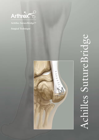
2. Surgical Technique
Achilles SutureBridge
1 2
A direct midline incision is made posteriorly with the patient in The Haglund’s prominence is removed using the micro-sagittal
the prone position. The incision is carried down to the calcaneus saw and osteotome. Care is taken to chamfer off the medial and
and calcaneal tendon insertion. The Achilles tendon is split at the lateral sides of the calcaneus so as not to leave a prominence that is
line incision, full thickness, from dorsal to ventral and is debrided palpable under the skin creating difficulties with footwear.
removing all tendinopathic tissue. The Achilles tendon is released
distally, and reflected medially and laterally, exposing the whole
calcaneal tuberosity with a Haglund's prominence. Care is taken
to maintain some medial and lateral attachments to assist with
the accurate restoration of the Achilles’ length. Complete tendon
debridement may require complete tendon detachment in some cases.
3 4
Prepare the bone for insertion of the two 5.5 mm Bio-Corkscrew® Insert the Arthrex 5.5 mm Bio-Corkscrew FT anchors into the
FT anchors by punching and tapping. Two holes are created about holes. The anchors are fully seated when inserted to the laser line
1 cm proximal to the distal insertion of the Achilles tendon and on the driver shaft. Slide the window open on the driver handle to
central to each half of the tendon. release the FiberWire sutures and needles. Pull the driver handles
out of the anchors. The needles may be cut from the suture, but
it is critical to leave sufficient suture tails for the SutureBridge
construct.
3. 5 6
The 5.5 mm Bio-Corkscrew FT anchors are double-loaded with Just distal to the end of the Achilles tendon insertion and directly
two #1 FiberWire sutures, one in blue and one in TigerWire® inferior to the Bio-Corkscrew FT anchor placements, the 3.5 mm
(black/white). For a simple, single stitch proximally, one suture PushLockTM anchor punch is used to create two holes for the
from each anchor is sufficient. The excess sutures are cut and distal row.
unloaded, leaving a blue FiberWire suture in one anchor and a
TigerWire suture in the other. The Achilles tendon is tensioned
proximally and approximated to its native location. Depending on
surgeon preference, either a Mason-Allen or mattress type stitch is
used to bring the tendon down to bone.
7 8
One blue FiberWire and one TigerWire suture from each of the With appropriate tension maintained on the sutures, the medial
proximal anchors are passed through the eyelet of the 3.5 mm button on the back of the handle is struck with a mallet to drive
Bio-PushLock anchor (a 4.5 mm PushLock may be substituted in the anchor into the bone and lock the sutures in place. The handle
softer bone). The 3.5 mm Bio-PushLock anchor is inserted up to is removed by turning counterclockwise until it releases from the
the laser line, just until the back tak portion of the anchor is even eyelet tip.
with the cortex. Suture tension is achieved by pulling one suture at
a time.
4. Optional:
Bio-TenodesisTM Screws may
be used for distal fixation.
Post-op Protocol
Postoperatively patients are treated with a below-knee
9 walking boot with or without a heel lift, depending on
surgeon preference - allowing them to weight-bear. The
SutureBridge construct can provide excellent security,
and avoiding the lift helps maximize flexibility and may
enhance rehabilitation. The patient should be protected
Steps 6-8 are followed for the other 3.5 mm Bio-PushLock anchor with crutches for approximately four weeks, at which
with the one remaining blue FiberWire and one TigerWire suture point physical therapy and range of motion is begun.
from each of the 5.5 mm Bio-Corkscrew FT anchors. Gradually wean your patients from the walking boot.
The resulting suture pattern should look similar to a capital 'M' or
sideways hourglass with the anchors at each corner. The FiberWire
suture is trimmed at the level of the cortex.
Implants
Achilles SutureBridge Convenience Pack AR-8927BNF-CP
contains the following, packaged for convenience and cost-effectiveness:
2 ea. Bio-Corkscrew FT, 5.5 mm x 15 mm,
w/two #1 FiberWire and Tapered Needles AR-8927BNF
2 ea. Bio-PushLock, 3.5 mm x 14 mm AR-1926B
Instruments
Bio-Corkscrew FT Punch, reusable AR-1927PB
Punch/Tap for Bio-Corkscrew FT AR-1927CTB
PushLock Punch, 3.5 mm AR-1926P
Bio-Corkscrew FT
Bio-PushLock
5. n ce
de
nfi
Co
Insertional calcific Achilles tendinosis is a
painful and frequently disabling condition.
While most patients with insertional
ith
Achilles tendinosis can be managed
nonoperatively, those patients who do not
kW
respond to conservative treatment may
al
require decompression and debridement
of the diseased tendon. The literature has
W described numerous operative approaches for
reattachment of the Achilles tendon and for
an associated tendon transfer of the flexor
hallucis longus (FHL) for augmentation.
While Arthrex provides means of fixation for
both, the reattachment of the tendon is the
focus of this technique guide.
The SutureBridge is a novel concept in
Achilles reattachment, following debridement.
While standard anchor fixation of the tendon
creates only a single point of compression
directly over the anchor, the SutureBridge
enables an hourglass pattern of FiberWire®
suture to be laid over the distal end of the
tendon. This four-anchor construct enables
a greater area of compression for the Achilles
tendon on the calcaneus, improving stability
and possibly allowing for earlier return to
normal activities.
Arthrex Achilles SutureBridge vs. Two-Anchor
Construct Peak Load Comparison*
Average Normalized Peak Load
3.5 Peak Load
3.0
2.5
2.0
2.05
1.5
1.0
1
0.5
0
SutureBridge Two-Anchor
*data on file