Coxa vara
•Transferir como PPTX, PDF•
43 gostaram•12,521 visualizações

A 3-year-old girl presented with a limp in her right leg and was diagnosed with coxa vara. Coxa vara is a decrease in the angle between the femoral neck and shaft to less than 120 degrees. It can be congenital, developmental, or secondary to other bone diseases. Surgical management involves subtrochanteric osteotomy to correct the angle, which is fixed internally with plates or screws. Postoperative care includes casting for 8-12 weeks until radiographic healing is seen. Complications can include recurrence, physeal injury, or leg length discrepancy.

Recomendados
Mais conteúdo relacionado
Mais procurados
Mais procurados (20)
Semelhante a Coxa vara
Semelhante a Coxa vara (20)
Coxa vara
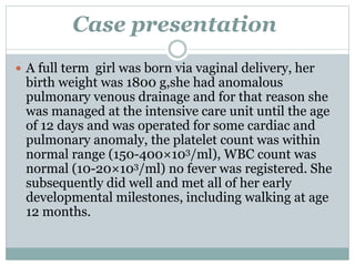
- 1. Case presentation A full term girl was born via vaginal delivery, her birth weight was 1800 g,she had anomalous pulmonary venous drainage and for that reason she was managed at the intensive care unit until the age of 12 days and was operated for some cardiac and pulmonary anomaly, the platelet count was within normal range (150-400×103/ml), WBC count was normal (10-20×103/ml) no fever was registered. She subsequently did well and met all of her early developmental milestones, including walking at age 12 months.
- 2. Case presentation cont.. She had no orthopedic issues until she was 3 year-10 months-old, when she presented with limp related to the right lower limb, with no pain. She had a leg-length discrepancy of 2 cm (right - left), limited right hip abduction to 25°, and internal rotation to 5°, also had a positive Trendelenburg test. No flexion/extension abnormalities.
- 3. DIAGNOSIS?
- 4. COXA VARA DR ISMAIL KHAN PG TRAINEE ORTHOPEDICS B UNIT HMC PESHAWAR.
- 5. INTRODUTION Coxa vara is progressive decrease in the angle between the femoral neck and shaft,less then 120 degrees Normal angle(120—135 degrees) Coxa vara is often bilateral A progressive shortening of the limb
- 6. Less then 120 degrees Less then 120
- 7. Epidemiology coxa vara(Congenital) is relatively rare condition incidence ranging from 1 per 13,000 population to 1 per 25,000 population Bilateral involvement seems to occur only half as often as unilateral involvement No clear pattern of inheritance But familial involvement in a number of cases has suggested an autosomal dominant genetic pattern of transmission.
- 8. CLASSIFICATION idiopathic : congenital : mild or severe coxa vara, with associated short or bowed femor. developmental : progressive, usually appearing between the ages of two and six years rachitic : usually associated with active rickets. adolescent : secondary to slipped capital femoral epiphysis. traumatic : usually following fracture of the femoral neck (rare in children). inflammatory : secondary to tuberculosis or other infection.
- 9. CLASSIFICATION cont.. secondary to other underlying bone diseases such as osteogenesis imperfecta cretinism dyschondroplasia Paget's disease osteoporosis capital coxa vara : occasionally seen in severe osteoarthritis and Legg-Perthes' disease
- 10. Etiology of congenital CV The exact cause of congenital coxa vara (CCV) remains unknown Many hypotheses have been proposed i. mechanical intrauterine stresses affecting hip development ii. avascular necrosis involving proximal femoral physis/head and neck; iii. Metabolic or endocrine abnormalities causing deficient or a delay in, the normal ossification process.
- 11. Pathophysiology Abnormal development of proximal femoral cartilaginous physis. Microscopically tissue in this defect consists of cartilage with irregular cell arrangement and ossification within it is atypical. Metaphyseal bone is osteoporotic Its trabeculae being atrophic. When walking is begun, the forces that the femoral neck must withstandare increased, and because the neck is weak, varus deformity gradually develops.
- 12. Presentation Affected children generally present between 1 to 6 years of age. usually present with gait abnormalities Limp(unilateral) ,weddling gait(bilateral) The Trendelenburg sign is commonly elicited in the affected hip or hips Shortenting of limb Trochanter is displaced upwards Decreased rotation and abduction of hip Pain stifness and flexion contracture
- 16. Figure . Abduction at the hip joint, viewed from behind. A, Normally, when weight is borne on one (e.g., the right) limb (during the stance phase of walking), the pelvis tends to sag on the free, or swing (left), side (because of gravity). This is counteracted by abduction of the hip on the stance (right) side, chiefly by strong contraction of the (right) gluteus medius, which acts on the pelvis from a fixed femur. B, When abduction of the (right) hip is interfered with on the supported side (positive Trendelenburg's sign)
- 17. Non surgical managment including spica cast immobilization and skeletal pin traction with bed rest, with generally unsatisfactory results
- 18. Surgical managment The treatment of choice is subtrochanteric osteotomy Surgery can be delayed until the child is 4 or 5 years old to make internal fixation easier Indications 1. femoral neck-shaft angle less than 90 - 100 degrees 2. Hilgenreiner's-epiphyseal angle greater than 45- 60 degrees 3. documented decrease in the femoral neck-shaft angle 4. Trendelenburg gait
- 20. Congenital coxa vara. Surgical methods of valgus-producing proximal femoral osteotomies. (A) Pauwels Y-shaped osteotomy. (B) Langenskiöld intertrochanteric osteotomy. (C) Borden subtrochanteric osteotomy.
- 27. The subtrochanteric osteotomy is fixed internally with either a) a blade plate or b) screw and plate combination Although biomechanically this may provide enough rigid internal fixation to eliminate the need for postoperative immobilization, a spica cast can be worn until union is complete.
- 28. TECHNIQUE ■ Perform an adductor tenotomy through a small medial incision. ■ Expose the greater trochanter and proximal shaft of the femur through an 8- to 10-cm lateral, longitudinal incision. ■ If a screw and side plate device is used for internal fixation, insert the screw in the midline of the femoral neck as determined by image intensification or anteroposterior and lateral radiographs. Insert the screw as close as possible to the trochanteric apophysis without entering it. If possible, center the screw in the femoral neck distal to the abnormal physis. If this is technically impossible, center the screw in the femoral head. ■ Make a transverse osteotomy slightly distal to the screw at about the level of the lesser trochanter. ■ If necessary, take a small lateral wedge of bone to correct the neck-shaft angle to 140 to 150 degrees. ■ Fix the side plate to the femoral shaft in the usual manner. ■ Irrigate the wound, and close it in layers, inserting irrigation-suction drainage if desired. ■ Apply a one and one-half spica cast
- 29. POSTOPERATIVE CARE The cast is removed at 8 to 12 weeks, when radiographic union of the osteotomy has occurred. Regular follow-up includes assessment of possible recurrence of the deformity and the development of progressive limb-length discrepancy.
- 30. Complications Recurrence of proximal femoral varus deformity. Premature physeal closurerelated to physeal injury at the time of surgery Greater trochanteric overgrowth-associated Acetabular dysplasia pseudarthrosis, avascular necrosis, leg-length discrepancy, and degenerative arthritis
- 31. THANKS