

Recomendados
Mais conteúdo relacionado
Mais procurados
Mais procurados (20)
Destaque
Destaque (20)
Semelhante a The excretory system
Semelhante a The excretory system (20)
Mais de DrChintansinh Parmar
Mais de DrChintansinh Parmar (20)
Último
Último (20)
The excretory system
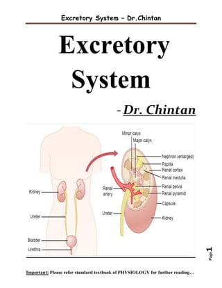
- 1. Excretory System – Dr.Chintan Important: Please refer standard textbook of PHYSIOLOGY for further reading… Page1 Excretory System -Dr. Chintan
- 2. Excretory System – Dr.Chintan Important: Please refer standard textbook of PHYSIOLOGY for further reading… Page2 INDEX 1. GFR ………………………………………………………………………………………………...03 2. Counter current Mechanism …………………………...…………...…………………..19 3. Micturition .……………………………………………………………………………………..32 4. PCT & DCT ………………..…………………………………………......................................41
- 3. Excretory System – Dr.Chintan Important: Please refer standard textbook of PHYSIOLOGY for further reading… Page3 Q-1. Describe the factors, regulation & measurement of GFR. Add a note on Nephritic and/or Nephrotic syndrome Urinary excretion = Glomerular filtration – tubular reabsorption + tubular secretion - GFR is determined by - (1) the balance of hydrostatic and colloid osmotic forces acting across the capillary membrane and - (2) The capillary filtration coefficient (Kf), the product of the permeability and filtering surface area of the capillaries. - In the average adult human, the GFR is about 125 ml/min, or 180 L/day.
- 4. Excretory System – Dr.Chintan Important: Please refer standard textbook of PHYSIOLOGY for further reading… Page4 - The fraction of the renal plasma flow that is filtered (the filtration fraction) averages about 0.2; - this means that about 20 per cent of the plasma flowing through the kidney is filtered - Filtration fraction = GFR/Renal plasma flow Glomerular Capillary Membrane - The capillary endothelium is perforated by thousands of small holes called fenestrae - Although the fenestrations are relatively large, endothelial cells are richly endowed with fixed negative charges that obstruct the passage of plasma proteins.
- 5. Excretory System – Dr.Chintan Important: Please refer standard textbook of PHYSIOLOGY for further reading… Page5 - The basement membrane consists of a meshwork of collagen and proteoglycan fibrillae that have large spaces through which large amounts of water and small solutes can filter. - The basement membrane effectively prevents filtration of plasma proteins because of strong negative electrical charges associated with the proteoglycans. - The final part of the glomerular membrane is a layer of epithelial cells that line the outer surface of the glomerulus. - These cells are not continuous but have long foot like processes (podocytes) that encircle the outer surface of the capillaries. - The foot processes are separated by gaps called slit pores through which the glomerular filtrate moves. - The epithelial cells also have negative charges, provide additional restriction to filtration of plasma proteins. Determinants of the GFR - The GFR is determined by - (1) the sum of the hydrostatic and colloid osmotic forces across the glomerular membrane, which gives the net filtration pressure,
- 6. Excretory System – Dr.Chintan Important: Please refer standard textbook of PHYSIOLOGY for further reading… Page6 - (2) The glomerular capillary filtration coefficient, Kf. - GFR = Kf X Net filtration pressure - The net filtration pressure represents the sum of the hydrostatic and colloid osmotic forces that either favor or oppose filtration across the glomerular capillaries - (1) hydrostatic pressure inside the glomerular capillaries (glomerular hydrostatic pressure, PG), which promotes filtration; - (2) the hydrostatic pressure in Bowman’s capsule (PB) outside the capillaries, which opposes filtration; - (3) the colloid osmotic pressure of the glomerular capillary plasma proteins (µG), which opposes filtration; and - (4) The colloid osmotic pressure of the proteins in Bowman’s capsule (µB), which promotes filtration. - (Under normal conditions, the concentration of protein in the glomerular filtrate is so low that the colloid osmotic pressure of the Bowman’s capsule fluid is considered to be zero.) - The GFR can therefore be expressed as - GFR = Kf X (PG – PB – µG + µB)
- 7. Excretory System – Dr.Chintan Important: Please refer standard textbook of PHYSIOLOGY for further reading… Page7 Determinants of the GFR - Increased Bowman’s Capsule Hydrostatic Pressure Decreases GFR - In certain pathological states associated with obstruction of the urinary tract, Bowman’s capsule pressure can increase markedly - serious reduction of GFR. - Precipitation of calcium or uric acid may lead to “stones” that lodge in the urinary tract, often in the ureter, thereby obstructing outflow of the urinary tract and raising Bowman’s capsule pressure. - Increased Glomerular Capillary Colloid Osmotic Pressure Decreases GFR - As blood passes from the afferent arteriole through the glomerular capillaries to the efferent arterioles, - the plasma protein concentration increases about 20 % - (1) the arterial plasma colloid osmotic pressure and
- 8. Excretory System – Dr.Chintan Important: Please refer standard textbook of PHYSIOLOGY for further reading… Page8 - (2) The fraction of plasma filtered by the glomerular capillaries (filtration fraction). - Increasing the arterial plasma colloid osmotic pressure raises the glomerular capillary colloid osmotic pressure - decreases GFR. - Increasing the filtration fraction also concentrates the plasma proteins and raises the glomerular colloid osmotic pressure - a greater rate of blood flow into the glomerulus tends to increase GFR, and a lower rate of blood flow into the glomerulus tends to decrease GFR. - Increased Glomerular Capillary Hydrostatic Pressure Increases GFR - Increased arterial pressure tends to raise glomerular hydrostatic pressure and so increases GFR. - Increased resistance of afferent arterioles reduces glomerular hydrostatic pressure and decreases GFR. - dilation of the afferent arterioles increases both glomerular hydrostatic pressure and GFR
- 9. Excretory System – Dr.Chintan Important: Please refer standard textbook of PHYSIOLOGY for further reading… Page9 - Constriction of the efferent arterioles increases the resistance to outflow from the glomerular capillaries. - This raises the glomerular hydrostatic pressure, and as long as the increase in efferent resistance does not reduce renal blood flow too much, GFR increases slightly - Efferent arteriolar constriction reduces renal blood flow - the filtration fraction and glomerular colloid osmotic pressure increase - So, if the constriction of efferent arterioles is severe, the rise in colloid osmotic pressure exceeds the increase in glomerular capillary hydrostatic pressure caused by efferent arteriolar constriction. - When this occurs, the net force for filtration actually decreases, causing a reduction in GFR. - Constriction of afferent arterioles reduces GFR. - the effect of efferent arteriolar constriction depends on the severity of the constriction;
- 10. Excretory System – Dr.Chintan Important: Please refer standard textbook of PHYSIOLOGY for further reading… Page10 - moderate efferent constriction raises GFR, - Severe efferent constriction reduces GFR. Physiologic Control of GFR and RBF - Sympathetic Nervous System Activation Decreases GFR - Strong activation of the renal sympathetic nerves can constrict the renal arterioles and decrease renal blood flow and GFR - Norepinephrine, Epinephrine and Endothelin Constrict Renal Blood Vessels and Decrease GFR - Angiotensin II Constricts Efferent Arterioles - raise glomerular hydrostatic pressure while reducing renal blood flow - helps prevent decreases in glomerular hydrostatic pressure and GFR
- 11. Excretory System – Dr.Chintan Important: Please refer standard textbook of PHYSIOLOGY for further reading… Page11 - Endothelial-Derived Nitric Oxide Decreases Renal Vascular Resistance and Increases GFR - Prostaglandins and Bradykinin Tend to Increase GFR Autoregulation of GFR and RBF - Feedback mechanisms intrinsic to the kidneys normally keep the renal blood flow and GFR relatively constant, despite marked changes in arterial blood pressure. - Role of Tubuloglomerular Feedback in Autoregulation of GFR - Decreased Macula Densa Sodium Chloride Causes Dilation of Afferent Arterioles and Increased Renin Release
- 12. Excretory System – Dr.Chintan Important: Please refer standard textbook of PHYSIOLOGY for further reading… Page12 Myogenic Autoregulation - The ability of individual blood vessels to resist stretching during increased arterial pressure. - Arterioles respond to increased wall tension or wall stretch by contraction of the vascular smooth muscle. - Stretch of the vascular wall allows increased movement of calcium ions from the ECF into the cells, causing them to contract - This contraction prevents overdistension of the vessel and raises vascular resistance - prevent excessive increases in RBF and GFR when arterial pressure increases.
- 13. Excretory System – Dr.Chintan Important: Please refer standard textbook of PHYSIOLOGY for further reading… Page13 Clearance Methods - The rates at which different substances are “cleared” from the plasma provide a useful way of quantitating the effectiveness with which the kidneys excrete various substances. - The renal clearance of a substance is the volume of plasma that is completely cleared of the substance by the kidneys per unit time - Renal clearance of a substance is calculated from the urinary excretion rate (U X V) of that substance divided by its plasma concentration (P). C = U X V / P Inulin Clearance - If a substance is freely filtered and is not reabsorbed or secreted by the renal tubules, - then the rate at which that substance is excreted in the urine is equal to the filtration rate of the substance by the kidneys - Inulin, a polysaccharide molecule with a molecular weight of about 5200. - Inulin is not produced in the body, is found in the roots of certain plants and must be administered intravenously to a patient to measure GFR.
- 14. Excretory System – Dr.Chintan Important: Please refer standard textbook of PHYSIOLOGY for further reading… Page14 - (1) if the clearance rate of the substance equals that of inulin, the substance is only filtered and not reabsorbed or secreted; - (2) if the clearance rate of a substance is less than inulin clearance, the substance must have been reabsorbed by the nephron tubules; and - (3) If the clearance rate of a substance is greater than that of inulin, the substance must be secreted by the nephron tubules. Creatinine Clearance - Creatinine is a by-product of muscle metabolism and is cleared from the body fluids almost entirely by glomerular filtration. - Because measurement of creatinine clearance does not require intravenous infusion into the patient, this method is much more widely used than inulin clearance for estimating GFR clinically. - creatinine clearance is not a perfect marker of GFR because a small amount of it is secreted by the tubules, so that the amount of creatinine excreted slightly exceeds the amount filtered GFR = CCreatinine = Ucreatinine X V / Pcreatinine - If GFR suddenly decreases by 50%, the kidneys will transiently filter and excrete only half as much creatinine, - causing accumulation of creatinine in the body fluids and raising plasma concentration
- 15. Excretory System – Dr.Chintan Important: Please refer standard textbook of PHYSIOLOGY for further reading… Page15 - plasma creatinine concentration is inversely proportional to GFR GLOMERULAR DISEASES - Glomerular diseases include a large and clinically significant group of renal diseases. - Glomerulonephritis (GN) is the term used for diseases that primarily involve the renal glomeruli. - I. Primary glomerulonephritis in which the glomeruli are the predominant site of involvement. - II. Secondary glomerular diseases include certain systemic and hereditary diseases which secondarily affect the glomeruli.
- 16. Excretory System – Dr.Chintan Important: Please refer standard textbook of PHYSIOLOGY for further reading… Page16 - The clinical presentation of glomerular disease is quite variable but in general four features—proteinuria, hematuria, hypertension and disturbed excretory function - nephritic and nephrotic syndromes; - acute and chronic renal failure; - asymptomatic proteinuria and haematuria ACUTE NEPHRITIC SYNDROME - This is the acute onset of haematuria, proteinuria, hypertension, oedema and oliguria following an infective illness about 10 to 20 days earlier. - 1. The haematuria is generally slight giving the urine smoky appearance and erythrocytes are detectable by microscopy - 2. The proteinuria is mild (less than 3 gm per 24 hrs) and is usually non-selective (nephritic range proteinuria). - 3. Hypertension is generally mild. - 4. Oedema in nephritic syndrome is usually mild and results from sodium and water retention. - 5. Oliguria is variable NEPHROTIC SYNDROME - it is characterised by findings of massive proteinuria, hypoalbuminaemia, oedema, hyperlipidaemia, lipiduria, and hypercoagulability. - 1. Heavy proteinuria (protein loss of more than 3 gm per 24 hrs) is the chief characteristic of nephrotic syndrome (nephrotic range proteinuria).
- 17. Excretory System – Dr.Chintan Important: Please refer standard textbook of PHYSIOLOGY for further reading… Page17 - A highly-selective proteinuria consists mostly of loss of low molecular weight proteins, while a poorly-selective proteinuria is loss of high molecular weight proteins in the urine. - In nephrotic syndrome, proteinuria mostly consists of loss of albumin (molecular weight 66,000) in the urine. - 2. Hypoalbuminaemia is produced primarily consequent to urinary loss of albumin, and partly due to increased renal catabolism and inadequate hepatic synthesis of albumin. - 3. Oedema in nephrotic syndrome appears due to fall in colloid osmotic pressure resulting from hypoalbuminaemia. - 4. Hyperlipidaemia - the liver faced with the stress of massive protein synthesis in response to heavy urinary protein loss, also causes increased synthesis of lipoproteins - 5. Lipiduria occurs following hyperlipidaemia due to excessive leakiness of glomerular filtration barrier. - 6. Hypercoagulability – - increased urinary loss of antithrombin III, - hyperfibrinogenaemia from increased synthesis in the liver, - decreased fibrinolysis, - increased platelet aggregation and - altered levels of protein C and S.
- 18. Excretory System – Dr.Chintan Important: Please refer standard textbook of PHYSIOLOGY for further reading… Page18
- 19. Excretory System – Dr.Chintan Important: Please refer standard textbook of PHYSIOLOGY for further reading… Page19 Q-2. Write in details about counter current mechanism & formation of concentrated urine by kidney Concentrated Urine The ability of the kidney to form a urine that is more concentrated than plasma is essential for survival of mammals that live on land, including humans. Water is continuously lost from the body through various routes, including the lungs by evaporation into the expired air, the gastrointestinal tract by way of the feces, the skin through evaporation and perspiration, and the kidneys through the excretion of urine. Fluid intake is required to match this loss, but the ability of the kidney to form a small volume of concentrated urine minimizes the intake of fluid required to maintain homeostasis, a function that is especially important when water is in short supply. When there is a water deficit in the body, the kidney forms a concentrated urine by continuing to excrete solutes while increasing water reabsorption and decreasing the volume of urine formed. The human kidney can produce a maximal urine concentration of 1200 to 1400 mOsm/L, four to five times the osmolarity of plasma. Some desert animals, such as the Australian hopping mouse, can concentrate urine to as high as 10,000 mOsm/L. This allows the mouse to survive in the desert without drinking water Obligatory Urine Volume The maximal concentrating ability of the kidney dictates how much urine volume must be excreted each day to rid the body of waste products of metabolism and ions that are ingested.
- 20. Excretory System – Dr.Chintan Important: Please refer standard textbook of PHYSIOLOGY for further reading… Page20 Minimal volume of urine that must be excreted, called the obligatory urine volume, - 0.5 L/Day (1) a high level of ADH, which increases the permeability of the distal tubules and collecting ducts to water, thereby allowing these tubular segments to avidly reabsorb water, and (2) a high osmolarity of the renal medullary interstitial fluid, which provides the osmotic gradient necessary for water reabsorption to occur in the presence of high levels of ADH. Concentrated Urine The renal medullary interstitium surrounding the collecting ducts normally is very hyperosmotic, so that when ADH levels are high, water moves through the tubular membrane by osmosis into the renal interstitium; from there it is carried away by the vasa recta back into the blood. The countercurrent mechanism depends on the special anatomical arrangement of the loops of Henle and the vasa recta. In the human, about 25 per cent of the nephrons are juxtamedullary nephrons, with loops of Henle and vasa recta that go deeply into the medulla before returning to the cortex. Countercurrent Mechanism The osmolarity of interstitial fluid in almost all parts of the body is about 300 mOsm/L, which is similar to the plasma osmolarity. The osmolarity of the interstitial fluid in the medulla of the kidney is much higher, increasing progressively to about 1200 to 1400 mOsm/L in the pelvic tip of the medulla.
- 21. Excretory System – Dr.Chintan Important: Please refer standard textbook of PHYSIOLOGY for further reading… Page21 This means that the renal medullary interstitium has accumulated solutes in great excess of water. Once the high solute concentration in the medulla is achieved, it is maintained by a balanced inflow and outflow of solutes and water in the medulla. 1. Active transport of sodium ions and co-transport of potassium, chloride, and other ions out of the thick portion of the ascending limb of the loop of Henle into the medullary interstitium 2. Active transport of ions from the collecting ducts into the medullary interstitium 3. Facilitated diffusion of large amounts of urea from the inner medullary collecting ducts into the medullary interstitium 4. Diffusion of only small amounts of water from the medullary tubules into the medullary interstitium
- 22. Excretory System – Dr.Chintan Important: Please refer standard textbook of PHYSIOLOGY for further reading… Page22 Because the thick ascending limb is almost impermeable to water, the solutes pumped out are not followed by osmotic flow of water into the interstitium. Thus, the active transport of sodium and other ions out of the thick ascending loop adds solutes in excess of water to the renal medullary interstitium. There is some passive reabsorption of sodium chloride from the thin ascending limb of Henle’s loop, which is also impermeable to water, adding further to the high solute concentration of the renal medullary interstitium. The descending limb of Henle’s loop, in contrast to the ascending limb, is very permeable to water, and the tubular fluid osmolarity quickly becomes equal to the renal medullary osmolarity. Therefore, water diffuses out of the descending limb of Henle’s loop into the interstitium, and the tubular fluid osmolarity gradually rises First, the loop of Henle is filled with fluid with a concentration of 300 mOsm/L, the same as that leaving the proximal tubule
- 23. Excretory System – Dr.Chintan Important: Please refer standard textbook of PHYSIOLOGY for further reading… Page23 Next, the active pump of the thick ascending limb on the loop of Henle is turned on, reducing the concentration inside the tubule raising the interstitial concentration; this pump establishes a 200-mOsm/L concentration gradient between the tubular fluid and the interstitial fluid Step 3 is that the tubular fluid in the descending limb of the loop of Henle and the interstitial fluid quickly reach osmotic equilibrium because of osmosis of water out of the descending limb. The interstitial osmolarity is maintained at 400 mOsm/L because of continued transport of ions out of the thick ascending loop of Henle.
- 24. Excretory System – Dr.Chintan Important: Please refer standard textbook of PHYSIOLOGY for further reading… Page24 Step 4 is additional flow of fluid into the loop of Henle from the proximal tubule, which causes the hyperosmotic fluid previously formed in the descending limb to flow into the ascending limb
- 25. Excretory System – Dr.Chintan Important: Please refer standard textbook of PHYSIOLOGY for further reading… Page25 Once the fluid is in the ascending limb, additional ions are pumped into the interstitium, with water remaining behind, until a 200-mOsm/L osmotic gradient is established, with the interstitial fluid osmolarity rising to 500 mOsm/L Then, once again, the fluid in the descending limb reaches equilibrium with the hyperosmotic medullary interstitial fluid as the hyperosmotic tubular fluid from the descending limb of the loop of Henle flows into the ascending limb, Still more solute is continuously pumped out of the tubules and deposited into the medullary interstitium
- 26. Excretory System – Dr.Chintan Important: Please refer standard textbook of PHYSIOLOGY for further reading… Page26 These steps are repeated over and over, with the net effect of adding more and more solute to the medulla in excess of water; this process gradually traps solutes in the medulla and multiplies the concentration gradient established by the active pumping of ions out of the thick ascending loop of Henle, eventually raising the interstitial fluid osmolarity to 1200 to 1400 mOsm/L
- 27. Excretory System – Dr.Chintan Important: Please refer standard textbook of PHYSIOLOGY for further reading… Page27 The repetitive reabsorption of NaCl by the thick ascending loop of Henle & Continued inflow of new NaCl from the proximal tubule into the loop of Henle is called the countercurrent multiplier. NaCl reabsorbed from the ascending loop of Henle keeps adding to the newly arrived NaCl, thus “multiplying” its concentration in the medullary interstitium. Role of Distal Tubule and Collecting Ducts When the tubular fluid leaves the loop of Henle and flows into the DCT in the renal cortex, the fluid is dilute, with an osmolarity of only about 100 mOsm/L The early distal tubule further dilutes the tubular fluid because this segment, like the ascending loop of Henle, actively transports NaCl out of the tubule but is relatively impermeable to water. As fluid flows into the cortical collecting tubule, the amount of water reabsorbed is critically dependent on the plasma concentration of ADH In the absence of ADH, this segment is almost impermeable to water and fails to reabsorb water but continues to reabsorb solutes and further dilutes the urine. When there is a high concentration of ADH, the cortical collecting tubule becomes highly permeable to water, so that large amounts of water are now reabsorbed from the tubule into the cortex interstitium, where it is swept away by the rapidly flowing peritubular capillaries. These large amounts of water are reabsorbed into the cortex, rather than into the renal medulla, helps to preserve the high medullary interstitial fluid osmolarity. As the tubular fluid flows along the medullary collecting ducts, there is further water reabsorption from the tubular fluid into the interstitium, but the
- 28. Excretory System – Dr.Chintan Important: Please refer standard textbook of PHYSIOLOGY for further reading… Page28 total amount of water is relatively small compared with that added to the cortex interstitium. The reabsorbed water is quickly carried away by the vasa recta into the venous blood. When high levels of ADH are present, the collecting ducts become permeable to water, so that the fluid at the end of the collecting ducts has essentially the same osmolarity as the interstitial fluid of the renal medulla — about 1200 mOsm/L Thus, by reabsorbing as much water as possible, the kidneys form highly concentrated urine. Urea Contributes When there is water deficit and blood concentrations of ADH are high, large amounts of urea are passively reabsorbed from the inner medullary collecting ducts into the interstitium. As water flows up the ascending loop of Henle and into the distal and cortical collecting tubules, little urea is reabsorbed because these segments are impermeable to urea.
- 29. Excretory System – Dr.Chintan Important: Please refer standard textbook of PHYSIOLOGY for further reading… Page29 In the presence of ADH, water is reabsorbed rapidly from the cortical collecting tubule and the urea concentration increases rapidly because urea is not very permeant in this part of the tubule. Then, as the tubular fluid flows into the inner medullary collecting ducts, still more water reabsorption takes place, causing an even higher concentration of urea in the fluid. This high concentration of urea in the tubular fluid of the inner medullary collecting duct causes urea to diffuse out of the tubule into the renal interstitium. This diffusion is greatly facilitated by specific urea transporters - UT-AI - activated by ADH, increasing transport of urea out of the inner medullary collecting duct people who ingest a high-protein diet, yielding large amounts of urea as a nitrogenous “waste” product, can concentrate their urine much better than people whose protein intake and urea production are low. Malnutrition is associated with a low urea concentration in the medullary interstitium and considerable impairment of urine concentrating ability.
- 30. Excretory System – Dr.Chintan Important: Please refer standard textbook of PHYSIOLOGY for further reading… Page30 Recirculation of Urea Countercurrent Exchange 1. The medullary blood flow is low, accounting for less than 5 per cent of the total renal blood flow. This sluggish blood flow is sufficient to supply the metabolic needs of the tissues & helps to minimize solute loss from the medullary interstitium. 2. The vasa recta serve as countercurrent exchangers, minimizing washout of solutes from the medullary interstitium. - As blood descends into the medulla, it becomes progressively more concentrated, partly by solute entry from the interstitium and partly by loss of water into the interstitium. - By the time the blood reaches the tips of the vasa recta, it has a concentration of about 1200 mOsm/L, the same as that of the medullary interstitium.
- 31. Excretory System – Dr.Chintan Important: Please refer standard textbook of PHYSIOLOGY for further reading… Page31 - As blood ascends back toward the cortex, it becomes progressively less concentrated as solutes diffuse back out into the medullary interstitium and as water moves into the vasa recta.
- 32. Excretory System – Dr.Chintan Important: Please refer standard textbook of PHYSIOLOGY for further reading… Page32 Q-3. Write in details about micturition & Cystometrogram. Add a note on applied aspect of micturition. • Micturition is the process by which the urinary bladder empties when it becomes filled. • This involves two main steps: • First, the bladder fills progressively until the tension in its walls rises above a threshold level; • this elicits the second step, which is a nervous reflex called the micturition reflex that empties the bladder or, if this fails, at least causes a conscious desire to urinate. • Although the micturition reflex is an autonomic spinal cord reflex, it can also be inhibited or facilitated by centers in the cerebral cortex or brain stem. Physiologic Anatomy • The urinary bladder is a smooth muscle chamber composed of two main parts: • (1) the body, which is the major part of the bladder in which urine collects, and • (2) the neck, which is a funnel-shaped extension of the body, passing inferiorly and anteriorly into the urogenital triangle and connecting with the urethra. • The smooth muscle of the bladder is called the detrusor muscle - contraction of the detrusor muscle is a major step in emptying the bladder
- 33. Excretory System – Dr.Chintan Important: Please refer standard textbook of PHYSIOLOGY for further reading… Page33 Innervation of the Bladder • The principal nerve supply of the bladder is by way of the pelvic nerves, which connect with the spinal cord through the sacral plexus, mainly connecting with cord segments S-2 and S-3. • Coursing through the pelvic nerves are both sensory nerve fibers and motor nerve fibers. The sensory fibers detect the degree of stretch in the bladder wall. • The motor nerves transmitted in the pelvic nerves are parasympathetic fibers. These terminate on ganglion cells located in the wall of the bladder. Short postganglionic nerves then innervate the detrusor muscle. • skeletal motor fibers transmitted through the pudendal nerve to the external bladder sphincter - somatic nerve fibers that innervate and control the voluntary skeletal muscle of the sphincter.
- 34. Excretory System – Dr.Chintan Important: Please refer standard textbook of PHYSIOLOGY for further reading… Page34 • The bladder receives sympathetic innervation from the sympathetic chain through the hypogastric nerves, connecting mainly with the L-2 segment of the spinal cord. • These sympathetic fibers stimulate mainly the blood vessels - sensory nerve fibers also pass by way of the sympathetic nerves and important in the sensation of fullness and pain Transport of Urine • Urine flowing from the collecting ducts into the renal calyces stretches the calyces and increases their intrinsic pacemaker activity, • which in turn initiates peristaltic contractions that spread to the renal pelvis and then downward along the length of the ureter • peristaltic contractions in the ureter are enhanced by parasympathetic stimulation and • inhibited by sympathetic stimulation
- 35. Excretory System – Dr.Chintan Important: Please refer standard textbook of PHYSIOLOGY for further reading… Page35 • The normal tone of the detrusor muscle in the bladder wall have a tendency to compress the ureter, • thereby preventing backflow of urine from the bladder when pressure builds up in the bladder during micturition or bladder compression • Vesicoureteral reflux – enlargement of the ureters - can increase the pressure in the renal calyces and structures of the renal medulla, causing damage - hydronephrosis The Cystometrogram • When there is no urine in the bladder, the intravesicular pressure is about 0, • but by the time 30 to 50 milliliters of urine has collected, the pressure rises to 5 to 10 centimeters of water. • Additional urine — 200 to 300 milliliters — can collect with only a small additional rise in pressure; this constant level of pressure is caused by intrinsic tone of the bladder wall. • Beyond 300 to 400 milliliters, collection of more urine in the bladder causes the pressure to rise rapidly.
- 36. Excretory System – Dr.Chintan Important: Please refer standard textbook of PHYSIOLOGY for further reading… Page36 • Superimposed on the tonic pressure changes during filling of the bladder are periodic acute increases in pressure that last from a few seconds to more than a minute. • The pressure peaks may rise only a few centimeters of water or may rise to more than 100 centimeters of water. • These pressure peaks are called micturition waves in the Cystometrogram and are caused by the micturition reflex. Micturition Reflex • micturition contractions are the result of a stretch reflex initiated by sensory stretch receptors in the bladder wall, • Especially by the receptors in the posterior urethra when this area begins to fill with urine at the higher bladder pressures. • Sensory signals from the bladder stretch receptors are conducted to the sacral segments of the cord through the pelvic nerves • and then reflexively back again to the bladder through the parasympathetic nerve fibers by way of these same nerves. • When the bladder is only partially filled, these micturition contractions usually relax spontaneously after a fraction of a minute, the detrusor muscles stop contracting, and pressure falls back to the baseline. • As the bladder continues to fill, the micturition reflexes become more frequent and cause greater contractions of the detrusor muscle. • Once a micturition reflex begins, it is “self-regenerative.” • initial contraction of the bladder activates the stretch receptors to cause a greater increase in sensory impulses to the bladder and posterior urethra, which causes a further increase in reflex contraction of the bladder
- 37. Excretory System – Dr.Chintan Important: Please refer standard textbook of PHYSIOLOGY for further reading… Page37 • Cycle is repeated again and again until the bladder has reached a strong degree of contraction. • Then, after a few seconds to more than a minute, the self-regenerative reflex begins to fatigue and the regenerative cycle of the micturition reflex stops, permitting the bladder to relax. • the micturition reflex is a single complete cycle of • (1) progressive and rapid increase of pressure, • (2) a period of sustained pressure, and • (3) return of the pressure to the basal tone of the bladder • Once a micturition reflex has occurred but has not succeeded in emptying the bladder, • the nervous elements of this reflex usually remain in an inhibited state for a few minutes to 1 hour or more before another micturition reflex occurs. • As the bladder becomes more and more filled, micturition reflexes occur more and more often and more and more powerfully. • Once the micturition reflex becomes powerful enough, it causes another reflex, which passes through the pudendal nerves to the external sphincter to inhibit it. • If this inhibition is more potent in the brain than the voluntary constrictor signals to the external sphincter, urination will occur. • If not, urination will not occur until the bladder fills still further and the micturition reflex becomes more powerful. Role of the Brain • The micturition reflex is a completely autonomic spinal cord reflex, but it can be inhibited or facilitated by centers in the brain.
- 38. Excretory System – Dr.Chintan Important: Please refer standard textbook of PHYSIOLOGY for further reading… Page38 • These centers include • (1) strong facilitative and inhibitory centers in the brain stem, located mainly in the pons, and • (2) Several centers located in the cerebral cortex that are mainly inhibitory but can become excitatory. • The micturition reflex is the basic cause of micturition, but the higher centers normally exert final control of micturition • 1. The higher centers keep the micturition reflex partially inhibited, except when micturition is desired. • 2. The higher centers can prevent micturition, even if the micturition reflex occurs, by continual tonic contraction of the external bladder sphincter until a convenient time presents itself. • 3. When it is time to urinate, the cortical centers can facilitate the sacral micturition centers to help initiate a micturition reflex • and at the same time inhibit the external urinary sphincter so that urination can occur. Voluntary urination • First, a person voluntarily contracts his or her abdominal muscles, which increases the pressure in the bladder • And allows extra urine to enter the bladder neck and posterior urethra under pressure, thus stretching their walls. • This stimulates the stretch receptors, which excites the micturition reflex and simultaneously inhibits the external urethral sphincter. • Ordinarily, all the urine will be emptied, with rarely more than 5 to 10 milliliters left in the bladder.
- 39. Excretory System – Dr.Chintan Important: Please refer standard textbook of PHYSIOLOGY for further reading… Page39 Abnormalities of Micturition • Atonic Bladder Caused by Destruction of Sensory Nerve Fibers - preventing transmission of stretch signals from the bladder. • Person loses bladder control, despite intact efferent fibers from the cord to the bladder and despite intact neurogenic connections within the brain. • Instead of emptying periodically, the bladder fills to capacity and overflows a few drops at a time through the urethra - overflow incontinence. • crush injury to the sacral region of the spinal cord - syphilis can cause constrictive fibrosis around the dorsal root nerve fibers (tabes dorsalis) • Automatic Bladder Caused by Spinal Cord Damage Above the Sacral Region. • If the sacral cord segments are still intact, typical micturition reflexes can still occur but they are no longer controlled by the brain. • first micturition reflexes are suppressed because of the state of “spinal shock” • If the bladder is emptied periodically by catheterization, the excitability of the micturition reflex gradually increases - then, periodic automatic bladder emptying occurs. • Some patients can still control urination in this condition by stimulating the skin (scratching or tickling) in the genital region, which sometimes elicits a micturition reflex.
- 40. Excretory System – Dr.Chintan Important: Please refer standard textbook of PHYSIOLOGY for further reading… Page40 • Uninhibited Neurogenic Bladder Caused by Lack of Inhibitory Signals from the Brain. • Frequent and relatively uncontrolled micturition. • Partial damage in the spinal cord or the brain stem that interrupts most of the inhibitory signals. • Facilitative impulses passing continually down the cord keep the sacral centers so excitable that even a small quantity of urine elicits an uncontrollable micturition reflex, thereby promoting frequent urination.
- 41. Excretory System – Dr.Chintan Important: Please refer standard textbook of PHYSIOLOGY for further reading… Page41 Q-4. Write in details about structure & reabsorption at Proximal Convoluted Tubule(PCT) & Distal Convoluted Tubule(DCT) Tubular Reabsorption • Glucose and amino acids, are almost completely reabsorbed from the tubules, so that the urinary excretion rate is essentially zero. • Many of the ions in the plasma, such as sodium, chloride, and bicarbonate, are also highly reabsorbed, but their rates of reabsorption and urinary excretion are variable, depending on the needs of the body. • Certain waste products, such as urea and creatinine, are poorly reabsorbed from the tubules and excreted in relatively large amounts • For a substance to be reabsorbed, it must first be transported • (1) across the tubular epithelial membranes into the renal interstitial fluid and then • (2) through the peritubular capillary membrane back into the blood • water and solutes can be transported either through the cell membranes themselves (transcellular route) or • Through the junctional spaces between the cells (paracellular route).
- 42. Excretory System – Dr.Chintan Important: Please refer standard textbook of PHYSIOLOGY for further reading… Page42 Active Transport • Active transport can move a solute against an electrochemical gradient and requires energy • Transport that is coupled directly to an energy source, such as the hydrolysis of ATP, is termed primary active transport. • A good example of this is the sodium-potassium ATPase pump that functions throughout most parts of the renal tubule. • Transport that is coupled indirectly to an energy source, such as that due to an ion gradient, is referred to as secondary active transport. • Reabsorption of glucose by the renal tubule is an example of secondary active transport. • Renal tubular cells held together by tight junctions. Lateral intercellular spaces lie behind the tight junctions and separate the epithelial cells of the tubule.
- 43. Excretory System – Dr.Chintan Important: Please refer standard textbook of PHYSIOLOGY for further reading… Page43 • Solutes can be reabsorbed or secreted across the cells by way of the transcellular pathway or between the cells by moving across the tight junctions and intercellular spaces by way of the paracellular pathway. • most of the sodium is transported through the transcellular pathway. • In proximal tubule, water is also reabsorbed across the paracellular pathway, and substances dissolved in the water are carried with the reabsorbed fluid between the cells. Primary Active Transport • Primary active transporters – sodium potassium ATPase, hydrogen ATPase, hydrogen potassium ATPase, and calcium ATPase. • reabsorption of sodium ions across the proximal tubular membrane • On the basolateral sides of the tubular epithelial cell, the cell membrane has an extensive sodium-potassium ATPase system • transport sodium ions out of the cell into the interstitium. And, potassium is transported from the interstitium to the inside of the cell.
- 44. Excretory System – Dr.Chintan Important: Please refer standard textbook of PHYSIOLOGY for further reading… Page44 • 1. Sodium diffuses across the luminal membrane into the cell down an electrochemical gradient established by the sodium-potassium ATPase pump on the basolateral membrane. • 2. Sodium is transported across the basolateral membrane against an electrochemical gradient by the sodium-potassium ATPase pump. • 3. Sodium, water, and other substances are reabsorbed from the interstitial fluid into the peritubular capillaries by ultrafiltration, a passive process driven by the hydrostatic and colloid osmotic pressure gradients. Secondary Active Reabsorption • In secondary active transport, two or more substances interact with a specific membrane protein (a carrier molecule) and are transported together across the membrane. • As one substance (sodium) diffuses down its electrochemical gradient, the energy released is used to drive another substance (glucose, amino acids) against its electrochemical gradient • After entry into the cell, glucose and amino acids exit across the basolateral membranes by facilitated diffusion
- 45. Excretory System – Dr.Chintan Important: Please refer standard textbook of PHYSIOLOGY for further reading… Page45 Pinocytosis • The proximal tubule, reabsorb large molecules such as proteins by pinocytosis. • In this process, the protein attaches to the brush border of the luminal membrane, and this portion of the membrane then invaginates to the interior of the cell until it is completely pinched off and a vesicle is formed containing the protein. • Once inside the cell, the protein is digested into its constituent amino acids, which are reabsorbed through the basolateral membrane into the interstitial fluid. • Because pinocytosis requires energy, it is considered a form of active transport.
- 46. Excretory System – Dr.Chintan Important: Please refer standard textbook of PHYSIOLOGY for further reading… Page46 Transport Maximum • For most substances that are actively reabsorbed or secreted, there is a limit to the rate at which the solute can be transported, often referred to as the transport maximum. • This limit is due to saturation of the specific transport systems involved when the amount of solute delivered to the tubule (referred to as tubular load) exceeds the capacity of the carrier proteins and specific enzymes involved in the transport process. • Normally, glucose does not appear in the urine because essentially all the filtered glucose is reabsorbed in the proximal tubule • In the adult human, the transport maximum for glucose averages about 375 mg/min, whereas the filtered load of glucose is only about 125 mg/min (GFR X plasma glucose = 125 ml/min X 1 mg/ml). • With large increases in GFR and/or plasma glucose concentration that increase the filtered load of glucose above 375 mg/min, the excess glucose filtered is not reabsorbed and passes into the urine. • when the plasma concentration of glucose rises above about 180 mg/100 ml, a small amount of glucose begins to appear in the urine - threshold for glucose • appearance of glucose in the urine (at the threshold) occurs before the transport maximum is reached. • One reason for the difference between threshold and transport maximum is that all nephrons do not have the same transport maximum for glucose, and • some of the nephrons excrete glucose before others have reached their transport maximum
- 47. Excretory System – Dr.Chintan Important: Please refer standard textbook of PHYSIOLOGY for further reading… Page47 Passive Water Reabsorption • When solutes are transported out of the tubule by either primary or secondary active transport, their concentrations tend to decrease inside the tubule while increasing in the renal interstitium. • This creates a concentration difference that causes osmosis of water in the same direction that the solutes are transported, from the tubular lumen to the renal interstitium. • As water moves across the tight junctions by osmosis, it can also carry with it some of the solutes - solvent drag. • In the proximal tubule, the water permeability is always high, and water is reabsorbed as rapidly as the solutes.
- 48. Excretory System – Dr.Chintan Important: Please refer standard textbook of PHYSIOLOGY for further reading… Page48 • Water permeability in — the distal tubules, collecting tubules, and collecting ducts — can be high or low, depending on the presence or absence of ADH • When sodium is reabsorbed through the tubular epithelial cell, negative ions such as chloride are transported along with sodium because of electrical potentials Proximal Tubular Reabsorption • Normally, about 65 per cent of the filtered load of sodium and water and a 60 per cent of filtered chloride are reabsorbed by the proximal tubule before the filtrate reaches the loops of Henle. • The proximal tubule epithelial cells are highly metabolic and have large numbers of mitochondria to support potent active transport processes.
- 49. Excretory System – Dr.Chintan Important: Please refer standard textbook of PHYSIOLOGY for further reading… Page49 • tubular cells have an extensive brush border on the luminal (apical) side of the membrane and an extensive labyrinth of intercellular and basal channels - extensive membrane surface area on the luminal and basolateral sides of the epithelium for rapid transport • Brush border is also loaded with protein carrier molecules that transport a large fraction of the sodium ions across the luminal membrane linked by way of the cotransport mechanism with multiple organic nutrients such as amino acids and glucose. • The remainder of the sodium is transported from the tubular lumen into the cell by counter-transport mechanisms, which reabsorb sodium while secreting other substances into the tubular lumen, especially hydrogen ions • In the first half of the proximal tubule, sodium is reabsorbed by co- transport along with glucose, amino acids, and other solutes.
- 50. Excretory System – Dr.Chintan Important: Please refer standard textbook of PHYSIOLOGY for further reading… Page50 • In the second half of the proximal tubule, sodium is reabsorbed mainly with chloride ions Distal Tubule • The very first portion of the distal tubule forms part of the juxtaglomerular complex that provides feedback control of GFR and blood flow. • The next part of the distal tubule is highly convoluted and reabsorbs most of the ions, including sodium, potassium, and chloride, but is virtually impermeable to water and urea. • For this reason, it is referred to as the diluting segment because it also dilutes the tubular fluid.
- 51. Excretory System – Dr.Chintan Important: Please refer standard textbook of PHYSIOLOGY for further reading… Page51 • Approximately 5 % of the filtered load of sodium chloride is reabsorbed in the early distal tubule.
- 52. Excretory System – Dr.Chintan Important: Please refer standard textbook of PHYSIOLOGY for further reading… Page52 Late Distal Tubule and Cortical Collecting Tubule • The principal cells reabsorb sodium and water from the lumen and secrete potassium ions into the lumen. • The intercalated cells reabsorb potassium ions and secrete hydrogen ions into the tubular lumen. • Sodium reabsorption and potassium secretion by the principal cells depend on the activity of a sodium potassium ATPase pump in each cell’s basolateral membrane • This pump maintains a low sodium concentration inside the cell and, therefore, favors sodium diffusion into the cell through special channels
- 53. Excretory System – Dr.Chintan Important: Please refer standard textbook of PHYSIOLOGY for further reading… Page53 Intercalated Cells • Hydrogen ion secretion by the intercalated cells is mediated by a hydrogen- ATPase transport mechanism • H2O + CO2 carbonic anhydrase H2CO3 • H2CO3 → H+ + HCO3- • The hydrogen ions are then secreted into the tubular lumen, and for each hydrogen ion secreted, a bicarbonate ion becomes available for reabsorption across the basolateral membrane. • The intercalated cells can also reabsorb potassium ions.
- 54. Excretory System – Dr.Chintan Important: Please refer standard textbook of PHYSIOLOGY for further reading… Page54 Late Distal Tubule and Cortical Collecting Tubule Proximal tubule, early distal tubule
- 55. Excretory System – Dr.Chintan Important: Please refer standard textbook of PHYSIOLOGY for further reading… Page55 Phosphate Buffer Proximal tubule
- 56. Excretory System – Dr.Chintan Important: Please refer standard textbook of PHYSIOLOGY for further reading… Page56 ADH • The permeability of the late distal tubule and cortical collecting duct to water is controlled by the concentration of ADH (vasopressin). • With high levels of ADH, these tubular segments are permeable to water, but in the absence of ADH, they are virtually impermeable to water. • This special characteristic provides an important mechanism for controlling the degree of dilution or concentration of the urine. Thank You…