TORCH
•Transferir como PPTX, PDF•
18 gostaram•3,047 visualizações

Hi everyone This presentation explains details about intrauterine infections also called TORCH. Thank you

Recomendados
Mais conteúdo relacionado
Mais procurados
Mais procurados (20)
Destaque
Destaque (20)
Semelhante a TORCH
Semelhante a TORCH (20)
Último
Último (20)
TORCH
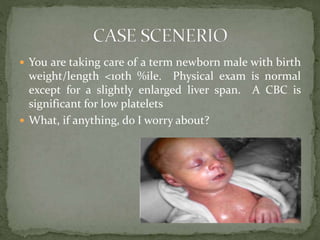
- 1. You are taking care of a term newborn male with birth weight/length <10th %ile. Physical exam is normal except for a slightly enlarged liver span. A CBC is significant for low platelets What, if anything, do I worry about?
- 2. DR ANKIT GUPTA KIMS KARAD
- 3. The original concept of the TORCH perinatal infections was to group five infections with similar presentations, including rash and ocular findings These five infections are: Toxoplasmosis Other (syphilis) Rubella Cytomegalovirus (CMV) Herpes simplex virus (HSV) Also described as STORCH
- 4. VIRUSES Varicella, Parvovirus B19, Hep B and C, HIV Enteroviruses (coxackie) BACTERIA Listeria, Neisseria Mycobacterium tuberculosis, salmonella, campylobater PARASITES Trepenoma pallidum, plasmodium FUNGAL Candida
- 5. ASYMPTOMATIC IN MOTHER HAVOC IN OFFSPRING
- 6. Fetus may get infected In utero During delivery Severity of manifestation depends on gestational age Less the age more the morbidity May lead to fetal loss or congenital malformations Neonate may be even asymptomatic at birth and may manifest symptoms later anytime during infancy or even upto first decade of life Therefore it is very important to recognize and manage the neonate at the earliest
- 7. WRONG CONCEPTS BASIC NOT CLEAR UNNECESSARY TREATMENT IMPROPER MANAGEMENT
- 8. COMMON PRESENTATION 1. Premature delivery 2. IUGR or intrauterine death 3. Jaundice, petechia or purpura 4. Hepatosplenomegaly, anemia,thrombocytopenia 5. Hydrocephaly, microcephaly,intracranial calcification 6. Chorioretinitis 7. Myocarditis & cardiac abnormalities But each infection have its peculiar presentation and management So, very important to differentiate and identify the particular infection
- 9. DNA virus and a member of the “herpesvirus group”. Most common congenital infection Severe sequalae are more common with infection in the 1st trimester, while the overall risk of infection is greatest in the 3rd trimester Congenital CMV affects about 0.2-2.5% of babies worldwide Of these, only 1-10% of the babies born with the CMV infection will have symptoms at birth and another 10- 15% may not show any symptoms at birth, but still may have long term affects such as hearing loss and learning disabilities
- 10. Secreted in all body fluids Close contact through saliva, blood, genital secretions, urine Neonatal infection Intrauterine or transplacental Intrapartum – cervical and vaginal secretions 2% to 28% of seropositive pregnant women shed CMV Approximately 50% of exposed infants become infected Develop clinical signs of CMV disease at about 4 to 6 weeks of age.
- 11. Postpartum period – Breast Feeding 9% to 88% of seropositive women shed CMV into their milk 50% to 60% of infants become infected Blood Transfusions 2 -20% from unscreened or unfiltered blood Especially preterms are more prone Source - Stehel EK and Sanchez PJ. Cytomegalovirus Infection in the Fetus and Neonate. NeoReviews 2005;6;e38-e45
- 14. BLUEBERRY MUFFIN SPOTS
- 15. COTTAGE CHEESE WITH KETCHUP TYPE OF CHORIORETINITIS NORMAL ABNORMAL
- 16. SENSINEURAL DEAFNESS Most commonly seen if infection occurs in first or second trimester Usually progressive, unilateral or bilateral and may even manifest later in life
- 17. ANTIBODY TITRE IgG and IgM by ELISA Not reliable VIRAL DETECTION (GOLD STANDARD) Neonatal body fluids – urine, blood, saliva by PCR or culture within first 3 weeks of life NEUROIMAGING Cranial Ultrasound or MRI OTHERS Ophthalmology and audiological evaluation
- 18. PREVENTION IS BETTER THAN CURE Mother should wash hands before and after handling the baby No donor breast milk High chances of transmission through breast milk especially in preterms Mother can continue breast feeding Avoid unnecessary blood transfusion
- 19. WHEN TO GIVE Specific indication Chorioretinitis Relative indication Hepatitis, menigioencephalitis, Rx Ganciclovir Induction - 5mg/kg/dose 12 hourly for 2-3 weeks Maintainence – 5mg/kg/dose once daily for 8-10 weeks Very toxic – Daily monitoring of blood counts and platelets Should be stopped if Neutrophils < 500/mm3 or platelets < 25000/mm3 May lead to testicular atrophy
- 20. Also known as German Measles Single stranded RNA virus, member of family Togavirus, and the genus, Rubivirus Vaccine preventable disease Effect on fetus 1st trimester- 80% infection, 90% malformation 2nd trimester- 25% infection, 30% malformation 3rd trimester- 100% infection, 10% malformation Spontaneous abortions occur in upto 20% of cases if infection occur within 20 wks of gestation.
- 21. MAY BE ASMPTOMATIC May present as acute viral illness Low grade fever, headache, swollen lymph nodes, joint pain runny nose and rash Contagious one week before and after the rash
- 22. Congenital rubella syndrome presents a classic triad EYE Cataract Glaucoma Chorioretinitis Microphthalmia EAR Sensorineural hearing loss is the most frequent sequelae -80% of infected children Bilateral and progressive HEART Congenital malformation in ~ 50% of children infected in first 8 wks of gestation and consist of PDA, Pulmonary artery stenosis
- 24. CHORIORETINITIS – Salt and Pepper type
- 25. ANTIBODY TITRE IgG and IgM by ELISA Reliable VIRAL DETECTION Pharyngeal secretions, urine, stool, eye discharge, blood and CSF upto 1 year of age
- 26. PRE-PREGNANCY Childhood vaccination – MMR Vaccination programs for girls in their teens PRE-NATAL Routine checkup of rubella immunity status at first visit Accidental vaccination in early pregnancy not an indication of termination
- 27. No specific treatment available Supportive management can be provided Self limiting disease Should be nursed in isolation Heart defects and cataracts can be corrected surgically, but damage to CNS is permanent Long term follow up important, as some abnormalities may develop beyond the first decade of life
- 28. Caused by Toxoplasma gondii – an obligate intracellular protozoan parasite Oocyst, excreted in cat feces – source of infection to humans In patients with an existing HIV infection, toxoplasmosis is an important opportunistic infection with considerable morbidity and mortality especially in the pregnant women
- 30. In most immunocompetent individuals, including children and pregnant women, the infection goes unnoticed •In approximately 10% of the patients it causes a self- limiting illness, most commonly in the 25-35 years age group Painless lymphadenopathy(Local or Generalised) is the most common presenting feature. Cervical lymph nodes are involved in particular. The mesenteric, mediastinal or the retroperitoneal nodes may also be involved •Other features include - Malaise, Fever, Fatigue, Muscle pain, Sore throat and headache
- 31. Clinical features vary widely and can manifest at different times before and after birth Most infected(70-90%) infants are asymptomatic at birth but up to 80% may develop learning and visual disabilities later Classic triad is found in < 10% Chorioretinitis Intrcranial calcification Hydrocephalus OTHERS Srabismus, nystagmus and visual impairment
- 32. Chorioretinitis – Central destructive type with involvement of macula
- 33. Hydrocephalus
- 34. Diffuse cerebral calcification NORMAL ABNORMAL
- 35. SEROLOGY BY ELISA (Enzyme linked immunosorbent essay) or ISAGA (Immunoglobulin M immunosorbent agglutination assay) Positive toxoplasma IgG in infant at 12 months of age – diagnostic (GOLD STANDARD) Positive IgM or IgA at 5-10 days of life ISAGA more sensitive than ELISA CSF analysis Positive T.Gondii specific IgM in CSF fluid diagnostic Can produce CSF eosinophilia or extremely high proteins level (> 1000mg/dl) PCR Positive in CSF, blood, or urine is diagnostic
- 36. MOTHER SPIRAMYCIN 2gm/day throughout pregnancy After 20 weeks gestation add sulfadiazine and pyrimethamine INFANT FIRST 6 MONTHS Sulfadiazine 100 mg/kg/day in 2 divided doses Pyrimethamine 1 mg/kg/day single dose NEXT 6 MONTHS Same therapy for 1 month alternated with spiramycin for 1 month(100 mg/kg/day in 2 divided doses) Precaution Sulfadiazine not to be used during 1st week Folinic acid (5mg twice weekly) to be given along with pyrimethamine
- 37. CORTICOSTEROIDS When toxoplasmosis associated with acute inflammatory response ( chorioretinitis, raised CSF proteins etc) Given for 8-12 weeks Eyes to be examined every 3 months till the age of 18 months and then yearly NEITHER MOTHER NOR INFANT IS INFECTIOUS AND CAN BE HANDLED NORMALLY
- 38. Endemic in cat friendly countries unlike INDIA Cats should be handled with gloves, kept indoors and hand washed after their handling Meat should be eat after thorough cooking Hands should be washed before and after eating
- 39. • DNA Virus • Two type – HSV 1 – HSV 2 • Type 1 is responsible for most non-genital infections. However, more than half of new cases of genital herpes in adolescents and young adults are caused by HSV-1 infection. • Type 2 HSV is recovered almost exclusively from the genital tract and is usually transmitted by sexual contact. Most recurrences—greater than 90 percent—are secondary to HSV-2.
- 40. HSV infection of the neonate can be acquired intrauterine, intrapartum, or postnatal Most infections are acquired in intrapartum (85%) period as ascending infections with rupture membranes or by delivery through an infected cervix or vagina RISK OF DEVELOPING INFECTION 25 % with HSV1 2% with HSV2
- 41. MAY BE ASMPTOMATIC Most infected women shed virus intermittently, and most HSV transmission to a partner occurs during periods of asymptomatic viral shedding Vesicles on genitilia, labia majora, introitus Both partners should be treated
- 42. Most asymptomatic at birth IUGR – Not common Peculiar presentation If In utero infection SKIN Scarring, vesicular lesions EYES Keratitis, Microophtalmia, conjuctivitis, chorioretinitis CNS Microcephaly, Menigio-encephalitis, intracranial calcification(rarely)
- 43. If Intrapartum or Postpartum DISSEMINATED DISEASE Involves multiple organs – liver, lungs, skin, eye, brain etc Poor feeding, lethargy, fever, apnea etc CNS Seizures, lethargy, bulging anterior fontanelle DISEASE LIMITED TO SKIN, EYES, OR MOUTH Vesicles, or zoster like eruptions
- 44. Presentations of congenital HSV PRESNTATION OF CONGENITAL HSV
- 46. Viral cultures Cultures obtained from conjunctiva, throat, faeces, urine, nasal pharynx, and CSF. The virus grows readily, with preliminary results available in 24-72 h Immunologic assays To detect HSV antigen in lesion scrapings, usually using monoclonal anti-HSV antibodies in either an ELISA or fluorescent microscopy assay, are very specific and 80-90% sensitive Tzanck smear Cytological examination of the base of skin vesicles, looking for characteristic but nonspecific giant cells is only about 50% sensitive Serologic tests Are not helpful in diagnosis of neonatal infection Lumbar puncture Should be performed in all suspected cases. Evidence of hemorrhagic CSF with increased white blood cells and protein may be found
- 47. PREVENTION If active genital HSV lesions or prodromal symptoms at the time of delivery Cesarean section should be performed Preferably within < 4 hours of rupture of membranes Breast feeding not contraindicated Unless active lesions present on breast Mother should wash hands before touching the baby and should avoid kissing the baby
- 48. TREATMENT ACYCLOVIR – Drug of choice Dose – 20 mg/kg/dose 8 hourly Disease localized to eyes, skin and mouth For 14 days Disseminated disease or CNS involvement For 21 days No serious side effects VIDARABINE – Alternative but more side effects Prevention and management of complications
- 49. Sexually transmitted disease caused by the spirochete, Treponema pallidum Early congenital syphilis is when clinical manifestation occurs before 2 years of age, late congenital syphilis is when manifestation occurs at more than 2 years of life As per WHO, in 2009 2.6 million stillbirths and 3.1 million neonatal deaths due to congenital syphilis Syphilis can cause: Preterm delivery Stillbirth Congenital infection Neonatal death
- 50. Mostly intrauterine during 16th to 28th weeks but may occur as early as 9th weeks If mother has untreated primary or secondary syphilis Risk of fetal infection 100% In late maternal syphilis of more than 2 years Risk of infection minimal After infection, it takes 10-45 days for blood tests to become positive So initial negative test does not rule out infection
- 51. If adequate treatment taken Mostly asymptomatic at birth If untreated may lead to Stillbirths Hydrops fetalis Premature delivery May develop symptoms between 3 weeks and 14 weeks after birth Early congenital syphilis – first 2 years Late congenital syphilis – after 2 years
- 52. MUCOCUTANEOUS Snuffles - Blood tinged nasal discharge Maculopapular rash involving palms and soles SKELETAL Symmetrical long bone lesions Osteochondritis – within 5 weeks of infection Periostitis - after 16 weeks of infection RETICUOENDOTHELIAL Generalized non tender lymphadenopathy Anemia , leukopenia
- 55. MANIFESTATION OF LATE CONGENITAL SYPHILIS HUTCHINSON INCISORS INTERSTITIAL KERATITIS EIGHTH NERVE DEAFNESS
- 56. TWO TYPES OF TESTS TREPONEMAL Fluoresent treponemal antobody absortion (FTA-ABS) Traponema pallidum partcile agglutition (TP-PA) NON-TREPONEMAL Veneral disease research laboratory (VDRL) Rapid plasma reagin (RPR) Automated reagin test (ART) VDRL is performed routinely in mothers during antenatal visits In high risk should be repeated at 28 weeks gestation
- 57. Any positive non-treponemal test in mother or infant must be confirmed with a treponemal test Blood, bones and CSF examination in all cases A positive VDRL on CSF is diagnostic of neurosyphilis To assess disease activity Non-treponemal antibodies disappear in unaffected infants by 6 months of age Treponemal antibodies disappear by 15-18 months
- 58. Aqueous or crystalline penicillin G Dose - 50000 IU/kg/dose Duration Without evidence of CNS infection – 10-14 days With evidence of CNS infection – 21 days Dosing < 1 week age – 12 hourly > 1 week age – 8 hourly Patient become non infectious after 24 hours of therapy
- 59. Non Treponemal antibody titer should decline by 3 months and become negative by 6 months If raised at 3 months or positive at 6 months – Retreatment Infant with neurosyphilis should be followed with CSF examination every 6 months for 3 years or until cell count is normal If CSF count still elevated at 6 months or VDRL in CSF is positive – Retreatment
- 60. Cause chicken pox – usually a mild disease If occurs in pregnancy may cause severe disease in mother and baby Congenital Varicella Syndrome Skin scarring, malformed extremities, cataracts and brain abnormalities If disease occurs in mother 5 days before or 2 days after delivery severe fatal neonatal disease may occur Management Vaccination of the neonate with varicella vaccine Varicella IVIG (25 IU/kg) for passive immunity within 96 hours Inj Acyclovir in affected neonates (doubtful utility)
- 61. Caused by Plasmodium Defined as presence of malarial parasites (ring forms) in the peripheral smear of the newborn within 7 days of life May be aquired In utero or during delivery Incidence is low due to Placenta acts as barrier to the parasite IgG antibodies transfer from immune mother Resistance of RBC’s containing fetal Hb to parasite
- 62. Effect on fetus Abortion, still birth, prematurity, low birth weight Clinical features in neonate Anemia, hepatosplenomegaly, jaundice, refusal to feed Treatment Oral Chloroquine phosphate 10 mg/kg loading 5 mg/kg at 6, 24 and 48 hours Intravenous chloroquine unsafe Radical therapy with primaquine unnecessary In chloroquine resistant oral quinine sulfate recommended
- 63. Tuberculosis continues to be a major health problem Congenital tuberculosis is fortunately rare (340 cases) Route of transmission Pulomonary tuberculosis not a threat to fetus Tubercular endometriosis, miliary Tb and genital Tb Post natal exposure from infected mother Clinical features Poor feeding, lethargy, skin papules, jaundice Hepatosplenomegaly, lymphadenopathy, tachypnea Meningitis or sudden death
- 64. Diagnosis Mantaux test is usually negative Chest X ray may show evidence of miliary Tb CSF should be examined for CNS involvement Acid fast stains and cultures of gastric aspirate
- 65. Treatment Rifampicin (10mg/kg/day), pyrizinimide (30mg/kg/day), isoniazid(INH) (5mg/kg/day) Pyrizinimide for first 2 months and rifampicin and isoniazid for 9- 12 months INH should be supplemented with pyridoxine Corticosteroids if CNS involvement Aminoglycosides for 2 months in HIV infected newborns INH for 3 months in asmptomatic infants of mother with active Tb
- 66. 25-35%risk of mother to child transmission(MTCT) during perinatal period 30% occur during pregnancy and 70% during labor and delivery Risk of infection through breast milk is 10-15% Risk factors Advanced disease, high viral load, low CD4 count Vaginal delivery, PROM, chorioamnionitis Breast feeding
- 67. Diagnosis ELISA AND WESTERN BLOT MAY BE FALSE POSITIVE Because of presence of maternal IgG antibodies Persistence beyond 15 months indicate infection Early detection can be done by HIV specific IgM antibodies Viral culture, p24 core anigen HIV DNA by PCR
- 68. Vertical transmission can be reduced to <2% by administration of antiretroviral therapy (ART) to mother NEWBORN BABY (NVP prophylaxis) <2kg – 2mg/kg 2-2.5kg – 10mg once daily > 2.5 kg – 15mg once daily Nevirapine – each ml = 10 mg Duration – 6 weeks After 6 weeks – Start Clotrimazole prophylactic therapy
- 69. Breastfeeding Should be avoided if formula feeding is acceptable, feasible, affordable, sustainable and safe (AFASS)
- 70. SCREENING MATERNAL HISTORY NEONATAL PRESENTATION WHICH CLINICAL INVESTIGATIONS WHICH MICROBILOGICAL TESTING
- 71. If suspected exposure in women or with clinical manifestations Confirm presence of TORCH specific IgG Confirm failure of appearance of IgM Reassure patient If maternal infection confirmed serologically Early Pregnancy Termination should be discussed Late Pregnancy Confirmation of fetal infection by invasive procedures Fetal growth and well being monitored if infection suspected or confirmed Labour, Delivery, and Postnatal If fetal infection suspected Blood for serologic investigation If fetal infection confirmed Careful pediatric assessment and follow up SOURCE- Dk james , infection in pregnancy in High Risk Pregnancy Management Option, 4th edition, elsevier , uk , 2011, pp-556.
- 72. ROUTINE SCREENING IN EVERY WOMAN NOT RECOMMENED Cost of the investigations Difficult to differentiate between primary infection and reinfection Sensitivity and specificity of the serological test variable Create unnecessary confusion and treatment
- 73. HISTORY INFECTION EXPOSURE 1. Handling or ingestion of raw meat 2. Contact with diapered children 3. Travel to certain regions 4.Kitten or cat at home 5. No of sexual partners, illicit drug use 6. Unimmunized Toxoplasmosis Cytomegalovirus Tuberculosis, malaria Toxoplasmosis Syphilis, herpes simplex, HepB, HIV Rubella ILLNESS 1. Rash 2. Arthritis Syphilis, rubella, parvovirus B19 Rubella Mononucleosis like fatigue, lymphadenopathy Toxoplasmosis, HIV Screening in pregnancy or previous history HBV, rubella, syphilis, HIV Fetal ultrasonography Variable
- 74. GENERAL FEATURES 1. Premature delivery 2. IUGR or intrauterine death 3. Jaundice, petechia or purpura 4. Hepatosplenomegaly, anemia,thrombocytopenia 5. Hydrocephaly, microcephaly,intracranial calcification 6. Chorioretinitis 7. Myocarditis & cardiac abnormalities With suggestive maternal history SUSPECT TORCH
- 75. CATARCT, MICROPHTALMIA, GLAUCOMA CONGENITAL HEART DISEASE DEAFNESS RUBELLA
- 76. BLUEBERRY MUFFIN SPOTS COTTAGE CHEESE IN KETCHUP CHORIORETINITIS PERIVENTRICULAR CALCIFICATION CYTOMEGALOVIRUS
- 77. HYDROCEPHALUS OCULAR DEFECTS CENTRALLY DESTRUCTIVE CHORIORETINITIS DIFFUSE CEREBRAL CALCIFICATION TOXOPLASMOSIS
- 78. SNUFFLES PERIOSTITIS OR OSTEOCHONDRITIS KERATITIS MACULOPAPULAR RASH SYPHILLIS
- 79. SKIN LESIONS CONJUCTIVITIS DISSEMINATED DISEASE HERPES SIMPLEX
- 80. PHYSICAL EXAMINATION Gestational age, height, weight, head circumference Liver/spleen size Skin lesions (petechie, purpura, rash etc) Ophthalmologic examination (pediatric expert) LABORATORY Complete blood count and smear with platelet count Liver function tests and bilirubin (direct and indirect) CSF examination ( as relevant) Maternal and infant sera for microbiological testing ( not cord blood) Hold pretransfusion blood for additional tests
- 81. OTHER INVESTIGATIONS Cranial ultrasound, Computed Tomographic (CT) scan or Magnetic resonance imaging (MRI) as relevant Long bone X-rays (if suspecting syphilis or rubella) Placenta pathology FOLLOW UP Audiology assessment Serology Prevention and management of complications
- 82. SPECIMEN TESTS INTERPRETATION Urine Viral culture or detection (CMV, HSV, Rubella) For CMV within 3 weeks should be done. If positive, diagnostic Throat swab Viral detection (CMV, HSV, Rubella) If positive, diagnostic Blood Viral detection If positive, diagnostic Serology of baby and mother IgG and IgM antibody detection Diffrentiates past and recent infection CSF Culture or detection (CMV, HSV, toxoplasmosis) Rubella sepcific IgM antibody VDRL If positive, diagnostic Nasopharngeal secretions Dark field for T pallidum If positive, diagnostic
- 83. Skin lesions Culture or detection of herpes, varicella zoster or dark field for T pallidum If positive, diagnostic Stool Culture for enterovirus If positve, diagnostic Placenta Patholgy Variable DETECTION REFERS TO CULTURE OR POLYMERASE CHAIN REACTION
- 84. The TORCH test is used to screen pregnant women and newborns for antibodies to the infectious diseases included in the panel, if either the mother or new born has symptoms The blood test can determine if the person has had a recent infection, a past infection, or has never been exposed. The test is ordered when a pregnant woman is suspected of having any of the TORCH infections These infections can be serious if they occur during pregnancy because they can cross the placenta from the mother to the developing fetus and can cause congenital defects in the newborn
- 85. TEST DONE - ELISA Results are usually given as positive or negative, indicating the presence or absence of IgG and IgM antibodies for each of the infectious agents tested for with the panel A "normal“ result is negative (undetectable) IgM antibody in the blood of the mother or newborn IgM antibodies produced in the mother cannot cross the placenta, so presence of this type of antibody strongly suggests an active infection in the infant Presence of IgG and absence of IgM antibody in an infant may reflect passive transfer of maternal antibody to the baby and does not indicate active infection in that infant
- 86. If IgG positive in neonate Compare with mother’s IgG titer > 4 times indicates recent infection Repeat after 3 months Same or rising titer indicates recent infection Presence of IgM and absence of IgG indicates false positive
- 87. Likewise, the presence of IgM antibody in a pregnant woman suggests a new infection with the virus or parasite Further testing must be done to confirm these results since IgM antibody may be present for other reasons IgG antibody in the pregnant woman may be a sign of past infection with one of these infectious agents. By testing a second blood sample drawn two weeks later, the level of antibody can be compared If the second blood draw shows an increase in IgG antibody, it may indicate a recent infection with the infectious agent
- 88. Acute maternal infection doesn’t imply fetal infection Early diagnosis and treatment can make a difference TORCH doesn’t cause recurrent abortion ELISA is not the only diagnostic test All infected neonates should be followed up closely for delayed sequalae Reconfirmation of the positive test must be done from a reference laboratory Counselling is important
- 89. PIYUSH GUPTA CARE OF THE NEWBORN – MEHERBAN SINGH O P GHAI VARIOUS SOURCES FROM INTERNET