2. DEFINITION
• A chronic inflammatory disorder of the airways in
which many cells and cellular elements play a role.
• The chronic inflammation is associated with airway
hyperresponsiveness that leads to recurrent episodes
of wheezing,breathlessness,chest tightness and
coughing,particularly at night or in the early morning.
• These episodes are usually associated with widespread
but variable,airflow obstruction within the lung that is
often reversible either spontaneously or with
treatment
3. • 300 million affected individuals
• 1% -18% - global prevalence
• 15 million DALY – global burden
4. GENETICS
Higher concordance in Monozygotic twins
↑ed incidence in primary relatives
• ADAM-33 1st gene identified as Asthma susceptibility gene
• 10 most common genes a/w Asthma
Innate immunity (CD-14,HLA DRB1,DQB1)
Th₂ cell signalling (IL-4,IL-13,IL-4Ra)
Cellular inflammation (TNF,FCEDR1B)
Lung development (ADAM33,ADRB2)
GWAS : 17 q 21,11 p 14,5 q 23,Chr 18
Environment : Epigenetic modifications
5. ATOPIC ASTHMA
Begins in childhood.
A positive family history of atopy is common,
Asthmatic attacks are often preceded by allergic
rhinitis, urticaria, or eczema.
The disease is triggered by environmental
antigens, such as dusts, pollen, animal dander, and
foods, but potentially any antigen is implicated.
A skin test with the offending antigen results in an
immediate wheal-and-flare reaction, a classic
example of the type I IgE-mediated
hypersensitivity reaction .
6.
7. MODELS OF MECHANISMS OF ASTHMA
• LATE PHASE ASTHMATIC RESPONSE MODEL
Inhalation of allergen
Acute phase response – immediate onset
Wheezing,cough,SOB
Resolves within 1 hour
Late phase response
4-6 hours after allergen challenge
Persists for 24- 48 hours
Isolated LPR rare,seen in Occupational Asthma
8. • Late asthmatic reactions ̴ chronic asthma
• Increased airway responsiveness
• Decreased response to BD therapy
• Bronchial inflammation
• Asthma pts,with Dual phase: LPR prolonged
and intense
• Previously Airway Eosinophilia,now Basophil
levels correlate with LPR
• APR BAL:histamine,tryptase,PGD₂(mast cell)
• LPR BAL :histame,tryptase no PGD₂(basophil)
• Basophil: release Th₂ cytokines IL-4,IL-5,IL-13
9.
10. Respiratory viruses and asthma
• Respiratory syncitial virus,Rhino virus
• Airaway hyperresponsiveness is increased
• Persist as long as 4 weeks
• Acute neutrophilic reponse
• Potentiates eosinophilic airway inflammation
• ↑ production of IL-8,GM-CSF,INFᵞ ,RANTES
• Modulate airway environment,components of
inflammation(cells and mediators)
11. NON ATOPIC ASTHMA
• The mechanism of bronchial inflammation and hyper-
responsiveness is much less clear in individuals with non-atopic
asthma.
• viral infections of the respiratory tract (most common) and
inhaled air pollutants such as sulfur dioxide, ozone, and nitrogen
dioxide.
• In asthmatic subjects however, the bronchial response,
manifested as spasm, is much more severe and sustained.
• A positive family history is uncommon
• serum IgE levels are normal
• there are no associated allergies
• virus-induced inflammation of the respiratory mucosa lowers the
threshold of the subepithelial vagal receptors to irritants.
• the ultimate humoral and cellular mediators of airway obstruction
(e.g., eosinophils) are common to both atopic and non-atopic
variants of asthma.
12.
13.
14. Sputum and BAL
Curschmann’s spiral
• Cork screw shaped twists of condensed mucus
Creola bodies
• Clusters of surface airway epithelial cells
Charcot leyden crystals
• Eosinophil cell and granule membrane
lysophospholipase
15. • Airflow limitation in Asthma is recurrent and are
caused by
Bronchoconstriction
IgE dependent mediators from Mast cells
Airway edema
Ìnflammation,mucus hypersecretion,mucus
plugs,SM thickness
Airway hyperresponsiveness
Airway remodelling
17. Eosinophils
• Granulocytes derived from CD 34 cells
• IL-5 development and terminal differentiation
• Exposure to allergen,recruited into airway by chemotactic
signals-chemokine EOTAXIN
• Migration into airway dependant on extravasation of
peripheral blood eosinophils
• adhesion molecules on endothelium (VCAM 1)
On eosinophils (VLA 4)
• Recruitment of eosinophils IL-5,GM-CSF,RANTES
• Upon entry into airway,release mediators granule
proteins,leukotrienes(C₄),PG,cytokines.
• Peripheral blood eosinophilia prominent feature of asthma
18.
19. LYMPHOCYTES
• Prominent source of cytokines
• Increased no of activated T cells(CD₄) in airway
• Th₁ - IL-12,IFN ᵞ
• Th₂ - IL-4,IL-5,IL-9,IL-13
• Th₂ predominant in asthma
• IgE production (IL-4,IL-13)
• Eosinophilia (IL-5)
• Mucus secretion(IL-13)
• Airway hyper responsiveness (IL-13)
20.
21.
22. MAST CELLS
• Leukocytes that are effectors of inflammatory process
• Immature form in peripheral circulation,differentiate upon
localisation to a tissue compartment
• Degranulation → inflammatory mediators
• MC ̞ type- alveoli,bronchi & bronchioles
• Tryptase : ↑ AR to histamine,stimulate fibroblast,↑collagen
• Express high affinity IgE receptor & constitutively bound
• Encountering Allergen,IgE molecules bind with allergen
activates Mast cell
• Immediate release of Histamine,tryptase,followed by LT,PG
24. Macrophage and Dendritic cells
• Phagocytic cells capable of Antigen presenting
• Critical role in clearing of microbes
• Low affinity IgE receptors
• Suppress inflammation by secretion of Th₁
cytokines(IL-12,IL-18,IFN ᵞ )
• Dendritic cells- key antigen presenting cell
• Migrate to regional LN,interact with regulatory
cells to stimulate Th₂ production
25. NEUTROPHILS
• Increased in airways and sputum during acute
exacerbations and in the presence of smoking
• Determinant of lack of response to CS
treatment
27. Chemokines
• Recruitment or chemotaxis of inflammatory
cells
• Additional signalling function
• Attractive target for therapy
• CCR5 inhibitor – currently in use
28. Cytokines involved in pathogenesis of
asthma
IL-4
• cross-linking of immunoglobulines in B
lymphocytes – production of IgE and IgG4
• increases of expression of VCAM-1 and
mucous secretion
• inhibits of activation of Th1 and production of
IFNγ
29. IL-13
• induces production of IgE a IgG4
• activates mast cells
• increases bronchial hyperreactivity and
contractility of smooth muscles, affects the
differentiation of cilia
• induces the production of eotaxin, VCAM-1
• supress production of pro-inflammatory
cytokines
30. IL-5
• produced by mast cells and Th2 lymphocytes,
epithelial cells and eosinophils
• affects the proliferation and the
differentiation of B lymphocytes
• induces expression of IL-2R
• proliferating and differentiating factor for
eosinophils
31. IL-12
• produced by macrophages, dendritic cells and
monocytes
• decreases production of Th2 cytokines and
then production of IgE and IgG1
• decreases number of eosinophils in peripheral
blood and in sputum
32. IL-10
• large immunosupressive and anti-
inflammatory effect
• decreases expression of iNOS, COX2
• decreases release of IL-2, expression of MHC
class II., CD80, CD86 and CD32 on the surface
of APC and then presentation of allergen,
RANTES, IL-5
• correlation with asthma severity
33. IFNγ
• low levels in atopic people
• stimulatory effects on Th1 cells, inhibitory
effects on Th2 cells
• the nebulissation of IFNγ decreases the
number of eosinophils in BAL but this effect is
not significant
34. TGF-β
• remodeling
• induction of expression of Fas receptor on the
surface of epithelial cells, activation of
apoptosis, fagocytosis by macrophages,
exsudation of plasma, fibrosis
35. IgE
• Allergic inflammation prominent role in
asthma
• Mast cell mediators –major role in Asthma
• IgE – Mast cell activation
• As target for therapy
• Omalizumab
36. Leukotrienes
• Arachidonic acid metabolites
• Rapidly synthesised within minutes,following
activation
• LT C4,D4,E4 potent bronchoconstrictors
• Produced by several cell types including
eosinophils,mast cells
• Also increase mucus secretion
• Facilitate plasma leak,generating airway edema
37. PROSTANOIDS
• Arachidonic acid metabolites via COX pathway
• PGD₂,PGF₂,TXA₂ potent bronchoconstrictors
• Produced by eosinophils,mast cells
• PGD₂ predominant prostanoid involved.
38.
39. NITRIC OXIDE
• Role unclear
• Low levels,a bronchodilator & vasodilator
• Higher levels of NO in asthma
• NO react with superoxide anion in inflamed
tissue to produce biologic oxidants
• Level of severity of airway inflammation
• Exhaled NO tool to reflect airway
inflammation
40. AIRWAY EPITHELIUM is central to
pathogenesis of ASTHMA
• Epithelial stimulation to epithelial
shedding,even extensive areas of denudation
• MBP ,EPO & ECP implicated in injury
• Injured & stimulated epithelial cells secrete
GM-CSF,IL-1,IL-8,RANTES.
• Significant denudation of epithelium itself
result in variety of secondary effects
41. • Loss of barrier function permit direct access of
allergens on tissue cells (eg; mast cells)
• Loss of epithelial cells reduces ability to degrade
peptide and kinin mediators and to secrete
EDRF(which maintain dilatation)
• Sensory nerve exposure promote inflammation
and bronchoconstriction
• Provoke proliferation of myofibroblasts,secretion
of extracellular matrix protein(collagen) leading
to thickened BM
42.
43. EXTRACELLULAR MATRIX
• Prominent structural feature in Asthma
• Thickening of lamina reticularis
• Denuded epithelium expose BM to airspace
• Sub BM is enlarged and dense by deposition
of collagen,fibronectin,laminin….
• Epithelial cells and myofibroblasts contribute
to thickening
• GF:TGF B,PDGF,FGF,endothelin
44. FIBROBLASTS AND MYOFIBROBLASTS
• Abnormal mesenchymal cell proliferation & no
of Fibroblasts,Myofibroblasts ↑ed.
• MFB- tissue remodelling by releasing ECM
components elastin,fibronectin,laminin.
• Allergen challenge ↑no of MFB
• Role : contractile
response,mitogenesis,synthetic and secretory.
• Release RANTES
45. SMOOTH MUSCLE CELLS
• Excess accumulation of bronchial smooth muscle cells
prominent feature of airway wall remodeling
• pro-activating signals for converting airway smooth
muscle cells into a proliferative and secretory cell in
asthma are unknown, but may include viruses and IgE
• Another mechanism regulating smooth muscle
proliferation is through production of
metalloproteinase (MMP)-2
• nonspecific BHR is a basic mechanism underlying the
excessive smooth muscle contraction and airway
narrowing
46. NONSPECIFIC BHR
• Major functional abnormality in asthma
• Related to severity of symptoms over long periods
• Response to wide range of stimuli
• Not completely related to bronchial eosinophilic
inflammation
• Easier access of stimulus to epithelial & submucosal
sites enhance BHR
• Loss of epithelial tight junctions α BHR
• Lamina reticularis thickness α BHR
• More prolonged exposure leads to fibronectin,collagen
deposition in the outer airway wall
47. NERVES
• Dysfunction of the airway innervation in asthma contributes
to its pathophysiology.
• β-Adrenergic blockers and cholinergic agonists are known
to induce bronchoconstriction and produce symptoms of
asthma.
• Nonadrenergic noncholinergic (NANC) neural pathways
involving new neuromediators, such as bradykinin,
neurokinin, vasoactive intestinal peptide (VIP), and
substance P.
• These neuromediators produce in vitro and in vivo features
of clinical asthma involving bronchoconstriction,
vasodilation, and inflammation.
• The NANC system has been proposed as an explanation for
bronchial hyperreactivity .
• ↓ VIP secreting neurons
48. BLOOD VESSELS
• Airway wall remodeling in asthma involves a
number of changes including increased
vascularity, vasodilation, and microvascular
leakage.
• number and size of bronchial vessels is
moderately increased.
• neovascularization or angiogenesis is still unclear.
• Vascular endothelial growth factor (VEGF) levels
are variable in asthmatic airways suggesting a low
degree of angiogenesis in patients with controlled
asthma.
49. GLANDS
• Bronchial hypersecretion is the consequence of
hypertrophy and hyperplasia of submucosal glands and
epithelial goblet cells.
• Increased mucus will certainly result in sputum
production and contribute to excessive airway
narrowing.
• The replacement of ciliated cells by goblet cells
contributes to airway remodeling in asthma.
• Impaired clearance of mucus is present during
exacerbations and is a potential important contributor
to fatal asthma.
50. AIRWAY HYPERRESPONSIVENESS
• Increased smooth muscle sensitivity and
contracture
• Dysfunctional neuroregulation
• Increased maximal contraction of bronchial
muscle as consequence of reduction/uncoupling
of opposing forces (elastic recoil)
Airway wall edema result in functional
detachment of alveolar walls
• Thickening of airway wall due to chronic
inflammation ,result in increased resistance to
airflow
52. SUMMARY
• Asthma is characterized by reversible bronchoconstriction
caused by airway hyper-responsiveness to a variety of stimuli.
• Atopic asthma is caused by a TH2 and IgE-mediated
immunologic reaction to environmental allergens and is
characterized by acute (immediate) and late-phase reactions.
The TH2 cytokines IL-4, IL-5, and IL-13 are important
mediators.
• Triggers for non-atopic asthma are less clear but include viral
infections and inhaled air pollutants.
• Eosinophils are key inflammatory cells found in all subtypes of
asthma; eosinophil products such as major basic protein are
responsible for airway damage.
• Airway remodeling (basement membrane thickening and
hypertrophy of bronchial smooth muscle) adds to the
element of obstructive disease.
A model for allergic asthma. A, Sensitization to allergen. Inhaled allergens (antigens) elicit a TH2-dominated response favoringIgE production and eosinophil recruitment (priming or sensitization). B, Allergen-triggered asthma. On re-exposure to antigen (Ag) the immediate reaction is triggered by Ag-induced cross-linking of IgE bound to IgE receptors on mast cells in the airways. These cells release preformed mediators that open tight junctions between epithelial cells. Antigen can then enter the mucosa to activate mucosal mast cells and eosinophils, which in turn release additional mediators. Collectively, either directly or through neuronal reflexes, the mediators induce bronchospasm, increased vascular permeability, and mucus production, besides recruiting additional mediator-releasing cells from the blood. C, Late phase (hours). The arrival of recruited leukocytes (neutrophils, eosinophils, basophils, and TH2 cells) signals the initiation of the late phase of asthma and a fresh round of mediator release from leukocytes, endothelium, and epithelial cells. Factors, particularly from eosinophils (e.g., major basic protein, eosinophil cationic protein), also cause damage to the epithelium
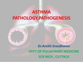
Comparison of a normal bronchiole with that in a person with asthma. Note the accumulation of mucus in the bronchial lumen resulting from an increase in the number of mucus-secreting goblet cells in the mucosa and hypertrophy of submucosal mucous glands. In addition, there is intense chronic inflammation caused by recruitment of eosinophils, macrophages, TH2 cells and other inflammatory cells. Basement membrane underlying the mucosal epithelium is thickened, and there is hypertrophy and hyperplasia of smooth muscle cells