1. RAK COLLEGE OF MEDICAL SCIENCES
DEPARTMENT OF ANATOMY
GROSS ANATOMY DISSECTION LAB HAND OUTS- RESPIRATORY SYSTEM
1
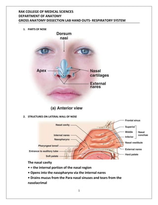
1. PARTS OF NOSE
2. STRUCTURES ON LATERAL WALL OF NOSE
The nasal cavity
• = the internal portion of the nasal region
• Opens into the nasopharynx via the internal nares
• Drains mucus from the Para nasal sinuses and tears from the
nasolacrimal
2. RAK COLLEGE OF MEDICAL SCIENCES
DEPARTMENT OF ANATOMY
GROSS ANATOMY DISSECTION LAB HAND OUTS- RESPIRATORY SYSTEM
2
Ducts
• Is divided in half midsagittally by the nasal septum, which is made up of
Cartilage and bones (the perpendicular plate of the ethmoid and the
Vomer)
• Its walls have 3 pairs of nasal conchae (superior, middle, and inferior).
3. NASOPHARYNX
The pharynx (throat)
• = a common passageway for air and
Food
• Extends from the internal nares to the
Junction of the esophagus and larynx
• Has 3 regions:
– 1. The nasopharynx (respiratory only) – from the internal nares to the uvula of the
soft palate
• It’s lined with PCCE
3. RAK COLLEGE OF MEDICAL SCIENCES
DEPARTMENT OF ANATOMY
GROSS ANATOMY DISSECTION LAB HAND OUTS- RESPIRATORY SYSTEM
3
• It contains the pharyngeal tonsil (adenoid) and the openings of the auditory
(Eustachian) tubes
– 2. The oropharynx (respiratory and digestive) – from the uvula of the soft palate to
the base of the tongue (or tip of the epiglottis)
• It’s lined with stratified squamous epithelium
• It contains the palatine and lingual tonsils
– 3. The laryngopharynx (respiratory and digestive) –from the base of the tongue (or
tip of the epiglottis) to the junctionof the esophagus and larynx.
The larynx (“voice box”)
• Connects the laryngopharynx with the trachea
• Is formed by 9 pieces of cartilage and some connecting ligaments and muscles
• The vocal folds and above are covered with stratified squamous epithelium
• Inferior to the vocal folds is covered with respiratory epithelium (PCCE)
• Epiglottis = an elastic cartilage plate that closes off the glottis (= the superior
opening of the larynx) during swallowing.
• The vestibular folds close off the glottis during breath holding, lifting heavy objects,
etc.
• The vocal folds = the vocal cords,
– Contain elastic fibers
– Vibrate to produce speech sounds
– Are attached anteriorly to the thyroid cartilage
– Are attached posteriorly to the arytenoid cartilages.
The trachea (“windpipe”)
= a short (4-5”) tube that carries air between the larynx and the junction of the
primary bronchi.
4. RAK COLLEGE OF MEDICAL SCIENCES
DEPARTMENT OF ANATOMY
GROSS ANATOMY DISSECTION LAB HAND OUTS- RESPIRATORY SYSTEM
4
The trachea divides at the sternal angle/T 4 vertebra into two primary bronchi, which
enters the hilum of lungs and further divides into secondary bronchi or lobar bronchi.
Each lobar bronchus further divides in to tertiary bronchi/bronchopulmonary
segmental bronchi. There are ten BP SEGMENTS in both lungs.
Left Lung
Superior (upper) lobe:
1. Apical
2. Posterior
3. Anterior .
5. RAK COLLEGE OF MEDICAL SCIENCES
DEPARTMENT OF ANATOMY
GROSS ANATOMY DISSECTION LAB HAND OUTS- RESPIRATORY SYSTEM
5
4. Superior lingular
5. Inferior lingular
Inferior (lower) lobe:
6. RAK COLLEGE OF MEDICAL SCIENCES
DEPARTMENT OF ANATOMY
GROSS ANATOMY DISSECTION LAB HAND OUTS- RESPIRATORY SYSTEM
6
6. Superior (apical) …………
7. Medial basal ………. .
7. RAK COLLEGE OF MEDICAL SCIENCES
DEPARTMENT OF ANATOMY
GROSS ANATOMY DISSECTION LAB HAND OUTS- RESPIRATORY SYSTEM
7
8. Anterior basal ……
9. Lateral basal …. .
10. Posterior basal ……
Right Lung
8. RAK COLLEGE OF MEDICAL SCIENCES
DEPARTMENT OF ANATOMY
GROSS ANATOMY DISSECTION LAB HAND OUTS- RESPIRATORY SYSTEM
8
Superior (upper) lobe:
1. Apical
2. Posterior
3. Anterior
Middle lobe
4. Lateral
5. Medial
Inferior (lower) lobe:
6. Superior (apical)
7. Medial basal
8. Anterior basal
9. Lateral basal
10. Posterior basal
9. RAK COLLEGE OF MEDICAL SCIENCES
DEPARTMENT OF ANATOMY
GROSS ANATOMY DISSECTION LAB HAND OUTS- RESPIRATORY SYSTEM
9
The pleura
• Each lung is surrounded by the pleura (= a double-layered serous membrane):
– 1. The parietal pleura lines the inside of the thoracic cavity
– 2. The visceral pleura cover the surface of the lungs
• The ultra-thin pleural cavity is between the two layers and contains pleural fluid,
which…
– Reduces the friction between layers during the movements of ventilation
– Causes the layers to adhere to one another, preventing the collapse of the lung.
10. RAK COLLEGE OF MEDICAL SCIENCES
DEPARTMENT OF ANATOMY
GROSS ANATOMY DISSECTION LAB HAND OUTS- RESPIRATORY SYSTEM
10
RESPIRATORY MUSCLES
11. RAK COLLEGE OF MEDICAL SCIENCES
DEPARTMENT OF ANATOMY
GROSS ANATOMY DISSECTION LAB HAND OUTS- RESPIRATORY SYSTEM
11
--------------------------------------------------------------------------------
Each lung has an apex, base, and hilum (hilus)
The Apex of the Lung
The rounded, tapered superior end or apex of the lung extends through the superior thoracic
aperture into the root of the neck. It lies in close contact to the dome or the copula of the
pleura. The apex of the lung is crossed by the subclavian artery, which produces a groove in
the mediastinal surface. The artery, however, is separated from the copula by the
suprapleural membrane.
The Base of the Lung
This is the concave diaphragmatic surface of the lung, which is related to the dome of the
diaphragm. The base of the right lung is deeper because the right dome rises to a more
superior level. Its inferior border is thin and sharp where it enters the cost diaphragmatic
recess.
12. RAK COLLEGE OF MEDICAL SCIENCES
DEPARTMENT OF ANATOMY
GROSS ANATOMY DISSECTION LAB HAND OUTS- RESPIRATORY SYSTEM
12
The Root of the Lung
The root serves as the attachment of the lung and is the "highway" for the transmission of the
structures entering and leaving the lung at the hilum. It connects the medial surface of the
lung to the heart and trachea and is surrounded by the reflection of parietal to visceral pleura.
The Hilum of the Lung
This is the where the root is attached to the lung. It contains the main bronchus, pulmonary vessels
(one artery and two veins), bronchial vessels, lymph vessels, and nerves entering and leaving the lung.
The Main Differences between the Right and Left Lungs
1. The right lung has 3 lobes while the left has 2 lobes.
2. The right lung is larger and heavier than the left lung, but is shorter and wider because the
right dome of the diaphragm is higher and the heart and pericardium bulge more to the
left.
3. The anterior margin of the right lung is straight, whereas the margin of the left lung has a
deep cardiac notch.
4. The right main bronchus is wider, shorter and more vertical (significant in inhaled
obstructions) than the left one.
Lobes and Fissures of the Lungs
The Left Lung
This is divided into superior (upper) and inferior (lower) lobes by a long deep oblique fissure.
This extends from its costal to its medial surface.
The superior lobe has a large cardiac notch on its anteriorborder, where the lung is deficient
owing to the bulge of the heart.
The anteroinferior part of the superior lobe has a small tongue-like projection called the
lingula.
The inferior lobe of the left lung is larger than the superior lobe and lies inferoposterior to the
oblique fissure.
The Right Lung
13. RAK COLLEGE OF MEDICAL SCIENCES
DEPARTMENT OF ANATOMY
GROSS ANATOMY DISSECTION LAB HAND OUTS- RESPIRATORY SYSTEM
13
This is divided into superior (upper), middle, and inferior (lower) lobes by horizontal and
oblique fissures.
The horizontal fissure separates the superior and middle lobes.
The oblique fissure separates the inferior lobe from the superior and middle lobes.
The superior lobe is smaller than in the left lung, and the middle lobe is wedge-shaped.
Surfaces of the Lung
The Costal Surface of the Lung
This surface is large, smooth, and convex. It is related to the costal pleura, which separates it
from the ribs, their costal cartilages, and the innermost intercostal muscles.
The posterior part of this surface is related to the thoracic vertebrae.
The Mediastinal Surface
This medial surface is concave because it is related to the middle mediastinum.
Because 2/3 of the heart is to the left, the pericardial concavity is deeper in the left lung.
The mediastinal surface of the embalmed lung shows a cardiac impression produced by the
heart and the great vessels. This surface also contains the root of the lung, around which the
pleura forms a "sleeve" or covering. The pulmonary ligament hangs inferiorly from the pleural
sleeve around the root of the lung.
The Diaphragmatic Surface
This is a deeply concave surface, often referred to as the base of the lung. It rests on the
convex dome of the diaphragm. The concavity is deeper in the right lung because of the higher
position of the dome. Laterally and posteriorly, the diaphragmatic surface is bound by a thin
sharp margin that projects into the cost diaphragmatic recess.
Borders of the Lungs
14. RAK COLLEGE OF MEDICAL SCIENCES
DEPARTMENT OF ANATOMY
GROSS ANATOMY DISSECTION LAB HAND OUTS- RESPIRATORY SYSTEM
14
The Anterior Border of the Lung
This border is thin and sharp and overlaps the pericardium. There is an indentation in the
anterior border of the left lung (cardiac notch). In each lung, the anterior border separates the
costal surface from the mediastinal surface. During deep inspiration, the lung projects into the
costomediastinal recess of the pleura.
The Posterior Border of the Lung
This border is broad and rounded and lies in the deep concavity at the side of the thoracic
region of the vertebral column, called the paravertebral gutter.
The Inferior Border of the Lung
This border circumscribes the diaphragmatic surface from the costal surface. It is thin and
sharp where it projects into the costodiaphragmatic recess. It is, however, blunt and rounded
medially, where it separates the diaphragmatic surface from the mediastinal surface.
Surface Markings of the Lungs
Apex of the Lung
This is represented by a line drawn superolaterally from the sternoclavicular joint to a point
2.5 cm superior to the medial 1/3 of the clavicle and then inferolaterally to the junction of the
middle and lateral thirds of the clavicle.
Anterior Border of the Right Lung
This corresponds to the anterior border of the right pleura. Between the level of the 2nd and
4th cartilages, its anterior border is near the median plane. Inferior to the 4th costal cartilage,
the surface of the right lung gradually diverges from this plane and leaves the sternum
posterior to the 6th costal cartilage.
15. RAK COLLEGE OF MEDICAL SCIENCES
DEPARTMENT OF ANATOMY
GROSS ANATOMY DISSECTION LAB HAND OUTS- RESPIRATORY SYSTEM
15
The Anterior Border of the Left Lung
This corresponds to the anterior border of the left pleura as far as the level of the 4th costal
cartilage. Here, the anterior border deviates laterally to a point about 2.5 cm lateral to the left
edge of the sternum to form the cardiac notch. It then turns inferiorly and slightly medially to
the 6th costal cartilage.
The Inferior Borders of the Lungs
These are indicated by a line drawn from the inferior end of the line representing the anterior
border that crosses the 6th rib at the midclavicular line, the 8th rib in the midaxillary line and
the 10th rib in the midscapular line.
These borders end about 2.5 cm lateral to the spinous process of T10 vertebra.
They lie two ribs superior to the pleura on each of three vertical lines just mentioned.