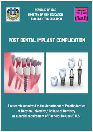
Post dental implant complication
•
2 gostaram•2,332 visualizações

http://t.me/dr_7li https://www.facebook.com/dr.7li

Recomendados
Recomendados
Mais conteúdo relacionado
Mais procurados
Mais procurados (20)
Semelhante a Post dental implant complication
Semelhante a Post dental implant complication (20)
Mais de Ali Khalaf
Último
Último (20)
Post dental implant complication
- 1. 0
- 2. 1 Submitted by: Ali Khalaf Anber Mahmood Abd Al-Sadah Majed Ali Nasser Zuwayd Supervised by: Lecturer Dr. Hanan Ali Hameed Assit.Prof. Dr. Malath Azeez Dedication To all those who are enlightened by the knowledge of the mind of othors , to candles that burn to light up for others , to everyone who taught me a letter , dedicate this humble research , asking god to find acceptance and success July 2020 A.D. 1441 A.H.
- 3. 2 Abstract The mainstream use of dental implants has allowed millions of patients to benefit from the predictability of dental implant therapy and, in many instances, dental implants have become the standard of care. Even though success rates in implant dentistry are well above 90 percent, complications do occur. Most complications are preventable with proper planning and execution. Others are inherent to the risks of surgery and may require intervention. The purpose of this review is to classify the possible complications that may occur and to discuss their prevention and management and compare it with different material (Zirconium , Titanium). This review started with a PubMed and Google Scholar search from 1969 to 2019. The search was conducted using key words: zirconia , titanium, dental, and implant. The full text of articles was obtained where possible. If it was not possible to obtain a full text, the electronically available abstracts were collected. General information about our review we found that: titanium abutments showed high resistance to screw loosening when compared to the zirconia abutments, zirconia and titanium abutment surfaces exhibit more or less similar clinical characteristics of peri-implant health and microbiological features, titanium abutment system was significantly more fracture resistant than the zirconia abutment, zirconia implants are better than titanium in the aesthetic zone. Key words: Implant complications, Implant failures, Implant, Zirconia, Titanium.
- 4. 3 Introduction Prosthetic dentistry, during the past decade, has significantly improved and developed to replacement missing parts of teeth, bone, gums, or facial structures, restoring functions and esthetic according to advancements in the science and patient’s demand and need. Conventional options in prosthodontics for substituting missing teeth include the removable partial and complete denture, partial and full coverage bridgework, and resin‑bonded bridgework [1]. An attractive alternative to conventional dentures and bridges became available with the introduction of implants into dental industry [2,3]. For so many years, virtually all dental implants were derived from one material called titanium. Since the 1960s, titanium has remained the industry standard for dental implants. In fact, titanium implants have become one of the most successful medical devices with a long-term medical success rate of 94-97%. But with the advent of technology, more and more dental implants are now made from different dental implant materials such as zirconium [4]. At present, titanium dental implant have side effect in discoloration when it placed inside the bone. Therefore studies and research have turned to the use of zirconium because it is more aesthetic. Zirconia implants show promise, their long-term success is not proven. If, aesthetics or allergy sensitivity is what is driving you towards Zirconia, a Zirconia abutment with a Titanium implant may be the best solution [4]. The basis for dental implants is osseointegration, where osteoblasts grow and directly integrate with surface of the implants surgically placed inside the alveolar bone [3]. Osseointegrated dental implants are an effective alternative in the rehabilitation of partial or total edentulous patients [5]. Several complications may affect the success of bone integrated implants in specific situations [6] which can be of a biological of biomechanical nature. According
- 5. 4 to El Askary et al [7] the signs of implant failure are: loosening or breaking of the screws that hold the crowns and abutments in place, edema or bleeding of soft tissue surrounding the implant, purulent exudate from the peri-implant sulcus, pain (rare), prosthesis fracture, angular bone loss and chronic infections. The most important factor in implant longevity, i.e. clinically successful implant treatment is the formation of a direct interface between the implant and the bone, without intervening soft tissue, a process called “osseointegration”. Osseointegrated dental implants represent an advance in modern odontology which has become a great option for the rehabilitation of missing single teeth in partially or completely edentulous patients. Despite the very high success rates, complications associated with implant treatment may occur [8]. Early loading failure may affect 2% to 6% of implants, and as many as 15% of restorations failure as a result of this problem [9,10]. Excess load on a final restoration after successful implant integration can result in failure of the implant itself. Therefore, it is important to clarify the risk factors for failure of implant prostheses in order to further improve the good success rate. The consequences of overload of dental implants can be divided into two groups: biological and biomechanical complications [11]. Biological complications can be divided into early failures and late failures [12]. In case of early failures, osseointegration was insufficient: the implant is lost before the first prosthetic loading. Late biological failures are characterized by pathological bone loss after full osseointegration was obtained at an earlier stage [13]. They are associated with overload. Some insight into bone physiology is needed for a proper understanding of these mechanisms [14]. In case of biomechanical complications, one or more components of an implant system failure are fracture of an implant itself, loosening or fracture of connecting screws or abutment screws, loosening or excessive wear of
- 6. 5 mesostructural components in overdentures and excessive wear or fracture of suprastructural porcelain or acrylic teeth [15,16]. In addition to biomechanical complications the bacterial infection is considered one of the rising complications after implant placement. The postoperative infection rate was reported to be 4-10% for patients receiving dental implants in spite of success rate of the dental implants was reported to be as high as 90- 95%[17,18] . The recurrent incidence of this infection is also a concern, which is about 5-8% and is even more difficult to control and treated by antibiotics. The implant materials placed within the oral cavity can interfere with the host defense mechanism and it might influence the required clinical dose of antibiotics to safeguard against infections[19,20]. Interestingly, no single complication has been closely associated with dental implant failure. Based on the above, the aim of the current review is to discuss specific complications associated with dental implants. Management protocols and possible means of avoiding certain complications are also briefly discussed. Post dental implants complications For the last years the usage of dental implants to replace missing teeth and restoration of masticatory function has been accepted as a routine method in clinical practice. Along with the high success rate, biological and biomechanical complications are observed in some cases [21-23]. Long term studies have demonstrated that loss of osseointegrated implant can be caused by secondary infections (peri-implantitis) functional overload and implant fracture [24,25]. Screw loosening It has been observed that screw loosening is a matter of concern for both manufacturers and dental professionals. Loosening of abutment screw is one of the most common mechanical complications breaking the integrity between the
- 7. 6 implant and abutment. The incidence varies between 4.3 and 10% and occurs in a relatively short period after functional loading of implants[26,27] Screw loosening leads to instability of the implant-abutment connection and the formation of a micro-gap, which may provoke the fracture of implant components [28]. The gap can cause the infiltration of microorganisms, which is harmful for surrounding tissues [29]. Additionally, loosening or failure of implant screws may result in component failure that may require more extensive repair [30,31]. The reasons for such loosening may turn out to be inadequate tightening torque, settling of implant components, inappropriate implant position, inadequate occlusal scheme or crown anatomy, poorly fitting frameworks, presence of microleakage at abutment-implant interface, improper screw design / material, and heavy occlusal forces [32,33]. Abutment screw loosening may result in mobility of prosthesis, which necessitates removing the prosthesis in order to tighten abutment. The most usual complications of abutment screw loosening include inflammation of gingival, failure of implant, and fracture of screws [34]. Sahin and Ayyildiz’s studies indicated that the loosening of abutment screw can be brought about by the presence of microleakage between the implant and abutment, permitting fluid penetration around the abutment screw. As a consequence of this the permeability is increased which provokes the occurrence of bacterial infection as well as peri-implantitis in some time [35]. The results of some researches showed that parafunctional activity, cantilevers and the time of complications setting in have a statistically significant impact with typical effect size in observed cases of abutment screw loosening [36]. Bruxism has a significant impact upon the incidence of biomechanical complications, including screw loosening due to repeated static and dynamic loading. Such loadings can be both along the axial axis in clenching the teeth
- 8. 7 and in less favorable lateral directions in grinding the teeth. The increased loading leads to fatigue and subsequent loosening and fracture of abutment screw [37-39]. The obtained results revealed also a strong connection between the cantilever extensions and cases of screw loosening. The patient may complain of soreness at the interface between soft tissue and fixture, swelling or fistula formation, and difficulty chewing. Prevention screw loosening To minimize screw loosening, various solutions are recommended, such as using diamond-like carbon coating over abutment screws, retightening screws after initial tightening, and increasing the torque value [40,41]. Preloading of the screw joint is essential for the prevention of screw loosening [42]. Many research found that the application of an adhesive material on the abutment screws is recommended to prevent or decrease screw loosening [43]. Graves et al reported a decrease of the force on a screw of 20%, and 33% when the diameter of an implant was increased from 3.75 mm to 5.0 mm, and 6.0 mm respectively. They postulated that this might indeed reduce the amount of screw loosening. So The diameter of the implant and the design of the implant (more surface area is better) are key factors and are paramount for implant success. The wider the diameter of the implant, the better the distribution of stresses will be [44]. Implant fracture Fracture of dental implants is a rare phenomenon. According to Balshi [45], only 0.2% of 4045 placed implants presented with fracture during 5 years of function. Data from a study carried out by Adell [46], in which 4636 implants were employed, revealed an average total fracture rate of less than 5%, 6% in
- 9. 8 the maxilla, and 3% in the mandible after 15 years of follow-up. Eckert [47] reported that among 4937 implants, the fracture rate was just 0.6% with no statistically significant difference noted between the arches. Causes of implant fracture may be divided into 3 categories:(1) defects in the design of the material, (2) non passive fit of the prosthetic structure, and (3) biomechanical or physiologic overload [47]. Possible causes of fracture include failure in the production and design of dental implants, bruxism or large occlusal forces, superstructure design, implant localization, implant diameter, metal fatigue, and bone resorption around the implant [48]. In addition, the galvanic activity of metals used in prosthetic restorations can cited as a cause [49]. Defects in the production and design of dental implants are very unlikely reasons for fracture. Microscopic analysis of fractured fixtures revealed no porosity or any other defects in the titanium structure, a finding that eliminated failure in the manufacturing process as causative [47,50]. Load factors are related to the magnitude and direction of occlusal forces. Ninety percent of dental implant fractures are located in the molar and premolar regions of the mouth, where chewing forces and lateral movements associated with cusp inclination generate undesirable forces [51]. Biomechanical and physiologic overload seems to be the most common cause of dental implant fracture; overload may be caused primarily by two factors: Parafunctional habits and prosthesis design [47]. Parafunctional habits such as bruxism or clenching may increase overload on the implant prosthesis system through the magnitude, duration, frequency, and direction of forces applied. According to Rangert [52], around 56% of patients with fractured dental implants presented with bruxism and marked occlusal forces. Parafunctional habits have been identified as the major causative factor associated with fixture fractures [47]. In any clinical situation, the presence of an extension or cantilever considerably increases the load on implants [53]. Shackleton and Slabbert [54] studied the existence of a relationship between survival time span of implant-supported prostheses and the length of their
- 10. 9 posterior cantilever extensions. They concluded that short cantilevers provide longer survival rates and recommended the use of mandibular extensions for a maximum of 15 mm. According to Rangert et al [51], good mandibular bone quality allows the use of cantilevers that measure 15-20 mm in length, whereas porous maxillary bone should not support cantilevers longer than 10 mm. It has been suggested that posterior cantilevers should be avoided or minimized, especially in partially edentulous patients. Implants with small diameters tend to fracture more easily than those with large diameters, especially when placed in the posterior region [48]. According to Krogh [55] among the causes of implant fracture are standard implants used in the molar region. In an implant of 3.75 mm diameter just 0.4 mm corresponds with the thickness of the titanium wall. Therefore, implants that are 5 and 6 mm in thickness are 3 and 6 times more resistant, respectively, than standard implants [56]. As well as the type of material can be one of influencing factors so the studies showed, zirconia implants exhibited high rates of fractures in preclinical animal studies using canine mandibles. In two different studies, Thoma et al [57,58] reported a higher incidence of zirconia implant fractures before and after loading in comparison with titanium implants. The localization position of dental implants also has a direct influence on the biomechanical distribution of forces. If the implant axis is placed at a certain distance from the center of the prosthetic crown, forces created by this distance from the occlusal contact point to the implant axle may cause screw loosening or component fracture. Management of implant fracture Balshi [45] suggested three methods for treating fractures of dental implants: (1) removal of the fractured implant (replace the implant and manufacture a new prosthesis), (2) alteration of the existing prosthesis and maintenance of the osseointegrated fractured part, and (3) alteration of the fractured implant and remanufacturing of the prosthetic portion. Treatment of fractured implants
- 11. 10 represents a clinical challenge. First, the fractured fragment must be atraumatically removed with minimum bone removal. A new fixture is placed and the time to osseointegration must pass; only after that, the prosthetic phase begins [49]. It is suggested that, for removal of the intraosseous portion of a dental implant, a trephine bur should be used and if possible, another implant with a larger diameter should be installed immediately [49][55]. Prevention of implant fracture A titanium alloy implant should ideally be used to reduce the possibility of implant body fracture. Parafunctional habits should be addressed with occlusal guards, narrow occlusal tables, no lateral contacts, and an ideal occlusal scheme. Peri-implantitis Peri-implant disease is defined as the inflammatory pathological change that takes place in the soft and hard tissues surrounding an osseointegrated implant[59]. When an implant is successfully osseointegrated, the peri‐implant disease that occurs is the consequence of disparity between the host defense and increasing bacterial load[60]. It usually takes about 5 years for the peri‐implant disease to progress and exhibit clinical signs and symptoms [61-63]. The incidence of peri‐implantitis and implant loss could be greater if the studies with longer follow‐up periods are evaluated[64]. In a healthy environment around the implant, the tissues play a pivotal role in preventing the spread of agents that can be pathognomonic, and if the biological barrier is breached, it could lead to bacterial contamination around the bone resulting in hasty destruction of the tissues surrounding the implant[65]. The peri‐ implant disease is also related to unequal occlusal load distribution, which may lead to loosening of the superstructure, infection of the surrounding area, eventually culminating into
- 12. 11 the inflammatory process [66]. Predisposing systemic conditions include uncontrolled diabetes mellitus, osteoporosis, smoking, long‐standing treatment with steroids, uncontrolled periodontitis, radiation therapy, and chemotherapeutics [60,67,68]. So far, the zirconia as an abutment material has offered superior esthetic and gingival healing advantages over titanium . Nevertheless, the biofilm formation on titanium and zirconium oxide surfaces is still a discussion point . Due to the lack of long-term studies, the zirconia has no proven advantage or disadvantage over titanium implants regarding peri-implant infection risk but probing depths around Ti abutments were slightly deeper than around ZrO2 abutments. The peri‐implant disease treatment strategies have been explored and employed to prevent failure of the implant treatment[64]. They include nonsurgical mechanical debridement, local antimicrobial delivery in periodontitis and peri‐implantitis, and surgical debridement with bone grafting. Implant removal is warranted if there is more than 60% of bone loss following peri‐implantitis, and there is an evidence of mobility[69]. Antibiotics for peri-implantitis The use of prophylactic antibiotics during implant placement remains controversial. A Cochrane review found insufficient evidence advocating or dissuading their use[70]. An update of the review, however, determined there was some evidence that 2 g of amoxicillin given orally 1 h preoperatively significantly reduced early failures of dental implants[71]. The review concluded by recommending the routine use of one dose of 2 g of prophylactic amoxicillin immediately prior to placing dental implants. However, it also stated that further research was required to confirm the findings . At present, there is no reliable evidence for the most successful method of treating peri- implantitis [72]. Despite a variety of therapeutic options infected implants are difficult to treat and usually require removal[73]. Some clinicians advise
- 13. 12 systemic antibiotics for the treatment of failing implants and a variety of drug regimens are described[74]. Oral agents such as doxycycline, clindamycin, co- amoxiclav, penicillin V, amoxicillin and a combination of amoxicillin and metronidazole have been recommended. Nevertheless, no double blind, randomised, placebo-controlled trial has been undertaken. Others strategies aimed at reducing bacterial adhesion and biofilm formation on implant abutment surfaces are of pertinent clinical interest and can be used for the maintenance of soft tissue health or possibly in the treatment of peri-implantitis. Studies have shown that antimicrobial (e.g. vancomycin or chitosan) derivatization of a Ti alloy surface renders it less susceptible for bacterial colonization in vitro [75]. Implant coatings that deliver antibiotics have been described as well, predominantly in the field of orthopaedics [76]. It was shown that the physical properties of the Ti surface can be adapted, for example by applying a coating of Ti-nitride through vapour deposition. This reduces plaque adhesion compared with uncoated Ti surfaces both in vitro and in vivo [77,78] and still facilitates cellular adhesion of human fibroblasts in vitro [79]. In addition, it has been observed that silver and zinc oxide-modified surfaces possess antibacterial properties as well so this can use to prevent the infection[80]. Discoloration Titanium is the gold standard material used to produce dental implants over more than 30 years [81], showing a high success rate in different clinical scenarios [82,83]. Nevertheless, titanium implants may present some esthetic issues: the gray color of titanium implant may be visible in the presence of thin peri-implant tissue, leading to esthetic concern, especially in the anterior area [84]. This aspect can get dramatically worse in case of peri-implant mucosa recedes over time. The
- 14. 13 availability of a “white” implant may be crucial in those clinical cases in which esthetic result is mandatory. Furthermore, titanium particles due to wear and corrosion products may be released in tissues close to implants, and they were found in regional lymph nodes [85]. In some cases, this may lead to host reaction or sensitization [86]. Some cases of allergic reaction to titanium are documented, even if rare [87,88]. So, using some nonmetallic material as an alternative to the titanium implant may be useful and, in some cases, critical. Last but not least, always more patients request completely metal-free prosthetic reconstructions. Ceramic implants were introduced to overwhelm some esthetic and biological problems that can arise from titanium. Today a new type of implant made out of Zirconia has recently been introduced into dental implantology as an alternative to titanium implants. Zirconia possesses many advantages over titanium in its biologic, esthetic, mechanical, and optical properties, as well as its inherent biocompatibility and low plaque affinity. This zirconia-based material has been shown to have improved flexural strength and fracture resistance over early versions of ceramic implants.[84] Zirconia implants have a definite aesthetic advantage over titanium implants (Healthy , Pink beautiful tissue around implant , No gum show through, like natural tooth , Resembles real tooth esthetics). Zirconia is advocated to have high biocompatibility and to have no adverse effect on the surrounding tissues [89]. Many studies evaluated tissue response to zirconia, concluding that zirconia has the ability to interact with peri-implant soft tissues [90]. The low bacterial colonization typical of the zirconia surface maybe plays a role in this high biocompatibility [91]. In a randomized- controlled trial (RCT), both titanium and zirconia one-piece implants supporting overdentures were evaluated [92]. Even if the crestal bone level changed
- 15. 14 greatly, no difference in clinical parameters (probing depth, bleeding index, plaque index, etc.) was found around the two types of implants after 12 months of function. Unfortunately, long-term follow-ups are missing, so no solid clinical evidence is currently available to recommend routine use of zirconia implants or to replace titanium implants, which is still found to be the gold standard for dental implantology. So, even if zirconia implants are a good option from theoretical and experimental point of view, the clinical long-term response is not yet available. Almost all the authors agree to be cautious for proposing zirconia implants as substitutes of titanium implants for replacing teeth. Long-term, well-designed perspective clinical studies are needed to address the missing aspects of this undoubtful promising alternative. Conclusions Our research concluded that titanium abutments showed high resistance to screw loosening when compared to the zirconia . Abutment screws that were tightened by the adhesive material showed a significant increase in removal torque value. Thus, the microleakage in abutment implant interface can be reduced not only by filling the gap exist in that interface, but also by improving the retention of screw retained abutments. Long screws have more threads that engage the implant and abutment. These features may help distribute the applied loads to the implants and surrounding bone more efficiently, making long screws more resistant to fracture. Also found that zirconia and titanium abutment surfaces exhibit more or less similar clinical characteristics of peri-implant health and microbiological features during the first 3 months could not be convincingly rejected for most
- 16. 15 parameters with the exception of the pocket probing depth somewhat shallower probing depths were observed around zirconia abutments after 3 months. But about the fracture titanium and zirconia abutments were tested in a stepped fatigue loading protocol. Within the limitations of this in vitro study, the titanium abutment system was significantly more fracture resistant than the zirconia abutment system. Researches do not support any obvious advantage of titanium or zirconia abutments over each other. However, there is a significant tendency in zirconia abutments evoking better color response of peri-implant mucosa and superior esthetic outcome. References 1. Hanif A, Qureshi S, Sheikh Z, Rashid H. Complications in implant dentistry. Eur J Dent 2017;11:135-40. 2. Chan R, Tseng T. Single tooth replacement‑expanded treatment options. Aust Dent J 1994;39:137‑49. 3. Zarb G, Albrektsson T, editors. Introduction to osseointegration. In: Branemark PI, editor. Tissue‑Integrated Prosthesis: Osseointegration in Clinical Dentistry. Chicago, Berlin: Quintessence; 1985. 4. Zeynep O¨ zkurt, Ender Kazazog˘ lu,Yeditepe University, Goztepe, Istanbul, Turkey. Zirconia Dental Implants: A Literature Review. Vol. XXXVII/No. Three/2011. 5. Santiago Júnior JF, Verri FR, Pellizzer EP, Moraes SLD, Carvalho BM. Implantes dentais curtos: alternativa conservadora na reabilitação bucal. Rev Cir Traumatol Bucomaxilofac. 2010 Apr-Jun;10(2):67-76.
- 17. 16 6. Naert I, van Steenberg D, Worthington P. Osseointegration in oral rehabilitation: an introduction. Quintessence: Chicago; 1993. 7. el Askary AS, Meffert RM, Griffin T. Why do dental implants fail? Part I. Implant Dent. 1999;8(2):173-85. 8. Lobbezoo F, Naeije M. Bruxism is mainly regulated centrally, not peripherally. J Oral Rehabil. 2001; 28: 1085-1091. 9. Lobbezoo F, Brouwers JEIG, Cune MS, Naeije M. Dental implants in patients with bruxing habits. J Oral Rehabil. 2006; 32: 152-159. 10. Kato T, Thie NM, Montplaisir JY, Lavigne GJ. Bruxism and orofacial movements during sleep. Dent Clin North Am. 2001; 45: 657-684. 11. Kato T, Dal-Fabbro C, Lavigne GJ. Current knowledge on awake and sleep bruxism: overview. Alpha Omegan. 2003; 96: 24-32. 12. Kato T, Thie NM, Huynh N, Miyawaki S, Lavigne GJ. Topical review: sleep bruxism and the role of peripheral sensory infl uences. J Orofac Pain. 2003; 17: 191-213. 13. De Laat A, Macaluso GM. Sleep bruxism as a motor disorder. MovDisord. 2002; 17: 67-69. 14. Lavigne GJ, Kato T, Kolta A, Sessle BJ. Neurobiological mechanisms involved in sleep bruxism. Crit Rev Oral Biol Med. 2003; 14: 30-46. 15. Sari S, Sonmez H. The relationship between occlusal factors and bruxism in permanent and mixed dentition in Turkish children. J Clin Pediatr Dent. 2001; 25: 191-194. 16. Giffi n KM. Mandibular adaptive reposturing: the aetiology of a common and multifaceted autodestructive syndrome. Gen Dent. 2003; 51: 62-67.
- 18. 17 17. Pye, A., Lockhart, D., Dawson, M., Murray, C. & Smith, A. (2009). A review of dental implants and infection. Journal of Hospital Infection, 72(2), 104-110. 18. Camps-Font, O., Figueiredo, R., Valmaseda-Castellón, E. & Gay-Escoda, C. (2015). Postoperative Infections After Dental Implant Placement: Prevalence, Clinical Features, and Treatment. Implant Dentistry, 24(6), 713-719. 19. A., Ergun, C. & Baydogan, M. (2009). Synthesis and characterization of Ag doped hydroxylapatite as an antibacterial scaffolds material. Proceedings from Defect and Diffusion Forum. 20. Stanić, V., Janaćković, D., Dimitrijević, S., Tanasković, S. B., Mitrić, M., Pavlović, M. S., Krstić, A., Jovanović, D. & Raičević, S. (2011). Synthesis of antimicrobial monophase silver-doped hydroxyapatite nanopowders for bone tissue engineering. Applied Surface Science, 257(9), 4510-4518. 21. Moraschini V, Poubel LA, Ferreira VF, Barboza Edos S. Evaluation of survival and success rates of dental implants reported in longitudinal studies with a follow-up period of at least 10 years: a systematic review. Int J Oral Maxillofac Surg. 2015 Mar;44(3):377-88. [PubMed] [CrossRef] 22. Tey VH, Phillips R, Tan K. Fiveyear retrospective study on success, survival and incidence of complications of single crowns supported by dental implants. Clin Oral Implants Res. 2016 Jun 22. [Epub ahead of print] [PubMed] [CrossRef] 23. Krishnan V, Tony Thomas C, Sabu I. Management of abutmentscrew loosening: review of literature and report of a case. J Indian Prosthodont Soc. 2014 Sep;14(3):208-14. [PubMed] [CrossRef] 24. Camps-Font O, Figueiredo R, Valmaseda-Castellón E, Gay-Escoda C. Postoperative Infections After Dental Implant Placement: Prevalence, Clinical
- 19. 18 Features, and Treatment. Implant Dent. 2015 Dec;24(6):713-9. [PubMed] [CrossRef] 25. Gupta S, Gupta H, Tandan A. Technical complications of implantcauses and management: A comprehensive review. Natl J Maxillofac Surg. 2015 Jan- Jun;6(1):3-8. [PubMed] [CrossRef] 26. Journal of Clinical and Diagnostic Research. 2019 Aug, Vol-13(8): ZE06- ZE09 27. Chaar MS, Att JR, Strub JR. Prosthetic outcome of cement-retained implant-supported fixed dental restorations: a systematic review. J Oral Rehabil. 2011 Sep;38(9):697- 711. [PubMed] [CrossRef] 28. Gupta S, Gupta H, Tandan A. Technical complications of implant- causes and management: A comprehensive review. Natl J Maxillofac Surg 2015;6:3-8. 29. do Nascimento C, Miani PK, Pedrazzi V, Gonçalves RB, Ribeiro RF, Faria AC, Macedo AP, de Albuquerque RF Jr. Leakage of saliva through the implant- abutment interface: in vitro evaluation of three different implant connections under unloaded and loaded conditions. Int J Oral Maxillofac Implants 2012;27:551-60. 30. Taylor TD. Prosthodontic problems and limitations associated with osseointegration. J Prosthet Dent 1998;79:74-8. 31. Stüker RA, Teixeira ER, Beck JC, da Costa NP. Preload and torque removal evaluation of three different abutment screws for single standing implant restorations. J Appl Oral Sci 2008;16:55-8. 32. Cho WR, Huh YH, Park CJ, Cho LR. Effect of cyclic loading and retightening on reverse torque value in external and internal implants. J Adv Prosthodont 2015;7:288-93.
- 20. 19 33. Sahin C, Ayyildiz S. Correlation between microleakage and screw loosening at implant-abutment connection. J Adv Prosthodont 2014;6:35-8. 34. Zeno HA, Buitrago RL, Sternberger SS, Patt ME, Tovar N, Coelho P, Kurtz KS, Tuminelli FJ. The effect of tissue entrapment on screw loosening at the implant/abutment interface of external- and internal-connection implants: An in vitro study. J Prosthodont 2016;25:216-23. 35. Canullo L, Penarrocha-Oltra D, Soldini C, Mazzocco F, Penarrocha M, Covani U. Microbiological assess-ment of the implant-abutment interface in different connections: crosssectional study after 5 years of functional loading. Clin Oral Implants Res. 2015 Apr;26(4):426-34. [PubMed] [CrossRef] 36. Komiyama O, Lobbezoo F, De Laat A, Iida T, Kitagawa T, Murakami H, et al. Clinicalmanagement of implantprostheses in patients with bruxism. Int J Biomater. 2012; 2012: 369063. [PubMed] [CrossRef] 37. Shemtov-Yona K, Rittel D, Levin L, Machtei EE. Effect of dental implant diameter on fatigue performance. Part I: mechanical behavior. Clin Implant Dent Relat Res. 2014 Apr;16(2):172-7. [PubMed] [CrossRef] 38. Marcelo CG, Filié Haddad M, Gennari Filho H, Marcelo Ribeiro Villa L, Dos Santos DM, Aldiéris AP. Dental implant fractures - aetiology, treatment and case report. J Clin Diagn Res. 2014 Mar;8(3):300-4. [PubMed] [CrossRef] 39. Sánchez-Pérez A, Moya- Villaescusa MJ, Jornet-Garcia A, Gomez S. Etiology, risk factors and management of implant fractures. Med Oral Patol Oral Cir Bucal. 2010 May 1;15(3):e504-8. [PubMed] 40. Diez JS, Brigagão VC, Cunha L, Neves AC, da Silva-Concilio LR. Influence of diamondlike carbon-coated screws on the implant-abutment interface. Int J Oral Maxillofac Implants 2012;27:1055-60.
- 21. 20 41. Siamos G, Winkler S, Boberick KG. Relationship between implant preload and screw loosening on implant-supported prostheses. J Oral Implantol 2002;28:67-73. 42. Dhingra A, Weiner S, Luke AC, Ricci JL. Analysis of dimensional changes in the screw and the surface topography at the interface of a titanium screw and a zirconia abutment under cyclic loading: an in vitro study. Int J Oral Maxillofac Implants 2013;28:661-9. 43. Mahnaz Arshad, Gholamreza Shirani , Sina Refoua , Mohammadreza Rahimi Yeganeh . J Adv Prosthodont 2017;9:99-103 44. Cavazos E, Bell FA. Preventing loosening of implant abutment screws. J ProsthetDent. 1996;75:566–569.doi:10.1016/S0022-3913(96)904643. [PubMed] [CrossRef] [Google Scholar] 45. Balshi TJ. An analysis and management of fractured implants: a clinical report. Int J Oral Maxillofac Implants. 1996;11:660–666. 46. Adell R, Eriksson B, Lekholm U, Branemark PI, Jemt T. A long-term follow-up study of osseointegrated implants in the treatment of totally edentulous jaws. Int J Oral Maxillofac Implants. 1990;5:347–358. 47. Eckert SE, Meraw SJ, Cal E, Ow RK. Analysis of incidence and associated factors with fractured im- plants: a retrospective study. Int J Oral Maxillofac Implants. 2000;15:662–667. 48. Green NT, Machtei EE, Horwitz J, Peled M. Fracture of dental implants: literature review and report of a case. Implant Dent. 2002;11:137–143. 49. Muroff FI. Removal and replacement of a fractured dental implant: case report. Implant Dent. 2003;12:206–210.
- 22. 21 50. Piatelli A, Piatelli M, Scarano A, Montesani L. Light and scanning electron microscopic report of four fractured implants. Int J Oral Maxillofac Implants. 1998;13:561–564. 51. Rangert B, Jemt T, Jorneus L. Forces and moments on Branemark Implants. Int J Oral Maxillofac Implants. 1989;4:241–247. 52. Rangert B, Krogh PHT, Langer B, Roekel NV. Bending overload and implant fracture: a retrospective clini- cal analysis. Int J Oral Maxillofac Implants. 1995;10:326–333. 53. Renouard F, Rangert B. Fatores de Risco em Implantodontia: Planejamento Cl ınico Simplificado para Progno stico e Tratamento. Sa ̃ oPaulo: Quintessence; 2001. [In Portuguese. 54. Shackleton J, Slabbert JCG. Survival of fixed implant supported prostheses related to cantilever lengths. J Prosthet Dent. 1994;71:23–26. 55. Krogh P. Surgical and biomechanical advantages of large-diameter implants. In: Siddiqui AA, Caudill R. Proceedings of the Fourth International Symposium on Implant Dentistry: Focus on Esthetics; January 27 through 29, 1994; San Diego, CA. J Prosthet Dent. 1994;72:623–634. 56. Graves S. Surgical and biomechanical advan- tages of large-diameter implants. In: Siddiqui AA, Caudill R. Proceedings of the Fourth International Symposium on Implant Dentistry: Focus on Esthetics; January 27 through 29, 1994; San Diego, CA. J Prosthet Dent. 1994;72:623–634. 57. Thoma DS, Benic GI, Muñoz F, et al. Histological analysis of loaded zirconia and titanium dental implants: an experimental study in the dog mandible. J Clin Periodontol. 2015;42:967–975.
- 23. 22 58.Thoma DS, Benic GI, Muñoz F, et al. Marginal bone-level alterations of loaded zirconia and titanium dental implants: an experimental study in the dog mandible. Clin Oral Implants Res. 2016;27:412–420. 59. Mombelli A, Lang NP. The diagnosis and treatment of peri-implantitis. Periodontol 2000 1998;17:63-76. 60. Heitz-Mayfield LJ. Peri-implant diseases: Diagnosis and risk indicators. J Clin Periodontol 2008;35 8 Suppl: 292-304. 61. Klinge B, Hultin M, Berglundh T. Peri-implantitis. Dent Clin North Am 2005;49:661-76, vii-viii. 62. Quirynen M, De Soete M, van Steenberghe D. Infectious risks for oral implants: A review of the literature. Clin Oral Implants Res 2002;13:1-19. 63. Mombelli A, Müller N, Cionca N. The epidemiology of peri-implantitis. Clin Oral Implants Res 2012;23 Suppl 6:67-76. 64. Rashid H, Sheikh Z, Vohra F, Hanif A, Glogauer M. Peri-implantitis: A re- view of the disease and report of a case treated with allograft to achieve bone regeneration. Dent Open J 2015;2:87-97. 65. Lang NP, Wilson TG, Corbet EF. Biological complications with dental implants: Their prevention, diagnosis and treatment. Clin Oral Implants Res 2000;11 Suppl 1:146-55. 66. Georgiev T. Kamen Nogalchev Local complications occurring during dental implantation. J IMAB: Annual Proceeding (Scientific Papers). 2010;16:35-7. 67. Prathapachandran J, Suresh N. Management of peri-implantitis. Dent Res J (Isfahan) 2012;9:516-21. 68. Albrektsson T, Isidor F. Consensus report: Implant therapy. In: Lang NP, Karring T, editors. Proceedings of the 1st European Workshop on Periodontology. Berlin: Quintessence; 1994. p. 365-9.
- 24. 23 69. Lang NP, Lindhe J. Clinical Periodontology and Implant Dentistry. Blackwell Munksgaard: Copenhagen, Denmark: John Wiley and Sons;2015 70. Esposito M, Coulthard P, Oliver R, Thomsen P, Worthington HV. Antibiotics to prevent complications fol- lowing dental implant treatment. Cochrane Database Syst Rev 2003 (3)CD004152. 71. Esposito M, Grusovin MG, Talati M, Coulthard P, Oliver R, Worthington HV. Interventions for replacing missing teeth: antibiotics at dental implant placement to prevent compli- cations. Cochrane Database Syst Rev 2008 (3)CD004152. 72. Esposito M, Grusovin MG, Coulthard P, Worthington HV. In- terventions for replacing missing teeth: treatment of peri- implantitis. Cochrane Database Syst Rev 2006 (3)CD004970. 73. Esposito M, Hirsch JM, Lekholm U, Thomsen P. Biological fac-tors contributing to failures of osseointegrated oral implants (II). Etiopathogenesis. Eur J Oral Sci 1998;106:721e764. 74. Yukna RA. Diagnosis and treatment of the ailing/failing den- tal implant. In: Block MS, Kent JN, editors. Endosseous im- plants for maxillofacial reconstruction. Philadelphia: WB Saunders; 1995. p. 602e607. 75. Antoci, V., Adams, C.S., Parvizi, J., Davidson, H.M., Composto, R.J., Freeman, T.A., Wickstrom, E., Ducheyne, P., Jungkind, D., Shapiro, I.M. & Hickok, N.J. (2008) The inhibition of Staphylococcus epider- midis biofilm formation by vancomycin-modified titanium alloy and implications for the treatment of periprosthetic infection. Biomaterials 29: 4684–4690. 76. Norowski, P.A. & Bumgardner, J.D. (2009b) Biomater- ial and antibiotic strategies for peri-implantitis: a review. Journal of Biomedical Materials Research B Applied Biomaterials 88: 530– 543.
- 25. 24 77. Grossner-Schreiber, B., Griepentrog, M., Haustein, I., Muller, W.D., Lange, K.P., Briedigkeit, H. & Gobel, U.B. (2001b) Plaque formation on surface modified dental implants. An in vitro study. Clinical Oral Implants Research 12: 543–551. 78. Scarano, A., Piattelli, M., Vrespa, G., Caputi, S. & Piattelli, A. (2003) Bacterial adhesion on titanium nitride-coated and uncoated implants: an in vivo hu- man study. Journal of Oral Implantology 29: 80–85. 79. Grossner-Schreiber, B., Herzog, M., Hedderich, J., Duck, A., Hannig, M. & Griepentrog, M. (2006) Focal adhesion contact formation by fibroblasts cul- tured on surface-modified dental implants: an in vitro study. Clinical Oral Implants Research 17: 736–745. 80. Norowski, P.A. & Bumgardner, J.D. (2009c) Biomater- ial and antibiotic strategies for peri-implantitis: a review. Journal of Biomedical Materials Research B Applied Biomaterials 88: 530– 543. 81. Brånemark P-I, Breine U, Adell R, Hansson BO, Lindström J, Ohlsson. Intra-osseous anchorage of dental prostheses: I. Experimental studies. Scand J Plast Reconstr Surg. 1969;3:81–100. 82. Adell R, Eriksson B, Lekholm U, Brånemark P-I, Jemt T. Long-term follow-up study of osseointegrated implants in the treatment of totally edentulous jaws. Int J Oral Maxillofac Implants. 1990;5:347–59. 83. Pjetursson BE, Thoma D, Jung R, Zwahlen M, Zembic A. A systematic review of the survival and complication rates of implant-supported fixed dental prostheses (fdps) after a mean observation period of at least 5 years. Clin Oral Implants Res. 2012;23 Suppl 6:22–38. DOI: 10.1111/j.1600-0501.2012.02546.x
- 26. 25 84. Pieralli S, Kohal RJ, Lopez Hernandez E, et al. Osseointegration of zirconia dental implants in animal investigations: a systematic review and meta-analysis. Dent Mater. 2018;34(2):171–182. 85. Weingart D, Steinemann S, Schilli W, Strub JR, Hellerich U, Assenmacher J, et al. Titanium deposition in regional lymph nodes after insertion of titanium screw implants in maxillofacial region. Int J Oral Maxillofac Surg. 1994;23:450–2. 86. Lalor PA, Revell PA, Gray AB, Wright S, Railton GT, Freeman MA. Sensitivity to titanium. A cause of implant failure? J Bone & Joint Surg, Br. 1991;73:25–8. 87. Siddiqi A, Payne AG, De Silva RK, Duncan WJ. Titanium allergy: Could it affect dental implant integration? Clin Oral Implants Res. 2011;22:673–80. DOI: 10.1111/j.1600-0501.2010.02081.x 88. Javed F, Al-Hezaimi K, Almas K, Romanos GE. Is titanium sensitivity associated with allergic reactions in patients with dental implants? A systematic review. Clin Implant Dent Relat Res. 2013;15:47–52. DOI: 10.1111/j.1708- 8208.2010.00330.x 89. Assal PA. The osseointegration of zirconia dental implants. Schweiz Monatsschr Zahnmed. 2013;123:644–54. 90. Hisbergues M, Vendeville S, Vendeville P. Zirconia: Established facts and perspectives for a biomaterial in dental implantology. J Biomed Mater Res B Appl Biomater. 2009;88:519–29. DOI: 10.1002/jbm.b.31147 91. Apratim A, Eachempati P, Krishnappa Salian KK, Singh V, Chhabra S, Shah S. Zirconia in dental implantology: A review. J Int Soc Prev Community Dent. 2015;5:147–56. DOI: 10.4103/2231-0762.158014
- 27. 26 92. Siddiqi A, Kieser JA, De Silva RK, Thomson WM, Duncan WJ. Soft and hard tissue response to zirconia versus titanium one-piece implants placed in alveolar and palatal sites: A randomized control trial. Clin Implant Dent Relat Res. 2015;17:483–96. DOI: 10.1111/cid.12159