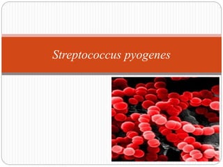
Streptococcus pyogens
•Transferir como PPTX, PDF•
237 gostaram•74,540 visualizações

Streptococcus pyogens

Recomendados
Mais conteúdo relacionado
Mais procurados
Mais procurados (20)
Destaque
Destaque (20)
Semelhante a Streptococcus pyogens
Semelhante a Streptococcus pyogens (20)
Mais de Deepa Devkota
Último
Último (20)
Streptococcus pyogens
- 2. Introduction Streptococci (streptos, twisted or coiled) Normal flora of humans upper respiratory tract and animals Some of them may be pathogens e.g. Streptococcus pyogenes causing pyogenic infections with a tendency to spread unlike staphylococcal infections It produces non-suppurative lesions, acute rheumatic fever and glomerulonephritis which occur as sequelae to infection
- 3. Morphology Ovoid to spherical in shape Gram-positive cocci arranged in chains or pairs Chain formation is due to the cocci dividing in one plane only and the daughter cells failing to separate completely Chains are longer in liquid than in solid media Non motile and non-sporing Capsulated (hyaluronic acid; non-immunogenic) Group A b-hemolytic streptococci
- 4. Gram staining of Streptococcus pyogenes
- 5. Cultural characteristics Aerobes and facultative anaerobes Optimum temperature: 37C Growth occurs only in media containing fermentable carbohydrates or enriched with blood or serum
- 6. i. Blood agar: Small (0.5-1mm), circular, semi-transparent colonies Produce wide zone of β- hemolysis Growth and hemolysis are promoted by 5-10% CO2 Virulent strains, on fresh isolation form lesions, produce a ‘matt’ (finely granular) colony while avirulent strains form ‘glossy’ colonies Mucoid colonies are formed by strains that produce large capsules ii. Liquid media: Glucose or serum broth Growth occurs as a granular turbidity with a powdery deposit No pellicle is formed
- 7. Biochemical reactions Catalase negative Bile insoluble Ferments sugars producing acid but no gas PYR test positive Hydrolyse pyrrolidonyl-beta-napthylamide (PYR) due to presence of peptidase, the resulting napthylamide produces a red colour upon the addition of 0.01% cinnamaldehyde reagent Faliure to ferment ribose Bile insoluble
- 8. Overview of reactions sensitive to S. pyogenes Fig. PYR test– positive for S. pyogenes
- 9. Resistance S. pyogenes is a delicate organism, easily destroyed by heat (54C for 30 min) Sensitive to bacitracin Has developed less resistance to drugs Dies in a few days in culture, unless stored at a low temperature (4C) preferably in Robertson’s cooked meat medium Rapidly inactivated by antiseptics and many antibiotics Fig. Zone of inhibition shown by S. pyogenes
- 10. Antigenic structure Capsular hyaluronic acid: • Non antigenic as hyaluronic acid is identical to that found in human connective tissue and hence bacteria can disguise themselves with an immunological self substance • Has weak anti-phagocytic activity but protects streptococci against immunological attacks
- 11. Antigenic structure A. Cell wall: 1. Outer layer: Protein and lipoteichoic acid 2. Middle layer: Group specific carbohydrate 3. Inner layer: Peptidoglycan (mucoprotein) Responsible for cell wall rigidity Enhances non specific resistance (pyrogenic and thrombolytic activity)
- 12. Virulence factor
- 13. Antigenic structure B. Group A-specific C-carbohydrates Serological grouping of streptococci is done on its basis Divided into 20 Lancefield groups (A to V) except I and J on the basis of group specific carbohydrates. All streptococci except viridans (a-hemolytic) group have a layer of carbohydrate
- 14. Antigenic structure C. Proteins Present in outermost layer Produces surface protein antigens (F,M, T and R) Useful in serological typing of S. pyogenes F-protein Recognizes host fibronectin, a matrix protein that is present in eukaryotic cells. Hence helps in attachment together with lipoteichoic acid and M protein
- 15. Antigenic structure M protein Most antigenic Acts as a virulence factor by inhibiting phagocytosis Covered with lipoteichoic acid that enable the organism to attach to epithelial cell M protein is heat and acid stable but susceptible to tryptic digestion On the basis of antigenic difference in M protein, S. pyogenes can be divided into about 100 types
- 16. Antigenic structure M protein The most distal part of M protein shows extensive variability among strains hence individual may suffer from recurrent S. pyogenes infections with strains expressing different versions of M protein
- 17. Antigenic structure T-Protein Common to many M- types Not associated with virulence and is not a protective antigen It is strongly antigenic R protein Non-type-specific and is associated with M- proteins of types 2,3 28 and 48 known as M-associated protein (MAP) Not associated to virulence and not a protective antigen Strongly antigenic
- 18. Antigenic structure D. Pili ( Fimbriae) Hair like and project from capsule Consist partly of M-protein Covered with lipoteichoic acid Important in the attachment of streptococci to epithelial cells
- 19. Structural components of S. pyogenes that cross react with human tissues Structural components of S. pyogenes Human tissue with which it cross reacts Capsular hyaluronic acid Synovial fluid Cell wall protein Myocardium Cell wall carbohydrtes Cardiac valves Cytoplasmic membrane antigens Vascular intima Peptidoglycans Skin antigens
- 20. Toxins and enzymes 1. Hemolysins (Streptolysins) 2. Erythrogenic toxin 3. Streptokinase (Fibrinolysin) 4. Deoxyribonucleases (Streptodornase, DNAase) 5. Hyaluronidase
- 21. Cytolytic toxins and other exo-enzymes produced by Streptococcus pyogenes
- 22. Hemolysins (Streptolysins) Produce complete disruption of RBC Contribute to tissue invasion and destruction There are two types of Streptolysins Streptolysin O Streptolysin S
- 23. Erythrogenic toxin Also known as pyrogenic exotoxin/ Dick/ Scarlatinal toxin Known as erythrogenic because its intradermal injection into susceptible individuals produced an erythematous reaction (Dick test) Dick test- Used to identify susceptibility to scarlet fever Primary effect of the toxin is production of fever hence also called Streptococcal pyrogenic exotoxin (SPE) Three types of SPE (A,B,C)
- 24. Mediate production of rash e.g. (scarlet fever) Superantigens hence massive release of cytokines occur that leads to variety of clinical signs including inflammation, shock and organ failure Fig. Rash in scarlet fever
- 25. Streptokinase (Fibrinolysin) Two types of streptokinase (A and B) Antigenic protein Promotes the lysis of human blood clot by converting plasminogen to plasmin Fibrinolysin facilitates spread of infection by breaking down the fibrin barrier around the lesions also known as spreading factor It is given intravenously for the treatment of early myocardial infarction and other thromboembolic disorders
- 26. Deoxyribonucleases (Streptodornase,DNAase) Degrades DNA Four antigenically distinct DNAases: A,B,C,D; B most antigenic Capable of liquefying DNA accumulated in thick pus derived from nuclei of necrotic cells, hence the exudate is thin in streptococcal infections Important therapeutically in liquefying localised collections of thick exudates (Empyema) Demonstration of anti-DNAase B antibody in the diagnosis of S. pyogenes infections when ASO titres is low
- 27. Hyaluronidase Breaks down hyaluronic acid of connective tissue and favors spread of infection Antigenic and specific antibodies are formed Degrades capsule Others are proteinase, phosphatase, amylase, esterases, NADase, C5a peptidase, lipase, Serum opacity factor (SOP) etc.
- 28. Pathogenicity Produces pyrogenic infection with a tendency to spread locally, along lymphatics and through blood stream Disease caused can be: Suppurative or Non suppurative
- 29. Pathogenicity
- 30. Pathogenicity
- 31. Suppurative complications 1. Respiratory infections Primary site of invasion is throat causing sore throat May be localized as tonsillitis or pharyngitis
- 32. Respiratory infections Lipoteichoic acid covering surface pili binds to the glycoprotein fibronectin on epithelial cells of pharynx From the throat, spreads to surrounding tissues leading to suppurative complications like Otitis media Mastoiditis Quinsy Suppurative adenitis Meningitis(rare)
- 33. 2. Skin and soft tissue infection S. pyogenes causes subcutaneous infections ranging cellutitis to necrotising fascilitis Include infections of wounds or burns, with a predilection to produce lymphangitis and cellulitis Infection of minor abrasions may lead to fatal septicemia S. pyogenes is also known as ‘flesh eating bacteria’ - extensive necrosis of subcutaneous and muscular tissue and adjacent fascia – causes Toxic shock like syndrome
- 34. a) Impetigo (Pyoderma) Pyo-purulent and derma-skin Caused by higher numbered M types S. pyrogen Superficial discrete crushed spot of less than one inch in diameter seen in children Lasts for 1-2 weeks and heals spontaneously without any scars
- 35. b) Erysipelas Erythros:red and pella:skin Hypersensitivity reaction to Streptococcal antigen Causes acute spreading lesions involving superficial lymphatics Affected skin is red, swollen, indurated and sharply demarcated from surrounding healthy skin Rare and seen only in older patients
- 36. c) Cellulitis and Necrotising fascilitis Involves deeper subcutaneous tissues Local inflammation and systemic signs like erysipelas are observed
- 37. 3. Genital infection Both aerobic and anaerobic Streptococci are normal habitat of female genitalia Causes puerperal sepsis with exogenous infection Puerperal fever is caused due to endogenous infection with anaerobic Streptococci Other suppurative infections: Abscesses in brain, lungs, kidney and liver causing septicemia and pyemia
- 38. Non suppurative complications After a latent period of 1-4 weeks Followed by rheumatic fever and acute glomerulonephritis
- 39. a) Rheumatic fever Complication of S.pyogenes pharyngitis due to specific I M protein types Characterized by aschoff nodules (sub cutaneous nodule) Causes inflammatory myocardial lesion of connective tissue degeneration of heart valves Results in chronic and progressive damage to heart valves, arthralgias to Frank arthritis Mimics epidemiologic character of streptococcal pharyngitis
- 40. b) Glomerulonephritis Caused by specific nephritogenic strains of group A streptococcus Characterized by acute inflammation of renal glomeruli with edema, hypertension, hematuria and proteinuria In contrast with rheumatic fever it is sequela of both pharyngeal and pyodermal streptococcal infection differing in nephrogenic M serotypes Mimics epidemiologic character of streptococcal infection Progressive, irreversible loss of renal function in young is common
- 41. Epidemiology Group A Streptococci causes transient asymptomatic colonization of oropharynx and skin Regulated by ability to mount specific immunity to M protein of colonizing strain and presence of competitive organism in oropharynx Pharyngitis is primarily disease of children(5-15 yrs.), infants and adults are also susceptible Pathogen spreads from respiratory droplets and direct contact especially in winter season
- 42. Epidemiology Soft tissue infections are proceeded by initial skin colonization to superficial or deep tissue through a break in the skin Re-infection occur due to multiplicity of M protein serotypes
- 43. Laboratory Diagnosis a. Throat swab culture: Detection of group A antigen b. Specific nucleic acid based test c. Elevation of anti hyaluronidase antibodies(strong evidence) 1. Specimen: Throat swab, pus swab or exudates are collected. 2. Microscopy: Gram-staining of pus can be examined Presence of Gram-positive cocci in chains can be indication.
- 44. 3. Culture: Swab from the affected area is collected and are either plated immediately or sent to laboratory in Pike’s medium. The specimen should be plated on blood agar and incubated at 37˚C anaerobically or under 5-10% CO2 ,as hemolysis develops better.
- 45. 4. Identification: Rapid diagnostic test kits are available for the detection of streptococcal group A antigen from throat swab Bacitracin sensitivity: Based on Maxted’s observation that they are more sensitive to bacitracin than other streptococci A filter paper disc of 0.04U is applied on the surface of an inoculated blood agar After incubation, a wide zone of inhibition is seen with S.pyrogenes but not with other streptococci
- 46. 5. Serology: a) Antistreptolysin O titration Standard test ASO titres higher than 200 are indicative of prior streptococcal infection. High levels are usually found in acute rheumatic fever but in glomerulonephritis, times are often low b) Antideoxyribonuclease B (anti-DNAase B): Commonly used Titres higher than 300 are taken c) Streptozyme test: A passive slide hemagglutination test using erthyrocytes sensitised with a crude preparation of streptococci It is a convenient, sensitive and specific screening test.
- 47. Treatment, prevention and control DRUGS USED: • For streptococcal pharyngitis: Oral penicillin V or amoxicillin • Oral cephalosporin or macrolides can be used for penicillin sensitive patients • For severe, systemic infection: Combined use of intravenous penicillin with protein synthesis inhibiting antibiotics(clindamycin) is recommended • Streptococcal pyogenes have developed resistance over tetracyclines and sulfonamides, newer macrolides • Antimicrobial drugs has no effect on glomerulonephritis and rheumatic fever
- 48. Prophylaxis Rheumatic fever requires long term antibiotic prophylaxis to prevent recurrence of disease Penicillin is used in patients who have developed early signs of rheumatic fever For acute glomerulonephritis no need of antibiotic therapy and prophylactic therapy(no re infection) For patients with serious soft tissue infection, drainage and aggressive surgical debridement must be initiated
- 49. References 1. Ananthanarayan R. & Paniker C. J. (2009). Textbook of Microbiology. Hyderabad: Universities Press (India) Private Limited. 2. Harvey Richard A. et all. Lippincott’s Illustrated Reviews Microbiology, Wolters Kluwer (India) Pvt. Ltd.,2nd edition. 3. Chakraborty P. (2009). A Textbook of Microbiology. Kolkata: New Central Book Agency (P) Ltd. 4. Murray et all. Medical microbiology, Elsevier Saunders, 7th edition.