SlideShare uma empresa Scribd logo
1 de 47
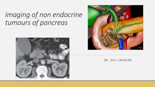
Imaging of non endocrine
tumours of pancreas
DR. DEV LAKHERA
Anatomy
L1 vertebral body
Pancreatic malignancy
2nd most common in GI malignancy
5 year - Survival rate <5%
Most are ductal in origin- pancreatic adenocarcinoma
Cystic pancreatic tumours and endocrine tumours rare.
Curative - surgical resection // partial pancreaticoduodenectomy - 'pylorus-
preserving’ Whipple
Classification of tumours
Pancreatic malignancies
Duct Cell Origin
• Solid neoplasms
1. Adenocarcinoma
2. Variant carcinomas
• Cystic neoplasms
1. Congenital cysts
2. Pseudocysts
3. Serous cystadenoma
4. Mucinous cystic neoplasm
5. Mucinous ductal ectasia
Acinar origin (1%)
1. Acinar cell carcinoma
2. Acinar cell cystadenocarcinoma
3. Pancreaticoblastoma
Rest are endocrine
Indeterminate Origin
1. Osteoclast-type giant cell carcinoma
2. Solid and papillary epithelial neoplasm
3. Mixed endocrine-exocrine neoplasm
4. Microadenocarcinoma.
Pancreatic cancers (85%) are
adenocarcinoma of ductal origin.
Pancreatic adenocarcinoma
 (M:F – 1.5:1)  60 - 70 years (60-70 male with painless jaundice)
Advanced local tumor extension (40%) / distant metastatic disease (40%)
Head - Painless jaundice
Body and tail - Pain and weight loss
Pancreatic tumor - Site
Mainly - Head of the pancreas (75%).
Minority - in the body (15%) and tail (10%).
At the time of diagnosis head malignancies - 2-3
cm - present earlier
Periampullary tumors (0.1%)
intense desmoplastic response
Tumors originating in the distal common bile duct or ampulla
may also grow into the pancreatic head and together with
pancreatic head carcinoma these tumors are often grouped
together under the name.
Imaging work up
Ultrasound – First line imaging test - Better detects masses >3 cm and mets >2 cm
CT
Higher diagnostic accuracy
Local and distant staging
Signs of unresectability
MRCP - sensitive for detecting a periampullary mass, but offers no significant additional staging
information
Clinical feature
painless obstructive jaundice
Ultrasonography (US)
 Hypoechoic
Necrotic tumors may show heterogenous
echopattern
Ductal obstruction and dilatation may also be
visualized.
Vascular involvement -thickening of
periarterial tissues
large, hypoechoic mass (M) obstructing the extrahepatic
common bile duct (CBD).
normally echogenic fat immediately adjacent to the artery is
replaced by tissue of lower echogenicity
hypoechoic mass in the pancreatic
head (A). On CDFI peritumoral
vascularity and absence of internal
vascularity seen. No vascular
invasion was seen in this case // A,
Transverse sonogram
reveals a subtle cancer in the body
of the pancreas (yellow arrows) with
a pseudocyst peripheral to the mass
(white arrow). B, CT image at a
similar level shows identical findings.
Endoscopic US
Second part of duodenum - pancreatic head, portal vein and papilla
Duodenal bulb - for head, neck and distal CBD
Stomach - for imaging of body, tail and the pancreatic duct
Sensitive for small (< 2 cm) solid tumors
Barium – head of pancreas
Widening of C loop of duodenum
Antral pad sign
effacement and distortion of the
mucosal pattern on the medial wall
Inverted 3 sign
Double contour of duodenal loop // Enlarged
duodenal loop with 'reversed 3' sign of Frostberg.
Earlier percutaneous transhepatic cholangiogram
shows characteristic ' gloved finger' obstruction of
intrapancreatic common bile duct pathognomonic
of carcinoma of the pancreatic head
first image is a prone view from UGI series showing mass effect on 2nd and 3rd portions of the duodenal sweep.
The second image is an RAO view from UGI series showing mass effect on 3rd portions of the duodenal sweep with
mild dilatation of proximal duodenum.
CT
Imaging modality of choice for the detection and presurgical staging.
High density oral contrast should be avoided and water should be used
Optimal imaging - early-portal phase 40-50 sec - pancreatic phase.
Late portal phase
Overall assessment of the abdomen to look for liver metastases, lymph nodes and peritoneal
implants.
Plain scan for pancreatic calcifications
When performing CT evaluation of pancreatic tumors, as oral
contrast.
Characteristic imaging features
Solid schirrous type
Reduced vascular perfusion
Ill defined margins
Not encapsulated
Desmoplastic reactionFibrous tissue, hard
slow growing / Rather than heterogenous
enhamcnet – soild hypoenehancing
masss with illdefined margins /
Fibrous tissue, hard slow growing / Rather than heterogenous enhancment – solid hypoenhancing masss
with illdefined margins /
.
During the early portal phase,
ill defined nonenhancing
hypodense area in tail of the
pancreas. B.. During the
venous phase, the mass is
almost isodense with the
remainder of the pancreas
(arrow)..
Adenocarcinoma, head of
pancreas and same patient as
C. The mass is unrecognizable
20 seconds later during the
portal phase. Both cases
illustrate the critical
importance of pancreatic
phase imaging
Strong tendency to obstruct ducts – with dilatation
Parenchymal thinning
Side branches may undergo cystic dilatation with
obstruction rupture causing pancreatitis and
extrapancreatic effusion / pseudocyst formation
CBD obstruction –compression by mass /
lymphadenopathy
CECT image showing an ill defined hypodense lesion in the pancreatic head which appears bulky.
There is also upstream dilatation of the main pancreatic duct// Metastatic lymphadenopathy
adjacent to suprapancreatic part of the bile duct can also cause ductal obstruction.
subtle hypodense hypoenhancing
area in uncinate process of
pancreas with dilatation of
pancreatic duct.
Double duct sign -
Concurrent dilatation of pancreatic duct and CBD
CECT images showing the “double duct sign”, dilated CBD and
MPD due to mass within the pancreatic head
Extension into adjacent structures
Portal, splenic and superior mesenteric vein (SMV) lying
adjacent to the pancreas and confluence .
Duodenum by head masses, stomach by body masses and
hilum of spleen by tail masses.
subcentimetric lymphnodes have to mentioned.
Perivascular and perineural tissues are particularly susceptible
to involvement
Metastatic dissemination
◦ regional lymph nodes, i.e. coeliac, common hepatic, superior mesenteric and para-aortic.
◦ hepatic via portal venous drainage,
◦ omental and peritoneal via intraperitoneal shedding of tumor cells.
Unresectability
Vascular ingrowth
Ingrowth into the celiac axis, hepatic artery or
superior mesenteric artery also preclude resection.
Vascular encasement
◦ obliteration of the normal fat between the pancreatic margin and the adjacent vessel
◦ more than 180-degree contact between the tumor and the vessel
◦ morphologic changes in the artery, including narrowing or encasement of the affected artery.
MDCT – more accurate description of local spread
Venous invasion – Criteria - > 180 o
Ill defined hypodense lesion in uncinate
process appearing to contact the
superior mesenteric artery over a greater
than 180-degree circumference (arrow).
B. coronal // mass (asterisk) in the body
of the pancreas that contacts but does
not surround the splenic artery. Note the
upstream atrophy and main pancreatic
duct dilation in the body and tail of the
pancreas (arrow). // The hepatic
metastasis is seen on the portal
acquisition; on the pancreatic phase,
there is only a vague region of low
attenuation. This case illustrates the
differing times of peak enhancement of
the pancreas and liver. The presence of
the hepatic metastasis makes this case
unresectable despite the fact that the
tumor is locally resectable.
Axial pancreatic-phase contrast-enhanced CT in a 78-yearold man with an adenocarcinoma of the midpancreatic
body (large arrow) with upstream pancreatic duct dilatation (small arrow) and peripancreatic extension of
tumor. The tumor has extended to encase the celiac axis (thin arrows), rendering the patient inoperable.// Axial
pancreatic-phase contrast-enhanced CT in a 73-yearold
woman with soft tissue encasement by pancreatic adenocarcinoma of the superior mesenteric artery (arrows),
but the superior mesenteric vein is unaffected (small arrow). This patient was inoperable.
Differential diagnosis
focal pancreatitis
lymphoma
metastasic disease.
MRI
Axial and coronal T1 weighted postcontrast images show ll defined hypointense non enhancing mass in uncinate
coronal image in a case of carcinoma pancreatic head showing an isointense mass causing obstructive
biliopathy. MRCP image in the case showing dilatation of the common duct proximal to the lesion and the
dilated main pancreatic duct (B)
Cystic tumours of pancreas
Most common cystic lesion benign – pseudocyst
Masses
◦ Mucinous cystic neoplasm
◦ Serous adenomas
Mucinous Cystic Neoplasms (MCNs)
Collective
◦ mucinous cystadenoma of pancreas
◦ mucinous cystadenocarcinoma of pancreas
◦ intraductal papillary neoplasms (IPMN) of the pancreas
Rare – body and tail
95% females
CT- Cystadenoma
Rounded or ovoid
Calcification when present more peripheral
Contents of the lesion may be heterogenous is
attenuation
Internal septations may be present and tend to
be linear or curvilinear
CECT - thin walled
multiloculated
hypodense mass arising
from pancreatic body
and tail
multilocular
MR image revealing a multilocular cyst in
the pancreatic head. The fluid component shows high signal
and
mural nodules show isointense signal –
Differential diagnosis
mucinous cystadenocarcinoma of the pancreas:
at times impossible to differentiate on ultrasound or CT from a mucinous cystadenoma
pancreatic pseudocyst (unilocular)
variant of serous cystadenoma of the pancreas
Intraductal papillary mucinous neoplasm (10-
15%)
Older patients
Difficult to differentiate from chronic pancreatitis
main duct branch duct type
chronic pancreatitis head and uncinate process
segmental or diffuse
distribution
more localised and mass-like
Highest malignant potential may be multifocal
indolent
CT - main duct IPMN
Segmental or entire duct dilatation >5mm
Thinning of overlying pancreatic parenchyma
solid mural nodules are concerning for malignant
transformation- hyperdense nodules protruding into
the mucin-filled dilated ducts
enhancing nodules
branch duct IPMN
single or multiple side branches demonstrating marked dilatation
cystic mass-like appearance which often mimicks cystic tumours of
the pancreas
bunch of grapes appearance
microcystic variety has appearances similar to serous
cystadenomas, but again communication with the main pancreatic
duct is the key to correct diagnosis
Axial MR image (A) and coronal TRUFISP MR image (B) showing grossly dilated pancreatic duct with
intraluminal filling defects and a non dilated biliary system
Serous cystadenoma
Imaging appearance
Multicystic, lobulated mass in the pancreatic head - 'bunch of grapes’
Individual cysts are typically <20 mm in size and greater than six in number (except for the
oligocystic variety
Characteristic enhancing central scar - which can show associated stellate calcification (~20%)
CECT images showing a bulky, multiloculated hypodense predominantly cystic mass of the
pancreatic head with a central calcified stellate scar – serous cystadenoma
Corronal t2 fat sat large
multiloculated
hyperitnersne cystic
lesion I tail of [pancreas
with multiple septation
Pancreatic cysts
1. Von Hippel Lindau syndrome
2. Autosomal dominant polycystic kidney disease (ADPKD)
3. Cystic fibrosis
Pancreatic metastases
Uncommon - patients with widespread metastasis
PRIMARIES
◦ renal cell carcinoma (RCC): one of commonest of tumours that metastatise to the pancreas
◦ melanoma
◦ breast cancer
◦ lung cancer
◦ gastric cancer
Imaging findings are non specific
Well circumscribed mass – iso to hypodense, heterogenous enhancement
Lymphoma
Involvement : -Direct extension from
peripancreatic B-cell type lymphadenopathy
due to non-Hodgkin’s lymphoma.
Presentation
◦ Focal mass
Diffuse enlargement
bulky lobulated centrally necrotic hypodense mass replacing
entire pancreas except uncinate process
(A) with extensive liver metastases (B)—Pancreatic lymphoma
THANK YOU

Mais conteúdo relacionado

Mais procurados

IMAGING OF CYSTIC PANCREATIC TUMOURS
IMAGING OF CYSTIC PANCREATIC TUMOURSIMAGING OF CYSTIC PANCREATIC TUMOURS
IMAGING OF CYSTIC PANCREATIC TUMOURSDr I Gurubharath .
 
Magnetic resonance cholangiopancreatography ppt
Magnetic resonance cholangiopancreatography pptMagnetic resonance cholangiopancreatography ppt
Magnetic resonance cholangiopancreatography pptAnjan Dangal
 
Presentation1, radiological imaging of endometrial carcinoma.
Presentation1, radiological imaging of endometrial carcinoma.Presentation1, radiological imaging of endometrial carcinoma.
Presentation1, radiological imaging of endometrial carcinoma.Abdellah Nazeer
 
KEYS OF RADIOLOGY SPOTTERS GIT
KEYS OF RADIOLOGY SPOTTERS GITKEYS OF RADIOLOGY SPOTTERS GIT
KEYS OF RADIOLOGY SPOTTERS GITAnish Choudhary
 
CT enhancement protocols
CT enhancement protocols  CT enhancement protocols
CT enhancement protocols Yanan Zhai
 
HEPATOCELLULAR CARCINOMA RADIOLOGY
HEPATOCELLULAR CARCINOMA RADIOLOGYHEPATOCELLULAR CARCINOMA RADIOLOGY
HEPATOCELLULAR CARCINOMA RADIOLOGYRMLIMS
 
Diagnostic Imaging of Renal Tumors
Diagnostic Imaging of Renal TumorsDiagnostic Imaging of Renal Tumors
Diagnostic Imaging of Renal TumorsMohamed M.A. Zaitoun
 
Imaging Acute Abdomen (Part 1)
Imaging Acute Abdomen (Part 1)Imaging Acute Abdomen (Part 1)
Imaging Acute Abdomen (Part 1)Rathachai Kaewlai
 
Imaging of Malignant Liver Lesions
Imaging of Malignant Liver LesionsImaging of Malignant Liver Lesions
Imaging of Malignant Liver LesionsSahil Chaudhry
 
Urinary bladder pathology radiology
Urinary bladder pathology radiologyUrinary bladder pathology radiology
Urinary bladder pathology radiologyDr pradeep Kumar
 
Unilateral small kidney
Unilateral small kidneyUnilateral small kidney
Unilateral small kidneymaimusirdan
 
Mrcp radiology
Mrcp radiologyMrcp radiology
Mrcp radiologyVidya TK
 
Imaging of the adrenal glands
Imaging of the adrenal glands Imaging of the adrenal glands
Imaging of the adrenal glands Satish Naga
 
Presentation1.pptx, radiological imaging of prostatic diseases
Presentation1.pptx, radiological imaging of prostatic diseasesPresentation1.pptx, radiological imaging of prostatic diseases
Presentation1.pptx, radiological imaging of prostatic diseasesAbdellah Nazeer
 
Pediatric liver masses
Pediatric liver massesPediatric liver masses
Pediatric liver massesSidra Afzal
 
Imaging in mediastinal masses by Dr. Milan Silwal
Imaging in mediastinal masses by Dr. Milan SilwalImaging in mediastinal masses by Dr. Milan Silwal
Imaging in mediastinal masses by Dr. Milan SilwalMilan Silwal
 

Mais procurados (20)

Benign focal lesions in liver
Benign focal lesions in liverBenign focal lesions in liver
Benign focal lesions in liver
 
IMAGING OF CYSTIC PANCREATIC TUMOURS
IMAGING OF CYSTIC PANCREATIC TUMOURSIMAGING OF CYSTIC PANCREATIC TUMOURS
IMAGING OF CYSTIC PANCREATIC TUMOURS
 
Magnetic resonance cholangiopancreatography ppt
Magnetic resonance cholangiopancreatography pptMagnetic resonance cholangiopancreatography ppt
Magnetic resonance cholangiopancreatography ppt
 
MRI carcinoma Uterus and Cervix
MRI carcinoma Uterus and  CervixMRI carcinoma Uterus and  Cervix
MRI carcinoma Uterus and Cervix
 
Malignant liver lesions
Malignant liver lesionsMalignant liver lesions
Malignant liver lesions
 
Presentation1, radiological imaging of endometrial carcinoma.
Presentation1, radiological imaging of endometrial carcinoma.Presentation1, radiological imaging of endometrial carcinoma.
Presentation1, radiological imaging of endometrial carcinoma.
 
KEYS OF RADIOLOGY SPOTTERS GIT
KEYS OF RADIOLOGY SPOTTERS GITKEYS OF RADIOLOGY SPOTTERS GIT
KEYS OF RADIOLOGY SPOTTERS GIT
 
CT enhancement protocols
CT enhancement protocols  CT enhancement protocols
CT enhancement protocols
 
HEPATOCELLULAR CARCINOMA RADIOLOGY
HEPATOCELLULAR CARCINOMA RADIOLOGYHEPATOCELLULAR CARCINOMA RADIOLOGY
HEPATOCELLULAR CARCINOMA RADIOLOGY
 
Diagnostic Imaging of Renal Tumors
Diagnostic Imaging of Renal TumorsDiagnostic Imaging of Renal Tumors
Diagnostic Imaging of Renal Tumors
 
Imaging Acute Abdomen (Part 1)
Imaging Acute Abdomen (Part 1)Imaging Acute Abdomen (Part 1)
Imaging Acute Abdomen (Part 1)
 
Imaging of Malignant Liver Lesions
Imaging of Malignant Liver LesionsImaging of Malignant Liver Lesions
Imaging of Malignant Liver Lesions
 
Urinary bladder pathology radiology
Urinary bladder pathology radiologyUrinary bladder pathology radiology
Urinary bladder pathology radiology
 
Unilateral small kidney
Unilateral small kidneyUnilateral small kidney
Unilateral small kidney
 
Mrcp radiology
Mrcp radiologyMrcp radiology
Mrcp radiology
 
Imaging of the adrenal glands
Imaging of the adrenal glands Imaging of the adrenal glands
Imaging of the adrenal glands
 
Presentation1.pptx, radiological imaging of prostatic diseases
Presentation1.pptx, radiological imaging of prostatic diseasesPresentation1.pptx, radiological imaging of prostatic diseases
Presentation1.pptx, radiological imaging of prostatic diseases
 
Pediatric liver masses
Pediatric liver massesPediatric liver masses
Pediatric liver masses
 
Imaging in mediastinal masses by Dr. Milan Silwal
Imaging in mediastinal masses by Dr. Milan SilwalImaging in mediastinal masses by Dr. Milan Silwal
Imaging in mediastinal masses by Dr. Milan Silwal
 
Acute abdomen
Acute abdomenAcute abdomen
Acute abdomen
 

Semelhante a Imaging of non endocrine tumour of pancreas

Case of pancreatic adenocarcinoma and other pancreatic tumors
Case of pancreatic adenocarcinoma and other pancreatic tumorsCase of pancreatic adenocarcinoma and other pancreatic tumors
Case of pancreatic adenocarcinoma and other pancreatic tumorsAlex Allu
 
Pancreatic neoplasms
Pancreatic neoplasmsPancreatic neoplasms
Pancreatic neoplasmsdrksreenath
 
Cysticlesionsofpancreas 131020083630-phpapp02
Cysticlesionsofpancreas 131020083630-phpapp02Cysticlesionsofpancreas 131020083630-phpapp02
Cysticlesionsofpancreas 131020083630-phpapp02danghaihvqy
 
Ultrasound of pancrease in Radiology
Ultrasound of pancrease in RadiologyUltrasound of pancrease in Radiology
Ultrasound of pancrease in RadiologyMahesh Kumar
 
Veeru ca pancreas
Veeru ca pancreasVeeru ca pancreas
Veeru ca pancreasVeeru Reddy
 
Colorectal cancer. Colorectal Symptoms
Colorectal cancer. Colorectal SymptomsColorectal cancer. Colorectal Symptoms
Colorectal cancer. Colorectal SymptomsEneutron
 
Pancreatic neoplasms
Pancreatic neoplasmsPancreatic neoplasms
Pancreatic neoplasmsAjai Sasidhar
 
Gall Bladder Carcinoma
Gall Bladder CarcinomaGall Bladder Carcinoma
Gall Bladder CarcinomaDr.Mohsin Khan
 
Imaging in small bowel tumors Dr. Muhammad Bin Zulfiqar
Imaging in small bowel tumors Dr. Muhammad Bin ZulfiqarImaging in small bowel tumors Dr. Muhammad Bin Zulfiqar
Imaging in small bowel tumors Dr. Muhammad Bin ZulfiqarDr. Muhammad Bin Zulfiqar
 
Pancreatic cystic lesion by xiu
Pancreatic cystic lesion by xiuPancreatic cystic lesion by xiu
Pancreatic cystic lesion by xiuXiu Srithammasit
 
Carcinomarectum 111113085726-phpapp01
Carcinomarectum 111113085726-phpapp01Carcinomarectum 111113085726-phpapp01
Carcinomarectum 111113085726-phpapp01Drrajan Paliwal
 
Presentation2, radiological imaging of gastrointestinal schwannoma.
Presentation2, radiological imaging of gastrointestinal schwannoma.Presentation2, radiological imaging of gastrointestinal schwannoma.
Presentation2, radiological imaging of gastrointestinal schwannoma.Abdellah Nazeer
 
Presentation1.pptx imaging of the peritoneum and mesentry.
Presentation1.pptx imaging of the peritoneum and mesentry.Presentation1.pptx imaging of the peritoneum and mesentry.
Presentation1.pptx imaging of the peritoneum and mesentry.Abdellah Nazeer
 
Cystic lesions of the pancreas
Cystic lesions of the pancreasCystic lesions of the pancreas
Cystic lesions of the pancreasAtit Ghoda
 
Cystic neoplasm of pancreas
Cystic neoplasm of pancreasCystic neoplasm of pancreas
Cystic neoplasm of pancreasDr Dipesh K.K
 

Semelhante a Imaging of non endocrine tumour of pancreas (20)

Case of pancreatic adenocarcinoma and other pancreatic tumors
Case of pancreatic adenocarcinoma and other pancreatic tumorsCase of pancreatic adenocarcinoma and other pancreatic tumors
Case of pancreatic adenocarcinoma and other pancreatic tumors
 
Pancreatic neoplasms
Pancreatic neoplasmsPancreatic neoplasms
Pancreatic neoplasms
 
Cysticlesionsofpancreas 131020083630-phpapp02
Cysticlesionsofpancreas 131020083630-phpapp02Cysticlesionsofpancreas 131020083630-phpapp02
Cysticlesionsofpancreas 131020083630-phpapp02
 
Ultrasound of pancrease in Radiology
Ultrasound of pancrease in RadiologyUltrasound of pancrease in Radiology
Ultrasound of pancrease in Radiology
 
Veeru ca pancreas
Veeru ca pancreasVeeru ca pancreas
Veeru ca pancreas
 
CA Pancreas- Niazi.pptx
CA Pancreas- Niazi.pptxCA Pancreas- Niazi.pptx
CA Pancreas- Niazi.pptx
 
Colorectal cancer. Colorectal Symptoms
Colorectal cancer. Colorectal SymptomsColorectal cancer. Colorectal Symptoms
Colorectal cancer. Colorectal Symptoms
 
Pancreatic neoplasms
Pancreatic neoplasmsPancreatic neoplasms
Pancreatic neoplasms
 
Gall Bladder Carcinoma
Gall Bladder CarcinomaGall Bladder Carcinoma
Gall Bladder Carcinoma
 
Imaging in small bowel tumors Dr. Muhammad Bin Zulfiqar
Imaging in small bowel tumors Dr. Muhammad Bin ZulfiqarImaging in small bowel tumors Dr. Muhammad Bin Zulfiqar
Imaging in small bowel tumors Dr. Muhammad Bin Zulfiqar
 
Pancreatic cystic lesion by xiu
Pancreatic cystic lesion by xiuPancreatic cystic lesion by xiu
Pancreatic cystic lesion by xiu
 
Carcinomarectum 111113085726-phpapp01
Carcinomarectum 111113085726-phpapp01Carcinomarectum 111113085726-phpapp01
Carcinomarectum 111113085726-phpapp01
 
Pancreas and spleen 04022021
Pancreas and spleen 04022021Pancreas and spleen 04022021
Pancreas and spleen 04022021
 
Presentation2, radiological imaging of gastrointestinal schwannoma.
Presentation2, radiological imaging of gastrointestinal schwannoma.Presentation2, radiological imaging of gastrointestinal schwannoma.
Presentation2, radiological imaging of gastrointestinal schwannoma.
 
Carcinoma gb
Carcinoma gbCarcinoma gb
Carcinoma gb
 
Presentation1.pptx imaging of the peritoneum and mesentry.
Presentation1.pptx imaging of the peritoneum and mesentry.Presentation1.pptx imaging of the peritoneum and mesentry.
Presentation1.pptx imaging of the peritoneum and mesentry.
 
SPEN PANCREASE CASE
SPEN PANCREASE CASESPEN PANCREASE CASE
SPEN PANCREASE CASE
 
Cystic lesions of the pancreas
Cystic lesions of the pancreasCystic lesions of the pancreas
Cystic lesions of the pancreas
 
Cystic neoplasm of pancreas
Cystic neoplasm of pancreasCystic neoplasm of pancreas
Cystic neoplasm of pancreas
 
8 bladder tumor
8 bladder tumor8 bladder tumor
8 bladder tumor
 

Mais de Dev Lakhera

Artificial intelligence in radiology
Artificial intelligence in radiologyArtificial intelligence in radiology
Artificial intelligence in radiologyDev Lakhera
 
Imaging of abdominal trauma
Imaging of abdominal traumaImaging of abdominal trauma
Imaging of abdominal traumaDev Lakhera
 
Bowel and mesenteric injury
Bowel and mesenteric injuryBowel and mesenteric injury
Bowel and mesenteric injuryDev Lakhera
 
Malignant tumors of esophagus
Malignant tumors of esophagusMalignant tumors of esophagus
Malignant tumors of esophagusDev Lakhera
 
Valvular heart diseases imaging
Valvular heart diseases imagingValvular heart diseases imaging
Valvular heart diseases imagingDev Lakhera
 
Approach to adrenal lesions
Approach to adrenal lesionsApproach to adrenal lesions
Approach to adrenal lesionsDev Lakhera
 
Covid-19 Xray findings
Covid-19 Xray findingsCovid-19 Xray findings
Covid-19 Xray findingsDev Lakhera
 
Benign hepatic masses
Benign hepatic massesBenign hepatic masses
Benign hepatic massesDev Lakhera
 
Imaging of benign focal hepatic lesions
Imaging of benign focal hepatic lesionsImaging of benign focal hepatic lesions
Imaging of benign focal hepatic lesionsDev Lakhera
 
Basic and advanced mri imaging sequences in brain
Basic and advanced mri imaging sequences in brainBasic and advanced mri imaging sequences in brain
Basic and advanced mri imaging sequences in brainDev Lakhera
 
Barium studies in git
Barium studies in gitBarium studies in git
Barium studies in gitDev Lakhera
 
Imaging of portal hypertension
Imaging of portal hypertensionImaging of portal hypertension
Imaging of portal hypertensionDev Lakhera
 
Principle of usg imaging, construction of transducers
Principle of usg imaging, construction of transducersPrinciple of usg imaging, construction of transducers
Principle of usg imaging, construction of transducersDev Lakhera
 
Ct mri urography
Ct mri urographyCt mri urography
Ct mri urographyDev Lakhera
 
Imaging of thyroid
Imaging of thyroidImaging of thyroid
Imaging of thyroidDev Lakhera
 
Radiological vascular anatomy of brain
Radiological vascular anatomy of brainRadiological vascular anatomy of brain
Radiological vascular anatomy of brainDev Lakhera
 
congenital brain anomalies
congenital brain anomalies congenital brain anomalies
congenital brain anomalies Dev Lakhera
 
Congenital renal anomalies
Congenital renal anomaliesCongenital renal anomalies
Congenital renal anomaliesDev Lakhera
 
Congenital gastrointestinal anomalies
Congenital gastrointestinal  anomaliesCongenital gastrointestinal  anomalies
Congenital gastrointestinal anomaliesDev Lakhera
 

Mais de Dev Lakhera (20)

Artificial intelligence in radiology
Artificial intelligence in radiologyArtificial intelligence in radiology
Artificial intelligence in radiology
 
Imaging of abdominal trauma
Imaging of abdominal traumaImaging of abdominal trauma
Imaging of abdominal trauma
 
Bowel and mesenteric injury
Bowel and mesenteric injuryBowel and mesenteric injury
Bowel and mesenteric injury
 
Malignant tumors of esophagus
Malignant tumors of esophagusMalignant tumors of esophagus
Malignant tumors of esophagus
 
Valvular heart diseases imaging
Valvular heart diseases imagingValvular heart diseases imaging
Valvular heart diseases imaging
 
Approach to adrenal lesions
Approach to adrenal lesionsApproach to adrenal lesions
Approach to adrenal lesions
 
Covid-19 Xray findings
Covid-19 Xray findingsCovid-19 Xray findings
Covid-19 Xray findings
 
Benign hepatic masses
Benign hepatic massesBenign hepatic masses
Benign hepatic masses
 
Imaging of benign focal hepatic lesions
Imaging of benign focal hepatic lesionsImaging of benign focal hepatic lesions
Imaging of benign focal hepatic lesions
 
Basic and advanced mri imaging sequences in brain
Basic and advanced mri imaging sequences in brainBasic and advanced mri imaging sequences in brain
Basic and advanced mri imaging sequences in brain
 
Barium studies in git
Barium studies in gitBarium studies in git
Barium studies in git
 
Imaging of portal hypertension
Imaging of portal hypertensionImaging of portal hypertension
Imaging of portal hypertension
 
Principle of usg imaging, construction of transducers
Principle of usg imaging, construction of transducersPrinciple of usg imaging, construction of transducers
Principle of usg imaging, construction of transducers
 
Ct mri urography
Ct mri urographyCt mri urography
Ct mri urography
 
Imaging of thyroid
Imaging of thyroidImaging of thyroid
Imaging of thyroid
 
Radiological vascular anatomy of brain
Radiological vascular anatomy of brainRadiological vascular anatomy of brain
Radiological vascular anatomy of brain
 
congenital brain anomalies
congenital brain anomalies congenital brain anomalies
congenital brain anomalies
 
Congenital renal anomalies
Congenital renal anomaliesCongenital renal anomalies
Congenital renal anomalies
 
Congenital gastrointestinal anomalies
Congenital gastrointestinal  anomaliesCongenital gastrointestinal  anomalies
Congenital gastrointestinal anomalies
 
Dev
DevDev
Dev
 

Último

All Time Service Available Call Girls Marine Drive 📳 9820252231 For 18+ VIP C...
All Time Service Available Call Girls Marine Drive 📳 9820252231 For 18+ VIP C...All Time Service Available Call Girls Marine Drive 📳 9820252231 For 18+ VIP C...
All Time Service Available Call Girls Marine Drive 📳 9820252231 For 18+ VIP C...Arohi Goyal
 
Call Girls Hosur Just Call 9630942363 Top Class Call Girl Service Available
Call Girls Hosur Just Call 9630942363 Top Class Call Girl Service AvailableCall Girls Hosur Just Call 9630942363 Top Class Call Girl Service Available
Call Girls Hosur Just Call 9630942363 Top Class Call Girl Service AvailableGENUINE ESCORT AGENCY
 
Call Girls Madurai Just Call 9630942363 Top Class Call Girl Service Available
Call Girls Madurai Just Call 9630942363 Top Class Call Girl Service AvailableCall Girls Madurai Just Call 9630942363 Top Class Call Girl Service Available
Call Girls Madurai Just Call 9630942363 Top Class Call Girl Service AvailableGENUINE ESCORT AGENCY
 
Russian Call Girls Lucknow Just Call 👉👉7877925207 Top Class Call Girl Service...
Russian Call Girls Lucknow Just Call 👉👉7877925207 Top Class Call Girl Service...Russian Call Girls Lucknow Just Call 👉👉7877925207 Top Class Call Girl Service...
Russian Call Girls Lucknow Just Call 👉👉7877925207 Top Class Call Girl Service...adilkhan87451
 
9630942363 Genuine Call Girls In Ahmedabad Gujarat Call Girls Service
9630942363 Genuine Call Girls In Ahmedabad Gujarat Call Girls Service9630942363 Genuine Call Girls In Ahmedabad Gujarat Call Girls Service
9630942363 Genuine Call Girls In Ahmedabad Gujarat Call Girls ServiceGENUINE ESCORT AGENCY
 
VIP Hyderabad Call Girls Bahadurpally 7877925207 ₹5000 To 25K With AC Room 💚😋
VIP Hyderabad Call Girls Bahadurpally 7877925207 ₹5000 To 25K With AC Room 💚😋VIP Hyderabad Call Girls Bahadurpally 7877925207 ₹5000 To 25K With AC Room 💚😋
VIP Hyderabad Call Girls Bahadurpally 7877925207 ₹5000 To 25K With AC Room 💚😋TANUJA PANDEY
 
Call Girls Rishikesh Just Call 8250077686 Top Class Call Girl Service Available
Call Girls Rishikesh Just Call 8250077686 Top Class Call Girl Service AvailableCall Girls Rishikesh Just Call 8250077686 Top Class Call Girl Service Available
Call Girls Rishikesh Just Call 8250077686 Top Class Call Girl Service AvailableDipal Arora
 
Mumbai ] (Call Girls) in Mumbai 10k @ I'm VIP Independent Escorts Girls 98333...
Mumbai ] (Call Girls) in Mumbai 10k @ I'm VIP Independent Escorts Girls 98333...Mumbai ] (Call Girls) in Mumbai 10k @ I'm VIP Independent Escorts Girls 98333...
Mumbai ] (Call Girls) in Mumbai 10k @ I'm VIP Independent Escorts Girls 98333...Ishani Gupta
 
Top Quality Call Girl Service Kalyanpur 6378878445 Available Call Girls Any Time
Top Quality Call Girl Service Kalyanpur 6378878445 Available Call Girls Any TimeTop Quality Call Girl Service Kalyanpur 6378878445 Available Call Girls Any Time
Top Quality Call Girl Service Kalyanpur 6378878445 Available Call Girls Any TimeCall Girls Delhi
 
💕SONAM KUMAR💕Premium Call Girls Jaipur ↘️9257276172 ↙️One Night Stand With Lo...
💕SONAM KUMAR💕Premium Call Girls Jaipur ↘️9257276172 ↙️One Night Stand With Lo...💕SONAM KUMAR💕Premium Call Girls Jaipur ↘️9257276172 ↙️One Night Stand With Lo...
💕SONAM KUMAR💕Premium Call Girls Jaipur ↘️9257276172 ↙️One Night Stand With Lo...khalifaescort01
 
Night 7k to 12k Navi Mumbai Call Girl Photo 👉 BOOK NOW 9833363713 👈 ♀️ night ...
Night 7k to 12k Navi Mumbai Call Girl Photo 👉 BOOK NOW 9833363713 👈 ♀️ night ...Night 7k to 12k Navi Mumbai Call Girl Photo 👉 BOOK NOW 9833363713 👈 ♀️ night ...
Night 7k to 12k Navi Mumbai Call Girl Photo 👉 BOOK NOW 9833363713 👈 ♀️ night ...aartirawatdelhi
 
Saket * Call Girls in Delhi - Phone 9711199012 Escorts Service at 6k to 50k a...
Saket * Call Girls in Delhi - Phone 9711199012 Escorts Service at 6k to 50k a...Saket * Call Girls in Delhi - Phone 9711199012 Escorts Service at 6k to 50k a...
Saket * Call Girls in Delhi - Phone 9711199012 Escorts Service at 6k to 50k a...BhumiSaxena1
 
Most Beautiful Call Girl in Bangalore Contact on Whatsapp
Most Beautiful Call Girl in Bangalore Contact on WhatsappMost Beautiful Call Girl in Bangalore Contact on Whatsapp
Most Beautiful Call Girl in Bangalore Contact on WhatsappInaaya Sharma
 
Call Girls Service Jaipur {9521753030 } ❤️VVIP BHAWNA Call Girl in Jaipur Raj...
Call Girls Service Jaipur {9521753030 } ❤️VVIP BHAWNA Call Girl in Jaipur Raj...Call Girls Service Jaipur {9521753030 } ❤️VVIP BHAWNA Call Girl in Jaipur Raj...
Call Girls Service Jaipur {9521753030 } ❤️VVIP BHAWNA Call Girl in Jaipur Raj...khalifaescort01
 
Night 7k to 12k Chennai City Center Call Girls 👉👉 7427069034⭐⭐ 100% Genuine E...
Night 7k to 12k Chennai City Center Call Girls 👉👉 7427069034⭐⭐ 100% Genuine E...Night 7k to 12k Chennai City Center Call Girls 👉👉 7427069034⭐⭐ 100% Genuine E...
Night 7k to 12k Chennai City Center Call Girls 👉👉 7427069034⭐⭐ 100% Genuine E...hotbabesbook
 
(Low Rate RASHMI ) Rate Of Call Girls Jaipur ❣ 8445551418 ❣ Elite Models & Ce...
(Low Rate RASHMI ) Rate Of Call Girls Jaipur ❣ 8445551418 ❣ Elite Models & Ce...(Low Rate RASHMI ) Rate Of Call Girls Jaipur ❣ 8445551418 ❣ Elite Models & Ce...
(Low Rate RASHMI ) Rate Of Call Girls Jaipur ❣ 8445551418 ❣ Elite Models & Ce...parulsinha
 
Trichy Call Girls Book Now 9630942363 Top Class Trichy Escort Service Available
Trichy Call Girls Book Now 9630942363 Top Class Trichy Escort Service AvailableTrichy Call Girls Book Now 9630942363 Top Class Trichy Escort Service Available
Trichy Call Girls Book Now 9630942363 Top Class Trichy Escort Service AvailableGENUINE ESCORT AGENCY
 
The Most Attractive Hyderabad Call Girls Kothapet 𖠋 9332606886 𖠋 Will You Mis...
The Most Attractive Hyderabad Call Girls Kothapet 𖠋 9332606886 𖠋 Will You Mis...The Most Attractive Hyderabad Call Girls Kothapet 𖠋 9332606886 𖠋 Will You Mis...
The Most Attractive Hyderabad Call Girls Kothapet 𖠋 9332606886 𖠋 Will You Mis...chandars293
 
Premium Bangalore Call Girls Jigani Dail 6378878445 Escort Service For Hot Ma...
Premium Bangalore Call Girls Jigani Dail 6378878445 Escort Service For Hot Ma...Premium Bangalore Call Girls Jigani Dail 6378878445 Escort Service For Hot Ma...
Premium Bangalore Call Girls Jigani Dail 6378878445 Escort Service For Hot Ma...tanya dube
 
Top Rated Hyderabad Call Girls Erragadda ⟟ 9332606886 ⟟ Call Me For Genuine ...
Top Rated  Hyderabad Call Girls Erragadda ⟟ 9332606886 ⟟ Call Me For Genuine ...Top Rated  Hyderabad Call Girls Erragadda ⟟ 9332606886 ⟟ Call Me For Genuine ...
Top Rated Hyderabad Call Girls Erragadda ⟟ 9332606886 ⟟ Call Me For Genuine ...chandars293
 

Último (20)

All Time Service Available Call Girls Marine Drive 📳 9820252231 For 18+ VIP C...
All Time Service Available Call Girls Marine Drive 📳 9820252231 For 18+ VIP C...All Time Service Available Call Girls Marine Drive 📳 9820252231 For 18+ VIP C...
All Time Service Available Call Girls Marine Drive 📳 9820252231 For 18+ VIP C...
 
Call Girls Hosur Just Call 9630942363 Top Class Call Girl Service Available
Call Girls Hosur Just Call 9630942363 Top Class Call Girl Service AvailableCall Girls Hosur Just Call 9630942363 Top Class Call Girl Service Available
Call Girls Hosur Just Call 9630942363 Top Class Call Girl Service Available
 
Call Girls Madurai Just Call 9630942363 Top Class Call Girl Service Available
Call Girls Madurai Just Call 9630942363 Top Class Call Girl Service AvailableCall Girls Madurai Just Call 9630942363 Top Class Call Girl Service Available
Call Girls Madurai Just Call 9630942363 Top Class Call Girl Service Available
 
Russian Call Girls Lucknow Just Call 👉👉7877925207 Top Class Call Girl Service...
Russian Call Girls Lucknow Just Call 👉👉7877925207 Top Class Call Girl Service...Russian Call Girls Lucknow Just Call 👉👉7877925207 Top Class Call Girl Service...
Russian Call Girls Lucknow Just Call 👉👉7877925207 Top Class Call Girl Service...
 
9630942363 Genuine Call Girls In Ahmedabad Gujarat Call Girls Service
9630942363 Genuine Call Girls In Ahmedabad Gujarat Call Girls Service9630942363 Genuine Call Girls In Ahmedabad Gujarat Call Girls Service
9630942363 Genuine Call Girls In Ahmedabad Gujarat Call Girls Service
 
VIP Hyderabad Call Girls Bahadurpally 7877925207 ₹5000 To 25K With AC Room 💚😋
VIP Hyderabad Call Girls Bahadurpally 7877925207 ₹5000 To 25K With AC Room 💚😋VIP Hyderabad Call Girls Bahadurpally 7877925207 ₹5000 To 25K With AC Room 💚😋
VIP Hyderabad Call Girls Bahadurpally 7877925207 ₹5000 To 25K With AC Room 💚😋
 
Call Girls Rishikesh Just Call 8250077686 Top Class Call Girl Service Available
Call Girls Rishikesh Just Call 8250077686 Top Class Call Girl Service AvailableCall Girls Rishikesh Just Call 8250077686 Top Class Call Girl Service Available
Call Girls Rishikesh Just Call 8250077686 Top Class Call Girl Service Available
 
Mumbai ] (Call Girls) in Mumbai 10k @ I'm VIP Independent Escorts Girls 98333...
Mumbai ] (Call Girls) in Mumbai 10k @ I'm VIP Independent Escorts Girls 98333...Mumbai ] (Call Girls) in Mumbai 10k @ I'm VIP Independent Escorts Girls 98333...
Mumbai ] (Call Girls) in Mumbai 10k @ I'm VIP Independent Escorts Girls 98333...
 
Top Quality Call Girl Service Kalyanpur 6378878445 Available Call Girls Any Time
Top Quality Call Girl Service Kalyanpur 6378878445 Available Call Girls Any TimeTop Quality Call Girl Service Kalyanpur 6378878445 Available Call Girls Any Time
Top Quality Call Girl Service Kalyanpur 6378878445 Available Call Girls Any Time
 
💕SONAM KUMAR💕Premium Call Girls Jaipur ↘️9257276172 ↙️One Night Stand With Lo...
💕SONAM KUMAR💕Premium Call Girls Jaipur ↘️9257276172 ↙️One Night Stand With Lo...💕SONAM KUMAR💕Premium Call Girls Jaipur ↘️9257276172 ↙️One Night Stand With Lo...
💕SONAM KUMAR💕Premium Call Girls Jaipur ↘️9257276172 ↙️One Night Stand With Lo...
 
Night 7k to 12k Navi Mumbai Call Girl Photo 👉 BOOK NOW 9833363713 👈 ♀️ night ...
Night 7k to 12k Navi Mumbai Call Girl Photo 👉 BOOK NOW 9833363713 👈 ♀️ night ...Night 7k to 12k Navi Mumbai Call Girl Photo 👉 BOOK NOW 9833363713 👈 ♀️ night ...
Night 7k to 12k Navi Mumbai Call Girl Photo 👉 BOOK NOW 9833363713 👈 ♀️ night ...
 
Saket * Call Girls in Delhi - Phone 9711199012 Escorts Service at 6k to 50k a...
Saket * Call Girls in Delhi - Phone 9711199012 Escorts Service at 6k to 50k a...Saket * Call Girls in Delhi - Phone 9711199012 Escorts Service at 6k to 50k a...
Saket * Call Girls in Delhi - Phone 9711199012 Escorts Service at 6k to 50k a...
 
Most Beautiful Call Girl in Bangalore Contact on Whatsapp
Most Beautiful Call Girl in Bangalore Contact on WhatsappMost Beautiful Call Girl in Bangalore Contact on Whatsapp
Most Beautiful Call Girl in Bangalore Contact on Whatsapp
 
Call Girls Service Jaipur {9521753030 } ❤️VVIP BHAWNA Call Girl in Jaipur Raj...
Call Girls Service Jaipur {9521753030 } ❤️VVIP BHAWNA Call Girl in Jaipur Raj...Call Girls Service Jaipur {9521753030 } ❤️VVIP BHAWNA Call Girl in Jaipur Raj...
Call Girls Service Jaipur {9521753030 } ❤️VVIP BHAWNA Call Girl in Jaipur Raj...
 
Night 7k to 12k Chennai City Center Call Girls 👉👉 7427069034⭐⭐ 100% Genuine E...
Night 7k to 12k Chennai City Center Call Girls 👉👉 7427069034⭐⭐ 100% Genuine E...Night 7k to 12k Chennai City Center Call Girls 👉👉 7427069034⭐⭐ 100% Genuine E...
Night 7k to 12k Chennai City Center Call Girls 👉👉 7427069034⭐⭐ 100% Genuine E...
 
(Low Rate RASHMI ) Rate Of Call Girls Jaipur ❣ 8445551418 ❣ Elite Models & Ce...
(Low Rate RASHMI ) Rate Of Call Girls Jaipur ❣ 8445551418 ❣ Elite Models & Ce...(Low Rate RASHMI ) Rate Of Call Girls Jaipur ❣ 8445551418 ❣ Elite Models & Ce...
(Low Rate RASHMI ) Rate Of Call Girls Jaipur ❣ 8445551418 ❣ Elite Models & Ce...
 
Trichy Call Girls Book Now 9630942363 Top Class Trichy Escort Service Available
Trichy Call Girls Book Now 9630942363 Top Class Trichy Escort Service AvailableTrichy Call Girls Book Now 9630942363 Top Class Trichy Escort Service Available
Trichy Call Girls Book Now 9630942363 Top Class Trichy Escort Service Available
 
The Most Attractive Hyderabad Call Girls Kothapet 𖠋 9332606886 𖠋 Will You Mis...
The Most Attractive Hyderabad Call Girls Kothapet 𖠋 9332606886 𖠋 Will You Mis...The Most Attractive Hyderabad Call Girls Kothapet 𖠋 9332606886 𖠋 Will You Mis...
The Most Attractive Hyderabad Call Girls Kothapet 𖠋 9332606886 𖠋 Will You Mis...
 
Premium Bangalore Call Girls Jigani Dail 6378878445 Escort Service For Hot Ma...
Premium Bangalore Call Girls Jigani Dail 6378878445 Escort Service For Hot Ma...Premium Bangalore Call Girls Jigani Dail 6378878445 Escort Service For Hot Ma...
Premium Bangalore Call Girls Jigani Dail 6378878445 Escort Service For Hot Ma...
 
Top Rated Hyderabad Call Girls Erragadda ⟟ 9332606886 ⟟ Call Me For Genuine ...
Top Rated  Hyderabad Call Girls Erragadda ⟟ 9332606886 ⟟ Call Me For Genuine ...Top Rated  Hyderabad Call Girls Erragadda ⟟ 9332606886 ⟟ Call Me For Genuine ...
Top Rated Hyderabad Call Girls Erragadda ⟟ 9332606886 ⟟ Call Me For Genuine ...
 

Imaging of non endocrine tumour of pancreas

  • 1. Imaging of non endocrine tumours of pancreas DR. DEV LAKHERA
  • 3. Pancreatic malignancy 2nd most common in GI malignancy 5 year - Survival rate <5% Most are ductal in origin- pancreatic adenocarcinoma Cystic pancreatic tumours and endocrine tumours rare. Curative - surgical resection // partial pancreaticoduodenectomy - 'pylorus- preserving’ Whipple
  • 4. Classification of tumours Pancreatic malignancies Duct Cell Origin • Solid neoplasms 1. Adenocarcinoma 2. Variant carcinomas • Cystic neoplasms 1. Congenital cysts 2. Pseudocysts 3. Serous cystadenoma 4. Mucinous cystic neoplasm 5. Mucinous ductal ectasia Acinar origin (1%) 1. Acinar cell carcinoma 2. Acinar cell cystadenocarcinoma 3. Pancreaticoblastoma Rest are endocrine Indeterminate Origin 1. Osteoclast-type giant cell carcinoma 2. Solid and papillary epithelial neoplasm 3. Mixed endocrine-exocrine neoplasm 4. Microadenocarcinoma. Pancreatic cancers (85%) are adenocarcinoma of ductal origin.
  • 5. Pancreatic adenocarcinoma  (M:F – 1.5:1)  60 - 70 years (60-70 male with painless jaundice) Advanced local tumor extension (40%) / distant metastatic disease (40%) Head - Painless jaundice Body and tail - Pain and weight loss
  • 6. Pancreatic tumor - Site Mainly - Head of the pancreas (75%). Minority - in the body (15%) and tail (10%). At the time of diagnosis head malignancies - 2-3 cm - present earlier Periampullary tumors (0.1%) intense desmoplastic response Tumors originating in the distal common bile duct or ampulla may also grow into the pancreatic head and together with pancreatic head carcinoma these tumors are often grouped together under the name.
  • 7. Imaging work up Ultrasound – First line imaging test - Better detects masses >3 cm and mets >2 cm CT Higher diagnostic accuracy Local and distant staging Signs of unresectability MRCP - sensitive for detecting a periampullary mass, but offers no significant additional staging information Clinical feature painless obstructive jaundice
  • 8. Ultrasonography (US)  Hypoechoic Necrotic tumors may show heterogenous echopattern Ductal obstruction and dilatation may also be visualized. Vascular involvement -thickening of periarterial tissues large, hypoechoic mass (M) obstructing the extrahepatic common bile duct (CBD). normally echogenic fat immediately adjacent to the artery is replaced by tissue of lower echogenicity
  • 9. hypoechoic mass in the pancreatic head (A). On CDFI peritumoral vascularity and absence of internal vascularity seen. No vascular invasion was seen in this case // A, Transverse sonogram reveals a subtle cancer in the body of the pancreas (yellow arrows) with a pseudocyst peripheral to the mass (white arrow). B, CT image at a similar level shows identical findings.
  • 10. Endoscopic US Second part of duodenum - pancreatic head, portal vein and papilla Duodenal bulb - for head, neck and distal CBD Stomach - for imaging of body, tail and the pancreatic duct Sensitive for small (< 2 cm) solid tumors
  • 11. Barium – head of pancreas Widening of C loop of duodenum Antral pad sign effacement and distortion of the mucosal pattern on the medial wall Inverted 3 sign Double contour of duodenal loop // Enlarged duodenal loop with 'reversed 3' sign of Frostberg. Earlier percutaneous transhepatic cholangiogram shows characteristic ' gloved finger' obstruction of intrapancreatic common bile duct pathognomonic of carcinoma of the pancreatic head
  • 12. first image is a prone view from UGI series showing mass effect on 2nd and 3rd portions of the duodenal sweep. The second image is an RAO view from UGI series showing mass effect on 3rd portions of the duodenal sweep with mild dilatation of proximal duodenum.
  • 13. CT Imaging modality of choice for the detection and presurgical staging. High density oral contrast should be avoided and water should be used Optimal imaging - early-portal phase 40-50 sec - pancreatic phase. Late portal phase Overall assessment of the abdomen to look for liver metastases, lymph nodes and peritoneal implants. Plain scan for pancreatic calcifications When performing CT evaluation of pancreatic tumors, as oral contrast.
  • 14. Characteristic imaging features Solid schirrous type Reduced vascular perfusion Ill defined margins Not encapsulated Desmoplastic reactionFibrous tissue, hard slow growing / Rather than heterogenous enhamcnet – soild hypoenehancing masss with illdefined margins / Fibrous tissue, hard slow growing / Rather than heterogenous enhancment – solid hypoenhancing masss with illdefined margins /
  • 15. . During the early portal phase, ill defined nonenhancing hypodense area in tail of the pancreas. B.. During the venous phase, the mass is almost isodense with the remainder of the pancreas (arrow).. Adenocarcinoma, head of pancreas and same patient as C. The mass is unrecognizable 20 seconds later during the portal phase. Both cases illustrate the critical importance of pancreatic phase imaging
  • 16. Strong tendency to obstruct ducts – with dilatation Parenchymal thinning Side branches may undergo cystic dilatation with obstruction rupture causing pancreatitis and extrapancreatic effusion / pseudocyst formation CBD obstruction –compression by mass / lymphadenopathy CECT image showing an ill defined hypodense lesion in the pancreatic head which appears bulky. There is also upstream dilatation of the main pancreatic duct// Metastatic lymphadenopathy adjacent to suprapancreatic part of the bile duct can also cause ductal obstruction.
  • 17. subtle hypodense hypoenhancing area in uncinate process of pancreas with dilatation of pancreatic duct.
  • 18. Double duct sign - Concurrent dilatation of pancreatic duct and CBD CECT images showing the “double duct sign”, dilated CBD and MPD due to mass within the pancreatic head
  • 19. Extension into adjacent structures Portal, splenic and superior mesenteric vein (SMV) lying adjacent to the pancreas and confluence . Duodenum by head masses, stomach by body masses and hilum of spleen by tail masses. subcentimetric lymphnodes have to mentioned. Perivascular and perineural tissues are particularly susceptible to involvement
  • 20. Metastatic dissemination ◦ regional lymph nodes, i.e. coeliac, common hepatic, superior mesenteric and para-aortic. ◦ hepatic via portal venous drainage, ◦ omental and peritoneal via intraperitoneal shedding of tumor cells.
  • 21. Unresectability Vascular ingrowth Ingrowth into the celiac axis, hepatic artery or superior mesenteric artery also preclude resection.
  • 22. Vascular encasement ◦ obliteration of the normal fat between the pancreatic margin and the adjacent vessel ◦ more than 180-degree contact between the tumor and the vessel ◦ morphologic changes in the artery, including narrowing or encasement of the affected artery. MDCT – more accurate description of local spread Venous invasion – Criteria - > 180 o
  • 23. Ill defined hypodense lesion in uncinate process appearing to contact the superior mesenteric artery over a greater than 180-degree circumference (arrow). B. coronal // mass (asterisk) in the body of the pancreas that contacts but does not surround the splenic artery. Note the upstream atrophy and main pancreatic duct dilation in the body and tail of the pancreas (arrow). // The hepatic metastasis is seen on the portal acquisition; on the pancreatic phase, there is only a vague region of low attenuation. This case illustrates the differing times of peak enhancement of the pancreas and liver. The presence of the hepatic metastasis makes this case unresectable despite the fact that the tumor is locally resectable.
  • 24. Axial pancreatic-phase contrast-enhanced CT in a 78-yearold man with an adenocarcinoma of the midpancreatic body (large arrow) with upstream pancreatic duct dilatation (small arrow) and peripancreatic extension of tumor. The tumor has extended to encase the celiac axis (thin arrows), rendering the patient inoperable.// Axial pancreatic-phase contrast-enhanced CT in a 73-yearold woman with soft tissue encasement by pancreatic adenocarcinoma of the superior mesenteric artery (arrows), but the superior mesenteric vein is unaffected (small arrow). This patient was inoperable.
  • 25.
  • 26.
  • 28. MRI Axial and coronal T1 weighted postcontrast images show ll defined hypointense non enhancing mass in uncinate
  • 29. coronal image in a case of carcinoma pancreatic head showing an isointense mass causing obstructive biliopathy. MRCP image in the case showing dilatation of the common duct proximal to the lesion and the dilated main pancreatic duct (B)
  • 30. Cystic tumours of pancreas Most common cystic lesion benign – pseudocyst Masses ◦ Mucinous cystic neoplasm ◦ Serous adenomas
  • 31. Mucinous Cystic Neoplasms (MCNs) Collective ◦ mucinous cystadenoma of pancreas ◦ mucinous cystadenocarcinoma of pancreas ◦ intraductal papillary neoplasms (IPMN) of the pancreas Rare – body and tail 95% females
  • 32. CT- Cystadenoma Rounded or ovoid Calcification when present more peripheral Contents of the lesion may be heterogenous is attenuation Internal septations may be present and tend to be linear or curvilinear
  • 33. CECT - thin walled multiloculated hypodense mass arising from pancreatic body and tail
  • 34. multilocular MR image revealing a multilocular cyst in the pancreatic head. The fluid component shows high signal and mural nodules show isointense signal –
  • 35. Differential diagnosis mucinous cystadenocarcinoma of the pancreas: at times impossible to differentiate on ultrasound or CT from a mucinous cystadenoma pancreatic pseudocyst (unilocular) variant of serous cystadenoma of the pancreas
  • 36. Intraductal papillary mucinous neoplasm (10- 15%) Older patients Difficult to differentiate from chronic pancreatitis main duct branch duct type chronic pancreatitis head and uncinate process segmental or diffuse distribution more localised and mass-like Highest malignant potential may be multifocal indolent
  • 37. CT - main duct IPMN Segmental or entire duct dilatation >5mm Thinning of overlying pancreatic parenchyma solid mural nodules are concerning for malignant transformation- hyperdense nodules protruding into the mucin-filled dilated ducts enhancing nodules
  • 38. branch duct IPMN single or multiple side branches demonstrating marked dilatation cystic mass-like appearance which often mimicks cystic tumours of the pancreas bunch of grapes appearance microcystic variety has appearances similar to serous cystadenomas, but again communication with the main pancreatic duct is the key to correct diagnosis
  • 39. Axial MR image (A) and coronal TRUFISP MR image (B) showing grossly dilated pancreatic duct with intraluminal filling defects and a non dilated biliary system
  • 40. Serous cystadenoma Imaging appearance Multicystic, lobulated mass in the pancreatic head - 'bunch of grapes’ Individual cysts are typically <20 mm in size and greater than six in number (except for the oligocystic variety Characteristic enhancing central scar - which can show associated stellate calcification (~20%)
  • 41. CECT images showing a bulky, multiloculated hypodense predominantly cystic mass of the pancreatic head with a central calcified stellate scar – serous cystadenoma
  • 42. Corronal t2 fat sat large multiloculated hyperitnersne cystic lesion I tail of [pancreas with multiple septation
  • 43. Pancreatic cysts 1. Von Hippel Lindau syndrome 2. Autosomal dominant polycystic kidney disease (ADPKD) 3. Cystic fibrosis
  • 44. Pancreatic metastases Uncommon - patients with widespread metastasis PRIMARIES ◦ renal cell carcinoma (RCC): one of commonest of tumours that metastatise to the pancreas ◦ melanoma ◦ breast cancer ◦ lung cancer ◦ gastric cancer Imaging findings are non specific Well circumscribed mass – iso to hypodense, heterogenous enhancement
  • 45.
  • 46. Lymphoma Involvement : -Direct extension from peripancreatic B-cell type lymphadenopathy due to non-Hodgkin’s lymphoma. Presentation ◦ Focal mass Diffuse enlargement bulky lobulated centrally necrotic hypodense mass replacing entire pancreas except uncinate process (A) with extensive liver metastases (B)—Pancreatic lymphoma

Notas do Editor

  1. detailed understanding Anatomy is important for understanding the extent of involvement of pancreatic carcinoma// located in reteroperitonuem in anterior pararenal space// obliquely positioned with head and uncinate process lie in the c loop and tail cranially placed reaching upto splenic hilum covered by peritoneum and mobile// 15 cm length// anteriorly body stomach and anteriorly transverse mesocolon posteriorly ligament of trietz / / neck of pancreas inferiorly by mesenteric vessels-superior mesenteric vein and the retropancreatic segment of portal vein and inferior vena cava (IVC). Posteriorly it is in contact with superior mesenteric artery, splenic vein, left renal vein in the aortomesenteric space and the splenic artery. The upper part of the body is in close contact with the coeliac trunk.
  2. Variant pleomorphic, adenosquamous, colloid, anaplastic small cell. In clinical practice (adenocarcinoma, cystic neoplasms, pancreatic neuroendocrine tumors, and metastases) are encountered // Acinar cells are the exocrine (exo=outward) cells of the pancreas that produce and transport enzymes 
  3. Tumors originating in the distal common bile duct or ampulla may also grow into the pancreatic head and together with pancreatic head carcinoma these tumors are often grouped together under the name. 
  4. Pancreatic and biliary ductal obstruction// Diagnosiis and staging //  // MRI - T2W-images en dynamic T1W-images after intravenous administration of gadolinium
  5. as compared to normal parenchyma // large, hypoechoic mass (M) obstructing the extrahepatic common bile duct (CBD). // normally echogenic fat immediately adjacent to the artery is replaced by tissue of lower echogenicity
  6. hypoechoic mass in the pancreatic head (A). On CDFI peritumoral vascularity and absence of internal vascularity seen. No vascular invasion was seen in this case // A, Transverse sonogram reveals a subtle cancer in the body of the pancreas (yellow arrows) with a pseudocyst peripheral to the mass (white arrow). B, CT image at a similar level shows identical findings.
  7. tip is placed in the // EUS has been found markedly superior to abdominal US and CT in determining tumor size, extent and LN status
  8. of the second part of the duodenum due to focal mass and local oedema Double contour of duodenal loop // Enlarged duodenal loop with 'reversed 3' sign of Frostberg. Earlier percutaneous transhepatic cholangiogram shows characteristic ' gloved finger' obstruction of intrapancreatic common bile duct pathognomonic of carcinoma of the pancreatic head
  9.  first image is a prone view from UGI series showing mass effect on 2nd and 3rd portions of the duodenal sweep. The second image is an RAO view from UGI series showing mass effect on 3rd portions of the duodenal sweep with mild dilatation of proximal duodenum.
  10. Plain scan for pancreatic calcifications When performing CT evaluation of pancreatic tumors, as oral contrast.
  11. Fibrous tissue, hard slow growing / Rather than heterogenous enhamcnet – soild hypoenehancing masss with illdefined margins /
  12. .
  13. Within paths of growth // CECT image showing an ill defined hypodense lesion in the pancreatic head which appears bulky. There is also upstream dilatation of the main pancreatic duct// Metastatic lymphadenopathy adjacent to suprapancreatic part of the bile duct can also cause ductal obstruction.
  14. subtle hypodense hypoenhancing area in uncinate process of pancreas with dilatation of pancreatic duct.
  15. CECT images showing the “double duct sign”, dilated CBD and MPD due to mass within the pancreatic head
  16. Perivascular and perineural tissues are particularly susceptible to involvement… Hallmark small size of individual lesions .. So
  17. No capsule – easy spread into adjacent structures//should always be proven by means of cytologic or histologic biopsy before refraining from exploratory laparotomy.  Lymphnodes not enlarged heterogenous ln,s along coeliac axias, paraoaortic / mesneteric
  18. Addition of CT angio – more accurate detection / describe the extent of vascular involvement – surgeon for neoadjuvant therapy before surgery
  19. Ill defined hypodense lesion in uncinate process appearing to contact the superior mesenteric artery over a greater than 180-degree circumference (arrow). B. coronal // mass (asterisk) in the body of the pancreas that contacts but does not surround the splenic artery. Note the upstream atrophy and main pancreatic duct dilation in the body and tail of the pancreas (arrow). // The hepatic metastasis is seen on the portal acquisition; on the pancreatic phase, there is only a vague region of low attenuation. This case illustrates the differing times of peak enhancement of the pancreas and liver. The presence of the hepatic metastasis makes this case unresectable despite the fact that the tumor is locally resectable.
  20. Axial pancreatic-phase contrast-enhanced CT in a 78-yearold man with an adenocarcinoma of the midpancreatic body (large arrow) with upstream pancreatic duct dilatation (small arrow) and peripancreatic extension of tumor. The tumor has extended to encase the celiac axis (thin arrows), rendering the patient inoperable.// Axial pancreatic-phase contrast-enhanced CT in a 73-yearold woman with soft tissue encasement by pancreatic adenocarcinoma of the superior mesenteric artery (arrows), but the superior mesenteric vein is unaffected (small arrow). This patient was inoperable.
  21. Axial (A) and coronal (B) contrast-enhanced CT in a 76-year-old man with a large pancreatic adenocarcinoma having a central hypodense necrotic area (large arrows). The tumor also obstructs the duodenum B(small arrows). // Axial contrast-enhanced CT (A) and ERCP (B) in a 53-year-old man with chronic pancreatitis and a new pancreatic head mass caused by adenocarcinoma (large arrow). A stricture is present in the lower bile duct (small arrow), as well as calcification (arrowheads).
  22. Axial and coronal T1 weighted postcontrast images show ll defined hypointense non enhancing mass in uncinate process of the pancreas
  23. coronal image in a case of carcinoma pancreatic head showing an isointense mass causing obstructive biliopathy. MRCP image in the case showing dilatation of the common duct proximal to the lesion and the dilated main pancreatic duct (B)
  24. Differentiation betweeb adenoma and adenocarcinoma is difficult
  25. MR image revealing a multilocular cyst in the pancreatic head. The fluid component shows high signal and mural nodules show isointense signal –
  26. Metastasis at the time of presentation
  27. Axial MR image (A) and coronal TRUFISP MR image (B) showing grossly dilated pancreatic duct with intraluminal filling defects and a non dilated biliary system
  28. hypodense, near cystic attenuation masses that frequently show central calcification
  29. CECT images showing a bulky, multiloculated hypodense predominantly cystic mass of the pancreatic head with a central calcified stellate scar – serous cystadenoma
  30. Corronal t2 fat sat large multiloculated hyperitnersne cystic lesion I tail of [pancreas with multiple septation
  31. occur in association
  32. optimal attenuation difference between the hypodense tumor and the normal enhancing pancreatic parenchyma. / /