SlideShare uma empresa Scribd logo
1 de 9
PHYSIOLOGY OF EAR
The Nature of Sound Waves:
Sound waves are alternating high and low pressure regions traveling in a direction
through some medium (such as air) like ripples arise and travel over the surface of a pond
while tossing a stone into it.
 The frequency of a sound vibration is its pitch.
 The higher the frequency of vibration, the higher is the pitch.
 The sounds heard most acutely by the human ear are at frequencies between 500 and
5000 hertz (Hz).
 1 Hz = 1 cycle per second.
 The entire audible range extends from 20 to 20,000 Hz. Sounds of speech primarily
contain frequencies between 100 and 3000 Hz, and the “high C” sung by a soprano
has a dominant frequency at 1048 Hz.
 The larger the intensity (size or amplitude) of the vibration, the louder is the sound.
Sound intensity is measured in units called decibels (dB).
 An increase of one decibel represents a tenfold increase in sound intensity. The
hearing threshold-the point at which an average young adult can just distinguish
sound from silence is defined as 0 dB at 1000 Hz.
 Whispered speech has a decibel level of 30; normal conversation, 60; shouting, 80;
and a nearby motorcycle, 90. Sound becomes uncomfortable to a normal ear at about
120 dB, and painful above 140 dB. Rock concerts and headphones can easily produce
sounds over 110 dB.
 Continued exposure to high-intensity sounds is a cause of deafness or total hearing
loss. Deafness usually begins with loss of sensitivity for high-pitched sounds.
 Most people fail to notice their progressive hearing loss until destruction is extensive
and they begin having difficulty understanding speech. Wearing earplugs with a
noise-reduction rating of 30 dB can protect the sensitivity of ears.
PHYSIOLOGY OF HEARING:
The events involved in hearing are:
1. The auricle directs sound waves into the external auditory canal. When sound waves
strike the tympanic membrane, the vibrations causes the tympanic membrane to
vibrate back and forth.
2. The distance moved depends on the intensity and frequency of the sound waves
(eardrum vibrates slowly to low-frequency/low-pitched sounds and rapidly to high-
frequency/high-pitched sounds).
3. The central area of the eardrum connected to the malleus starts to vibrate and
vibration is transmitted to incus and to the stapes. The stapes moves back and forth,
and pushes the membrane of the oval window in and out.
4. The oval window vibrates about 20 times more vigorously than the eardrum because
the ossicles efficiently transmit small vibrations spread over a large surface area
(eardrum) into larger vibrations of a smaller surface (oval window).
5. The movement of the oval window sets up fluid pressure waves in the perilymph of
the cochlea. As the oval window bulges inward, it pushes on the perilymph of the
scala vestibuli.
6. Pressure waves are transmitted from the scala vestibuli to the scala tympani and
eventually to the round window, causing it to bulge outward into the middle ear.
7. As the pressure waves deform the walls of the scala vestibuli and scala tympani, they
also push the vestibular membrane back and forth, creating pressure waves in the
endolymph inside the cochlear duct.
8. The pressure waves in the endolymph cause the basilar membrane to vibrate, which
moves the hair cells of the spiral organ against the tectorial membrane. This leads to
bending of the hair cell stereocilia, which produces receptor potentials that ultimately
lead to the generation of nerve impulses.
 Sound waves of various frequencies cause regions of the basilar membrane to vibrate
more intensely. Membrane is narrower and stiffer at the base of the cochlea (portion
closer to the oval window), high-frequency (high-pitched) sounds near 20,000 Hz
induce maximum vibrations in this region.
 At the apex of the cochlea near the helicotrema, the basilar membrane is wider and
more flexible; low-frequency (low-pitched) sounds near 20 Hz cause maximum
vibration of the basilar membrane in this region.
 Loudness is determined by the intensity of sound waves. High-intensity sound waves
cause larger vibrations of the basilar membrane, which leads to a higher frequency of
nerve impulses reaching the brain. The hair cells transduce mechanical vibrations into
electrical signals.
 A tip link protein connects the tip of each stereocilium to a mechanically gated ion
channel called the transduction channel. As the stereocilia bend in the direction of
the taller stereocilia, the tip links tug on the transduction channels and open them.
 These channels allow cations in the endolymph, primarily K+, to enter the hair cell
cytosol. As cations enter, they produce a depolarizing receptor potential.
Depolarization quickly spreads along the plasma membrane and opens voltage-gated
Ca2+ channels in the base of the hair cell.
 The resulting inflow of Ca2+ triggers exocytosis of synaptic vesicles containing a
neurotransmitter, glutamate. Neurotransmitter is released, in the sensory neurons that
synapse with the base of the hair cell.
 Bending of the stereocilia in the opposite direction closes the transduction channels,
allows hyperpolarization to occur, and reduces neurotransmitter release from the hair
cells.
 This decreases the frequency of nerve impulses in the sensory neurons.
 The cochlea has the surprising ability to produce inaudible sounds, called otoacoustic
emissions and can be picked up by placing a sensitive microphone next to the
eardrum. They are caused by vibrations of the outer hair cells that occur in response
to sound waves and to signals from motor neurons.
 As they depolarize and repolarize, the outer hair cells rapidly shorten and lengthen.
The outer hair cell vibrations set up a traveling wave that goes back toward the stapes
and leaves the ear as an otoacoustic emission.
 Detection of these inner ear-produced sounds is a fast, inexpensive, and noninvasive
way to screen newborns for hearing defects.
THE AUDITORY PATHWAY:
 Bending of the stereocilia of the hair cells of the spiral organ causes the release of a
neurotransmitter (glutamate), which generates nerve impulses in the sensory neurons
that innervate the hair cells.
 The cell bodies of the sensory neurons are located in the spiral ganglia. Nerve
impulses pass along stimulate the axons of these neurons, forming the cochlear branch
of the vestibulocochlear (VIII) nerve. These axons synapse with neurons in the
cochlear nuclei in the medulla oblongata on the same side.
 Some of the axons from the cochlear nuclei decussate (cross over) in the medulla,
ascend in a tract called the lateral meniscus on the opposite side, and terminate in the
inferior colliculus in the midbrain. Other axons from the cochlear nuclei end in the
superior olivary nucleus in the pons on each side.
 Slight differences in the timing of nerve impulses arriving from the two ears at the
superior olivary nuclei allow us to locate the source of a sound.
 Axons from the superior olivary nuclei also ascend in the lateral meniscus tracts on
both sides and end in the inferior colliculi.
 From each inferior colliculus, nerve impulses are conveyed to the medial geniculate
nucleus in the thalamus and finally to the primary auditory area of the cerebral
cortex in the temporal lobe of the cerebrum.
 Because many auditory axons decussate in the medulla while others remain on the
same side, the right and left primary auditory areas receive nerve impulses from both
ears.
PHYSIOLOGY OF EQUILIBRIUM:
There are two types of equilibrium (balance).
 Static equilibrium refers to the maintenance of the position of the body (mainly the
head) relative gravity. Body movements of static equilibrium include tilting the head
and linear acceleration or deceleration.
 Dynamic equilibrium is the maintenance of body position (mainly the head) in
response to rotational acceleration or deceleration.
 The receptor organs for equilibrium are called the vestibular apparatus; these include
the saccule, utricle, and semicircular ducts.
 Otolithic Organs: Saccule and Utricle. The walls of both the utricle and the saccule
contain a small, thickened region called a macula. The two maculae (plural), which
are perpendicular to one another, are the receptors for static equilibrium. They provide
sensory information on the position of the head in space and are essential for
maintaining appropriate posture and balance.
 The maculae also detect linear acceleration and deceleration. The maculae consist of
two kinds of cells: hair cells, which are the sensory receptors, and supporting cells.
Hair cells have stereocilia (microvilli) of graduated height and 1 kinocilium (a cilium
anchored firmly to its basal body and extending beyond the longest stereocilium).
 The stereocilia are connected by tip links. The stereocilia and kinocilium forms hair
bundle. The columnar supporting cell secretes the thick, gelatinous, lycoprotein layer,
called the otolithic membrane that rests on the hair cells.
 A layer of dense calcium carbonate crystals (otoliths) spread over the entire otolithic
membrane on top of the macula. While tilting the head forward, the otolithic
membrane and the otoliths are pulled by gravity. It slides “downhill” over the hair
cells bending the hair bundles.
 As the hair cells depolarize and repolarize, they release neurotransmitter at a faster or
slower rate. The hair cells synapse with sensory neurons in the vestibular branch of
the vestibulocochlear (VIII) nerve. These neurons fire impulses at a slow or rapid rate
depending on the amount of neurotransmitter present.
 Motor neurons also synapse with the hair cells and sensory neurons and regulate the
sensitivity of the hair cells and sensory neurons.
 Semicircular Ducts: The three semicircular ducts function in dynamic equilibrium.
The ducts lie at right angles to one another in three planes: The two vertical ducts are
the anterior and posterior semicircular ducts, and the horizontal one is the lateral
semicircular duct. This positioning permits detection of rotational acceleration or
deceleration.
 In the ampulla, the dilated portion of each duct, is a small elevation called the crista
(plural is cristae). Each crista contains a group of hair cells and supporting cells.
Covering the crista is a mass of gelatinous material called the cupula.
 When head moves, the attached semicircular ducts and hair cells move with it. The
endolymph within the ampulla, is not attached and lags behind. As the moving hair
cells drag along the stationary endolymph, the hair bundles bend.
 Bending of the hair bundles produces receptor potentials and lead to nerve impulses
that pass along the vestibular branch of the vestibulocochlear (VIII) nerve.
EQUILIBRIUM PATHWAYS:
 Bending of hair cells in the semicircular ducts, utricle, or saccule causes the release
of a neurotransmitter (glutamate), which generates nerve impulses in the sensory
neurons that innervate the hair cells.
 The cell bodies of sensory neurons are located in the vestibular ganglia. Nerve
impulses pass along the axons of these neurons, which form the vestibular branch of
the vestibulocochlear (VIII) nerve. Most of these axons synapse with sensory neurons
in vestibular nuclei, the major integrating centers for equilibrium, in the medulla
oblongata and pons.
 The vestibular nuclei also receive input from the eyes and somatic receptors,
especially proprioceptors in the neck muscles that indicate the position of the head.
The remaining axons enter the cerebellum through the inferior cerebellar peduncles.
Bidirectional pathways connect the cerebellum and vestibular nuclei.
 The vestibular nuclei integrate information from vestibular visual, and somatic
receptors and then send commands to:
 The nuclei of cranial nerves-oculomotor (III), trochlear (IV), and abducens (VI)-that
control coupled movements of the eyes with those of the head to help maintain focus
on the visual field;
 Nuclei of the accessory (XI) nerves to help control head and neck movements to assist
in maintaining equilibrium;
 The vestibulospinal tract, which conveys impulses down the spinal cord to maintain
muscle tone in skeletal muscles to help maintain equilibrium; and
 The ventral posterior nucleus in the thalamus and the vestibular area in the parietal
lobe of the cerebral cortex provide us with the conscious awareness of the position
and movements of the head.
DISEASES OF THE EAR:
1. External otitis:
 Staphylococcus aureus is the usual cause of localised inflammation (boils) in the
external auditory meatus.
 The inflammation may be caused by bacteria or fungi or by an allergic reaction to
dandruff, soaps, hair sprays, hair dyes etc.
2. Acute otitis media:
 It is inflammation of the middle ear cavity caused by spread of microbes from an
upper respiratory tract infection via the auditory tube. Common in children with
severe earache.
 It may spread from the outer ear through perforation in the tympanic membrane
leading to the accumulation of pus and the outward bulging of the tympanic
membrane.
 Sometimes rupture the tympanic membrane and result in purulent discharge from
the ear (otorrhoea) and cause mastoiditis and labyrinthitis.
 The petrous portion of the temporal bone is very thin; the infection may spread
causing meningitis and brain abscess.
3. Serous otitis media:
'Glue ear', or secretory otitis media, is the collection of fluid (effusion) in the middle
ear cavity. Causes include:
a) Obstruction of the auditory tube by pharyngeal swelling enlarged adenoids or
tumour.
b) Barotrauma (usually caused by descent in an aeroplane when suffering from a
cold).
c) Untreated acute otitis media.
 Adults suffer from deafness and painless blockage of the ear. Children show delay
in speech development and behavioural disorders owing to deafness.
 Air present in the middle ear is absorbed and a negative pressure develops
resulting in retraction of the tympanic membrane.
 Fluid is drawn into the low-pressure cavity from surrounding blood vessels.
Conductive hearing loss occurs and may lead to secondary infection. (Common
cause for hearing impairment in children).
4. Chronic otitis media:
 Permanent perforation of the tympanic membrane due to acute otitis media (with
recurrent, persistent or untreated) and mechanical or blast injuries.
 During the healing process stratified epithelium from the outer ear grows into the
middle ear, forming a cholesteatoma (It is a collection of scaling epithelial cells
and pus forming material).
Continued development of cholesteatoma may lead to:
a. Destruction of the ossicles and conduction deafness.
b. Erosion of the roof of the middle ear and meningitis.
c. Spread of infection to the inner ear that may cause labyrinthitis.
5. Otosclerosis:
 A common cause of progressive conductive hearing loss in young adults affecting
one ear but is more commonly bilateral.
 It is usually hereditary, more common in females than males and often worsens
during pregnancy.
 Abnormal bone develops around the footplate of the stapes fusing it to the oval
window, reducing the ability to transmit sound waves across the tympanic cavity.
6. Presbycusis:
 This form of hearing impairment commonly accompanies the ageing process.
 Degenerative changes in the sensory cells of the spiral organ of Corti resulting in
sensorineural (perceptive) deafness.
 Perception of high-frequency sound is impaired first and later low-frequency
sound may also be affected.The individual develops difficulty in discrimination,
e.g. following a conversation in the presence of background noise.
7. Meniere's disease:
 It is a condition in which accumulation of endolymph cause distension and
increased pressure in the membranous labyrinth with destruction of the sensory
cells in the ampulla and cochlea.
 It is usually unilateral but both ears may be affected later. The cause unknown.
 Meniere's disease is associated with recurrent episodes of dizziness (vertigo),
nausea and vomiting, lasting for several hours. There may be continuous ringing
in the affected ear (tinnitus).
 Permanent hearing impairment develop as the spiral organ of Corti is destroyed.
8. Labyrinthitis:
 Caused by development of a fistula from a cholesteatoma or by spread of infection
from the middle ear.
 The spiral organ may be destroyed, causing sudden total nerve deafness in the
affected ear.
9. Motion sickness:
 Repetitive motion causes excessive stimulation of the semicircular canals and
vestibular apparatus and results in nausea and vomiting in some people.
10. Deafness:
 Hearing impairment can be classified in to 2 as conductive and sensorineural and
mixed when there is a combination of both types of deafness in one ear.
a. Conductive deafness:
 This is due to impaired transmission of sound waves from the outside to the
oval window, i.e. an abnormality of the outer or middle ear.
b. Sensorineural (perceptive) deafness:
 This is due to disease of the cochlea, the cochlear branch of the vestibular
nerve or the hearing area of the brain. The individual perceives noise but
cannot discriminate/understand between sounds.
Risk factors for congenital deafness include:
• Family history of hereditary deafness
• Viruses, e.g. maternal rubella during the first 3 months of pregnancy.
• Acute hypoxia at birth.

Mais conteúdo relacionado

Mais procurados (20)

Structure and function of neuron
Structure and function of neuronStructure and function of neuron
Structure and function of neuron
 
Anatomy of Nephron
Anatomy of NephronAnatomy of Nephron
Anatomy of Nephron
 
Internal Ear & Mechanism of Hearing
Internal Ear & Mechanism of HearingInternal Ear & Mechanism of Hearing
Internal Ear & Mechanism of Hearing
 
Assessment of hearing
Assessment of hearing Assessment of hearing
Assessment of hearing
 
Physiology of respiration
Physiology of respirationPhysiology of respiration
Physiology of respiration
 
Physiology of Ear
Physiology of EarPhysiology of Ear
Physiology of Ear
 
Hearing
HearingHearing
Hearing
 
Central nervous system
Central nervous systemCentral nervous system
Central nervous system
 
ppt on Brain
ppt on Brainppt on Brain
ppt on Brain
 
CEREBROSPINAL FLUID
CEREBROSPINAL FLUIDCEREBROSPINAL FLUID
CEREBROSPINAL FLUID
 
Cardiac cycle
Cardiac cycleCardiac cycle
Cardiac cycle
 
01 introduction and development
01 introduction and development01 introduction and development
01 introduction and development
 
Blood pressure
Blood pressureBlood pressure
Blood pressure
 
Hair cell function and purpose
Hair cell function and purposeHair cell function and purpose
Hair cell function and purpose
 
Cardiac Cycle.pptx
Cardiac Cycle.pptxCardiac Cycle.pptx
Cardiac Cycle.pptx
 
Neurons
NeuronsNeurons
Neurons
 
Cerebrospinal fluid(csf)
Cerebrospinal fluid(csf)Cerebrospinal fluid(csf)
Cerebrospinal fluid(csf)
 
HIS 120 The Basilar Membrane and the Traveling Wave
HIS 120 The Basilar Membrane and the Traveling WaveHIS 120 The Basilar Membrane and the Traveling Wave
HIS 120 The Basilar Membrane and the Traveling Wave
 
Electrocardiography
ElectrocardiographyElectrocardiography
Electrocardiography
 
The Cardiac Cycle
The Cardiac CycleThe Cardiac Cycle
The Cardiac Cycle
 

Semelhante a Physiology of ear

Semelhante a Physiology of ear (20)

SINH LÝ NGHE BÌNH THƯỜNG
SINH LÝ NGHE BÌNH THƯỜNGSINH LÝ NGHE BÌNH THƯỜNG
SINH LÝ NGHE BÌNH THƯỜNG
 
physiology of hearing and balance.pdf
physiology of hearing and balance.pdfphysiology of hearing and balance.pdf
physiology of hearing and balance.pdf
 
Hearing
HearingHearing
Hearing
 
hearing.pptx
hearing.pptxhearing.pptx
hearing.pptx
 
12. hearing system-08-09
12. hearing system-08-0912. hearing system-08-09
12. hearing system-08-09
 
Hearing and vestibular system - simple basics
Hearing and vestibular system - simple basicsHearing and vestibular system - simple basics
Hearing and vestibular system - simple basics
 
Auditory System
Auditory SystemAuditory System
Auditory System
 
Auditory pathways
Auditory pathwaysAuditory pathways
Auditory pathways
 
MECHANISM OF HEARING.pptx
MECHANISM OF HEARING.pptxMECHANISM OF HEARING.pptx
MECHANISM OF HEARING.pptx
 
Ear
EarEar
Ear
 
Psychology 001
Psychology 001Psychology 001
Psychology 001
 
Human ear
Human earHuman ear
Human ear
 
Hearing
Hearing Hearing
Hearing
 
Physiology of hearing and balance
Physiology of hearing and balancePhysiology of hearing and balance
Physiology of hearing and balance
 
Anatomy and physiology of ear
Anatomy and physiology of earAnatomy and physiology of ear
Anatomy and physiology of ear
 
Process of Hearing
Process of HearingProcess of Hearing
Process of Hearing
 
NEUROANATOMY 010 CR N 8, 9 Vestibulocochlear nerve (VIII) Glossopharyngeal (I...
NEUROANATOMY 010 CR N 8, 9 Vestibulocochlear nerve (VIII) Glossopharyngeal (I...NEUROANATOMY 010 CR N 8, 9 Vestibulocochlear nerve (VIII) Glossopharyngeal (I...
NEUROANATOMY 010 CR N 8, 9 Vestibulocochlear nerve (VIII) Glossopharyngeal (I...
 
Hearing
HearingHearing
Hearing
 
SENSORY ORGAN..(THE EAR).pptx
SENSORY ORGAN..(THE EAR).pptxSENSORY ORGAN..(THE EAR).pptx
SENSORY ORGAN..(THE EAR).pptx
 
Mechanism of Hearing (presentation)
Mechanism of Hearing (presentation)Mechanism of Hearing (presentation)
Mechanism of Hearing (presentation)
 

Mais de DAWN V TOMY

INFLAMMATION PATHOPHYSIOLOGY
INFLAMMATION PATHOPHYSIOLOGYINFLAMMATION PATHOPHYSIOLOGY
INFLAMMATION PATHOPHYSIOLOGYDAWN V TOMY
 
CELL INJURY AND ADAPTIONS
CELL INJURY AND ADAPTIONSCELL INJURY AND ADAPTIONS
CELL INJURY AND ADAPTIONSDAWN V TOMY
 
INTRACELLULAR ACCUMULATIONS
INTRACELLULAR ACCUMULATIONSINTRACELLULAR ACCUMULATIONS
INTRACELLULAR ACCUMULATIONSDAWN V TOMY
 
Hepatic disorders
Hepatic disordersHepatic disorders
Hepatic disordersDAWN V TOMY
 
Endocrine system
Endocrine systemEndocrine system
Endocrine systemDAWN V TOMY
 
Respiratory system
Respiratory systemRespiratory system
Respiratory systemDAWN V TOMY
 
3 autonomic nervous system
3 autonomic nervous system3 autonomic nervous system
3 autonomic nervous systemDAWN V TOMY
 
2 the central nervous system
2 the central nervous system2 the central nervous system
2 the central nervous systemDAWN V TOMY
 
1 the nervous system
1 the nervous system1 the nervous system
1 the nervous systemDAWN V TOMY
 
Respiratory system
Respiratory systemRespiratory system
Respiratory systemDAWN V TOMY
 
The central nervous system presentation dawn part 1
The central nervous system presentation dawn part 1The central nervous system presentation dawn part 1
The central nervous system presentation dawn part 1DAWN V TOMY
 
The nervous system presentation dawn part 2
The nervous system presentation dawn part 2The nervous system presentation dawn part 2
The nervous system presentation dawn part 2DAWN V TOMY
 
The nervous system presentation dawn part 1
The nervous system presentation dawn part 1The nervous system presentation dawn part 1
The nervous system presentation dawn part 1DAWN V TOMY
 
Types of movements at synovial joints
Types of movements at synovial jointsTypes of movements at synovial joints
Types of movements at synovial jointsDAWN V TOMY
 
The human body an orientation.
The human body an orientation.The human body an orientation.
The human body an orientation.DAWN V TOMY
 
The central nervous system presentation dawn part 2
The central nervous system presentation dawn part 2The central nervous system presentation dawn part 2
The central nervous system presentation dawn part 2DAWN V TOMY
 
Physiology of drug addiction
Physiology of drug addictionPhysiology of drug addiction
Physiology of drug addictionDAWN V TOMY
 

Mais de DAWN V TOMY (20)

INFLAMMATION PATHOPHYSIOLOGY
INFLAMMATION PATHOPHYSIOLOGYINFLAMMATION PATHOPHYSIOLOGY
INFLAMMATION PATHOPHYSIOLOGY
 
CELL INJURY AND ADAPTIONS
CELL INJURY AND ADAPTIONSCELL INJURY AND ADAPTIONS
CELL INJURY AND ADAPTIONS
 
INTRACELLULAR ACCUMULATIONS
INTRACELLULAR ACCUMULATIONSINTRACELLULAR ACCUMULATIONS
INTRACELLULAR ACCUMULATIONS
 
Hepatic disorders
Hepatic disordersHepatic disorders
Hepatic disorders
 
Eye
EyeEye
Eye
 
Endocrine system
Endocrine systemEndocrine system
Endocrine system
 
Anatomy of ear
Anatomy of earAnatomy of ear
Anatomy of ear
 
Respiratory system
Respiratory systemRespiratory system
Respiratory system
 
3 autonomic nervous system
3 autonomic nervous system3 autonomic nervous system
3 autonomic nervous system
 
2 the central nervous system
2 the central nervous system2 the central nervous system
2 the central nervous system
 
1 the nervous system
1 the nervous system1 the nervous system
1 the nervous system
 
Respiratory system
Respiratory systemRespiratory system
Respiratory system
 
The central nervous system presentation dawn part 1
The central nervous system presentation dawn part 1The central nervous system presentation dawn part 1
The central nervous system presentation dawn part 1
 
Spinal cord
Spinal cordSpinal cord
Spinal cord
 
The nervous system presentation dawn part 2
The nervous system presentation dawn part 2The nervous system presentation dawn part 2
The nervous system presentation dawn part 2
 
The nervous system presentation dawn part 1
The nervous system presentation dawn part 1The nervous system presentation dawn part 1
The nervous system presentation dawn part 1
 
Types of movements at synovial joints
Types of movements at synovial jointsTypes of movements at synovial joints
Types of movements at synovial joints
 
The human body an orientation.
The human body an orientation.The human body an orientation.
The human body an orientation.
 
The central nervous system presentation dawn part 2
The central nervous system presentation dawn part 2The central nervous system presentation dawn part 2
The central nervous system presentation dawn part 2
 
Physiology of drug addiction
Physiology of drug addictionPhysiology of drug addiction
Physiology of drug addiction
 

Último

On National Teacher Day, meet the 2024-25 Kenan Fellows
On National Teacher Day, meet the 2024-25 Kenan FellowsOn National Teacher Day, meet the 2024-25 Kenan Fellows
On National Teacher Day, meet the 2024-25 Kenan FellowsMebane Rash
 
SOC 101 Demonstration of Learning Presentation
SOC 101 Demonstration of Learning PresentationSOC 101 Demonstration of Learning Presentation
SOC 101 Demonstration of Learning Presentationcamerronhm
 
Kodo Millet PPT made by Ghanshyam bairwa college of Agriculture kumher bhara...
Kodo Millet  PPT made by Ghanshyam bairwa college of Agriculture kumher bhara...Kodo Millet  PPT made by Ghanshyam bairwa college of Agriculture kumher bhara...
Kodo Millet PPT made by Ghanshyam bairwa college of Agriculture kumher bhara...pradhanghanshyam7136
 
Plant propagation: Sexual and Asexual propapagation.pptx
Plant propagation: Sexual and Asexual propapagation.pptxPlant propagation: Sexual and Asexual propapagation.pptx
Plant propagation: Sexual and Asexual propapagation.pptxUmeshTimilsina1
 
On_Translating_a_Tamil_Poem_by_A_K_Ramanujan.pptx
On_Translating_a_Tamil_Poem_by_A_K_Ramanujan.pptxOn_Translating_a_Tamil_Poem_by_A_K_Ramanujan.pptx
On_Translating_a_Tamil_Poem_by_A_K_Ramanujan.pptxPooja Bhuva
 
Fostering Friendships - Enhancing Social Bonds in the Classroom
Fostering Friendships - Enhancing Social Bonds  in the ClassroomFostering Friendships - Enhancing Social Bonds  in the Classroom
Fostering Friendships - Enhancing Social Bonds in the ClassroomPooky Knightsmith
 
HMCS Max Bernays Pre-Deployment Brief (May 2024).pptx
HMCS Max Bernays Pre-Deployment Brief (May 2024).pptxHMCS Max Bernays Pre-Deployment Brief (May 2024).pptx
HMCS Max Bernays Pre-Deployment Brief (May 2024).pptxEsquimalt MFRC
 
Holdier Curriculum Vitae (April 2024).pdf
Holdier Curriculum Vitae (April 2024).pdfHoldier Curriculum Vitae (April 2024).pdf
Holdier Curriculum Vitae (April 2024).pdfagholdier
 
Key note speaker Neum_Admir Softic_ENG.pdf
Key note speaker Neum_Admir Softic_ENG.pdfKey note speaker Neum_Admir Softic_ENG.pdf
Key note speaker Neum_Admir Softic_ENG.pdfAdmir Softic
 
80 ĐỀ THI THỬ TUYỂN SINH TIẾNG ANH VÀO 10 SỞ GD – ĐT THÀNH PHỐ HỒ CHÍ MINH NĂ...
80 ĐỀ THI THỬ TUYỂN SINH TIẾNG ANH VÀO 10 SỞ GD – ĐT THÀNH PHỐ HỒ CHÍ MINH NĂ...80 ĐỀ THI THỬ TUYỂN SINH TIẾNG ANH VÀO 10 SỞ GD – ĐT THÀNH PHỐ HỒ CHÍ MINH NĂ...
80 ĐỀ THI THỬ TUYỂN SINH TIẾNG ANH VÀO 10 SỞ GD – ĐT THÀNH PHỐ HỒ CHÍ MINH NĂ...Nguyen Thanh Tu Collection
 
Google Gemini An AI Revolution in Education.pptx
Google Gemini An AI Revolution in Education.pptxGoogle Gemini An AI Revolution in Education.pptx
Google Gemini An AI Revolution in Education.pptxDr. Sarita Anand
 
Understanding Accommodations and Modifications
Understanding  Accommodations and ModificationsUnderstanding  Accommodations and Modifications
Understanding Accommodations and ModificationsMJDuyan
 
How to Create and Manage Wizard in Odoo 17
How to Create and Manage Wizard in Odoo 17How to Create and Manage Wizard in Odoo 17
How to Create and Manage Wizard in Odoo 17Celine George
 
ICT role in 21st century education and it's challenges.
ICT role in 21st century education and it's challenges.ICT role in 21st century education and it's challenges.
ICT role in 21st century education and it's challenges.MaryamAhmad92
 
Towards a code of practice for AI in AT.pptx
Towards a code of practice for AI in AT.pptxTowards a code of practice for AI in AT.pptx
Towards a code of practice for AI in AT.pptxJisc
 
Wellbeing inclusion and digital dystopias.pptx
Wellbeing inclusion and digital dystopias.pptxWellbeing inclusion and digital dystopias.pptx
Wellbeing inclusion and digital dystopias.pptxJisc
 
REMIFENTANIL: An Ultra short acting opioid.pptx
REMIFENTANIL: An Ultra short acting opioid.pptxREMIFENTANIL: An Ultra short acting opioid.pptx
REMIFENTANIL: An Ultra short acting opioid.pptxDr. Ravikiran H M Gowda
 
General Principles of Intellectual Property: Concepts of Intellectual Proper...
General Principles of Intellectual Property: Concepts of Intellectual  Proper...General Principles of Intellectual Property: Concepts of Intellectual  Proper...
General Principles of Intellectual Property: Concepts of Intellectual Proper...Poonam Aher Patil
 
HMCS Vancouver Pre-Deployment Brief - May 2024 (Web Version).pptx
HMCS Vancouver Pre-Deployment Brief - May 2024 (Web Version).pptxHMCS Vancouver Pre-Deployment Brief - May 2024 (Web Version).pptx
HMCS Vancouver Pre-Deployment Brief - May 2024 (Web Version).pptxmarlenawright1
 
How to setup Pycharm environment for Odoo 17.pptx
How to setup Pycharm environment for Odoo 17.pptxHow to setup Pycharm environment for Odoo 17.pptx
How to setup Pycharm environment for Odoo 17.pptxCeline George
 

Último (20)

On National Teacher Day, meet the 2024-25 Kenan Fellows
On National Teacher Day, meet the 2024-25 Kenan FellowsOn National Teacher Day, meet the 2024-25 Kenan Fellows
On National Teacher Day, meet the 2024-25 Kenan Fellows
 
SOC 101 Demonstration of Learning Presentation
SOC 101 Demonstration of Learning PresentationSOC 101 Demonstration of Learning Presentation
SOC 101 Demonstration of Learning Presentation
 
Kodo Millet PPT made by Ghanshyam bairwa college of Agriculture kumher bhara...
Kodo Millet  PPT made by Ghanshyam bairwa college of Agriculture kumher bhara...Kodo Millet  PPT made by Ghanshyam bairwa college of Agriculture kumher bhara...
Kodo Millet PPT made by Ghanshyam bairwa college of Agriculture kumher bhara...
 
Plant propagation: Sexual and Asexual propapagation.pptx
Plant propagation: Sexual and Asexual propapagation.pptxPlant propagation: Sexual and Asexual propapagation.pptx
Plant propagation: Sexual and Asexual propapagation.pptx
 
On_Translating_a_Tamil_Poem_by_A_K_Ramanujan.pptx
On_Translating_a_Tamil_Poem_by_A_K_Ramanujan.pptxOn_Translating_a_Tamil_Poem_by_A_K_Ramanujan.pptx
On_Translating_a_Tamil_Poem_by_A_K_Ramanujan.pptx
 
Fostering Friendships - Enhancing Social Bonds in the Classroom
Fostering Friendships - Enhancing Social Bonds  in the ClassroomFostering Friendships - Enhancing Social Bonds  in the Classroom
Fostering Friendships - Enhancing Social Bonds in the Classroom
 
HMCS Max Bernays Pre-Deployment Brief (May 2024).pptx
HMCS Max Bernays Pre-Deployment Brief (May 2024).pptxHMCS Max Bernays Pre-Deployment Brief (May 2024).pptx
HMCS Max Bernays Pre-Deployment Brief (May 2024).pptx
 
Holdier Curriculum Vitae (April 2024).pdf
Holdier Curriculum Vitae (April 2024).pdfHoldier Curriculum Vitae (April 2024).pdf
Holdier Curriculum Vitae (April 2024).pdf
 
Key note speaker Neum_Admir Softic_ENG.pdf
Key note speaker Neum_Admir Softic_ENG.pdfKey note speaker Neum_Admir Softic_ENG.pdf
Key note speaker Neum_Admir Softic_ENG.pdf
 
80 ĐỀ THI THỬ TUYỂN SINH TIẾNG ANH VÀO 10 SỞ GD – ĐT THÀNH PHỐ HỒ CHÍ MINH NĂ...
80 ĐỀ THI THỬ TUYỂN SINH TIẾNG ANH VÀO 10 SỞ GD – ĐT THÀNH PHỐ HỒ CHÍ MINH NĂ...80 ĐỀ THI THỬ TUYỂN SINH TIẾNG ANH VÀO 10 SỞ GD – ĐT THÀNH PHỐ HỒ CHÍ MINH NĂ...
80 ĐỀ THI THỬ TUYỂN SINH TIẾNG ANH VÀO 10 SỞ GD – ĐT THÀNH PHỐ HỒ CHÍ MINH NĂ...
 
Google Gemini An AI Revolution in Education.pptx
Google Gemini An AI Revolution in Education.pptxGoogle Gemini An AI Revolution in Education.pptx
Google Gemini An AI Revolution in Education.pptx
 
Understanding Accommodations and Modifications
Understanding  Accommodations and ModificationsUnderstanding  Accommodations and Modifications
Understanding Accommodations and Modifications
 
How to Create and Manage Wizard in Odoo 17
How to Create and Manage Wizard in Odoo 17How to Create and Manage Wizard in Odoo 17
How to Create and Manage Wizard in Odoo 17
 
ICT role in 21st century education and it's challenges.
ICT role in 21st century education and it's challenges.ICT role in 21st century education and it's challenges.
ICT role in 21st century education and it's challenges.
 
Towards a code of practice for AI in AT.pptx
Towards a code of practice for AI in AT.pptxTowards a code of practice for AI in AT.pptx
Towards a code of practice for AI in AT.pptx
 
Wellbeing inclusion and digital dystopias.pptx
Wellbeing inclusion and digital dystopias.pptxWellbeing inclusion and digital dystopias.pptx
Wellbeing inclusion and digital dystopias.pptx
 
REMIFENTANIL: An Ultra short acting opioid.pptx
REMIFENTANIL: An Ultra short acting opioid.pptxREMIFENTANIL: An Ultra short acting opioid.pptx
REMIFENTANIL: An Ultra short acting opioid.pptx
 
General Principles of Intellectual Property: Concepts of Intellectual Proper...
General Principles of Intellectual Property: Concepts of Intellectual  Proper...General Principles of Intellectual Property: Concepts of Intellectual  Proper...
General Principles of Intellectual Property: Concepts of Intellectual Proper...
 
HMCS Vancouver Pre-Deployment Brief - May 2024 (Web Version).pptx
HMCS Vancouver Pre-Deployment Brief - May 2024 (Web Version).pptxHMCS Vancouver Pre-Deployment Brief - May 2024 (Web Version).pptx
HMCS Vancouver Pre-Deployment Brief - May 2024 (Web Version).pptx
 
How to setup Pycharm environment for Odoo 17.pptx
How to setup Pycharm environment for Odoo 17.pptxHow to setup Pycharm environment for Odoo 17.pptx
How to setup Pycharm environment for Odoo 17.pptx
 

Physiology of ear

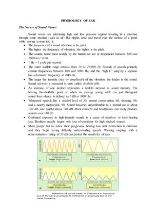
  • 1. PHYSIOLOGY OF EAR The Nature of Sound Waves: Sound waves are alternating high and low pressure regions traveling in a direction through some medium (such as air) like ripples arise and travel over the surface of a pond while tossing a stone into it.  The frequency of a sound vibration is its pitch.  The higher the frequency of vibration, the higher is the pitch.  The sounds heard most acutely by the human ear are at frequencies between 500 and 5000 hertz (Hz).  1 Hz = 1 cycle per second.  The entire audible range extends from 20 to 20,000 Hz. Sounds of speech primarily contain frequencies between 100 and 3000 Hz, and the “high C” sung by a soprano has a dominant frequency at 1048 Hz.  The larger the intensity (size or amplitude) of the vibration, the louder is the sound. Sound intensity is measured in units called decibels (dB).  An increase of one decibel represents a tenfold increase in sound intensity. The hearing threshold-the point at which an average young adult can just distinguish sound from silence is defined as 0 dB at 1000 Hz.  Whispered speech has a decibel level of 30; normal conversation, 60; shouting, 80; and a nearby motorcycle, 90. Sound becomes uncomfortable to a normal ear at about 120 dB, and painful above 140 dB. Rock concerts and headphones can easily produce sounds over 110 dB.  Continued exposure to high-intensity sounds is a cause of deafness or total hearing loss. Deafness usually begins with loss of sensitivity for high-pitched sounds.  Most people fail to notice their progressive hearing loss until destruction is extensive and they begin having difficulty understanding speech. Wearing earplugs with a noise-reduction rating of 30 dB can protect the sensitivity of ears.
  • 2. PHYSIOLOGY OF HEARING: The events involved in hearing are: 1. The auricle directs sound waves into the external auditory canal. When sound waves strike the tympanic membrane, the vibrations causes the tympanic membrane to vibrate back and forth. 2. The distance moved depends on the intensity and frequency of the sound waves (eardrum vibrates slowly to low-frequency/low-pitched sounds and rapidly to high- frequency/high-pitched sounds). 3. The central area of the eardrum connected to the malleus starts to vibrate and vibration is transmitted to incus and to the stapes. The stapes moves back and forth, and pushes the membrane of the oval window in and out. 4. The oval window vibrates about 20 times more vigorously than the eardrum because the ossicles efficiently transmit small vibrations spread over a large surface area (eardrum) into larger vibrations of a smaller surface (oval window). 5. The movement of the oval window sets up fluid pressure waves in the perilymph of the cochlea. As the oval window bulges inward, it pushes on the perilymph of the scala vestibuli. 6. Pressure waves are transmitted from the scala vestibuli to the scala tympani and eventually to the round window, causing it to bulge outward into the middle ear. 7. As the pressure waves deform the walls of the scala vestibuli and scala tympani, they also push the vestibular membrane back and forth, creating pressure waves in the endolymph inside the cochlear duct. 8. The pressure waves in the endolymph cause the basilar membrane to vibrate, which moves the hair cells of the spiral organ against the tectorial membrane. This leads to bending of the hair cell stereocilia, which produces receptor potentials that ultimately lead to the generation of nerve impulses.
  • 3.  Sound waves of various frequencies cause regions of the basilar membrane to vibrate more intensely. Membrane is narrower and stiffer at the base of the cochlea (portion closer to the oval window), high-frequency (high-pitched) sounds near 20,000 Hz induce maximum vibrations in this region.  At the apex of the cochlea near the helicotrema, the basilar membrane is wider and more flexible; low-frequency (low-pitched) sounds near 20 Hz cause maximum vibration of the basilar membrane in this region.  Loudness is determined by the intensity of sound waves. High-intensity sound waves cause larger vibrations of the basilar membrane, which leads to a higher frequency of nerve impulses reaching the brain. The hair cells transduce mechanical vibrations into electrical signals.  A tip link protein connects the tip of each stereocilium to a mechanically gated ion channel called the transduction channel. As the stereocilia bend in the direction of the taller stereocilia, the tip links tug on the transduction channels and open them.  These channels allow cations in the endolymph, primarily K+, to enter the hair cell cytosol. As cations enter, they produce a depolarizing receptor potential. Depolarization quickly spreads along the plasma membrane and opens voltage-gated Ca2+ channels in the base of the hair cell.  The resulting inflow of Ca2+ triggers exocytosis of synaptic vesicles containing a neurotransmitter, glutamate. Neurotransmitter is released, in the sensory neurons that synapse with the base of the hair cell.  Bending of the stereocilia in the opposite direction closes the transduction channels, allows hyperpolarization to occur, and reduces neurotransmitter release from the hair cells.  This decreases the frequency of nerve impulses in the sensory neurons.  The cochlea has the surprising ability to produce inaudible sounds, called otoacoustic emissions and can be picked up by placing a sensitive microphone next to the eardrum. They are caused by vibrations of the outer hair cells that occur in response to sound waves and to signals from motor neurons.  As they depolarize and repolarize, the outer hair cells rapidly shorten and lengthen. The outer hair cell vibrations set up a traveling wave that goes back toward the stapes and leaves the ear as an otoacoustic emission.  Detection of these inner ear-produced sounds is a fast, inexpensive, and noninvasive way to screen newborns for hearing defects. THE AUDITORY PATHWAY:  Bending of the stereocilia of the hair cells of the spiral organ causes the release of a neurotransmitter (glutamate), which generates nerve impulses in the sensory neurons that innervate the hair cells.
  • 4.  The cell bodies of the sensory neurons are located in the spiral ganglia. Nerve impulses pass along stimulate the axons of these neurons, forming the cochlear branch of the vestibulocochlear (VIII) nerve. These axons synapse with neurons in the cochlear nuclei in the medulla oblongata on the same side.  Some of the axons from the cochlear nuclei decussate (cross over) in the medulla, ascend in a tract called the lateral meniscus on the opposite side, and terminate in the inferior colliculus in the midbrain. Other axons from the cochlear nuclei end in the superior olivary nucleus in the pons on each side.  Slight differences in the timing of nerve impulses arriving from the two ears at the superior olivary nuclei allow us to locate the source of a sound.  Axons from the superior olivary nuclei also ascend in the lateral meniscus tracts on both sides and end in the inferior colliculi.  From each inferior colliculus, nerve impulses are conveyed to the medial geniculate nucleus in the thalamus and finally to the primary auditory area of the cerebral cortex in the temporal lobe of the cerebrum.  Because many auditory axons decussate in the medulla while others remain on the same side, the right and left primary auditory areas receive nerve impulses from both ears. PHYSIOLOGY OF EQUILIBRIUM: There are two types of equilibrium (balance).  Static equilibrium refers to the maintenance of the position of the body (mainly the head) relative gravity. Body movements of static equilibrium include tilting the head and linear acceleration or deceleration.  Dynamic equilibrium is the maintenance of body position (mainly the head) in response to rotational acceleration or deceleration.  The receptor organs for equilibrium are called the vestibular apparatus; these include the saccule, utricle, and semicircular ducts.  Otolithic Organs: Saccule and Utricle. The walls of both the utricle and the saccule contain a small, thickened region called a macula. The two maculae (plural), which
  • 5. are perpendicular to one another, are the receptors for static equilibrium. They provide sensory information on the position of the head in space and are essential for maintaining appropriate posture and balance.  The maculae also detect linear acceleration and deceleration. The maculae consist of two kinds of cells: hair cells, which are the sensory receptors, and supporting cells. Hair cells have stereocilia (microvilli) of graduated height and 1 kinocilium (a cilium anchored firmly to its basal body and extending beyond the longest stereocilium).  The stereocilia are connected by tip links. The stereocilia and kinocilium forms hair bundle. The columnar supporting cell secretes the thick, gelatinous, lycoprotein layer, called the otolithic membrane that rests on the hair cells.  A layer of dense calcium carbonate crystals (otoliths) spread over the entire otolithic membrane on top of the macula. While tilting the head forward, the otolithic membrane and the otoliths are pulled by gravity. It slides “downhill” over the hair cells bending the hair bundles.  As the hair cells depolarize and repolarize, they release neurotransmitter at a faster or slower rate. The hair cells synapse with sensory neurons in the vestibular branch of the vestibulocochlear (VIII) nerve. These neurons fire impulses at a slow or rapid rate depending on the amount of neurotransmitter present.  Motor neurons also synapse with the hair cells and sensory neurons and regulate the sensitivity of the hair cells and sensory neurons.  Semicircular Ducts: The three semicircular ducts function in dynamic equilibrium. The ducts lie at right angles to one another in three planes: The two vertical ducts are the anterior and posterior semicircular ducts, and the horizontal one is the lateral semicircular duct. This positioning permits detection of rotational acceleration or deceleration.  In the ampulla, the dilated portion of each duct, is a small elevation called the crista (plural is cristae). Each crista contains a group of hair cells and supporting cells. Covering the crista is a mass of gelatinous material called the cupula.  When head moves, the attached semicircular ducts and hair cells move with it. The endolymph within the ampulla, is not attached and lags behind. As the moving hair cells drag along the stationary endolymph, the hair bundles bend.  Bending of the hair bundles produces receptor potentials and lead to nerve impulses that pass along the vestibular branch of the vestibulocochlear (VIII) nerve.  In the ampulla, the dilated portion of each duct, is a small elevation called the crista (plural is cristae). Each crista contains a group of hair cells and supporting cells. Covering the crista is a mass of gelatinous material called the cupula.
  • 6.  When head moves, the attached semicircular ducts and hair cells move with it. The endolymph within the ampulla, is not attached and lags behind. As the moving hair cells drag along the stationary endolymph, the hair bundles bend.  Bending of the hair bundles produces receptor potentials and lead to nerve impulses that pass along the vestibular branch of the vestibulocochlear (VIII) nerve. EQUILIBRIUM PATHWAYS:  Bending of hair cells in the semicircular ducts, utricle, or saccule causes the release of a neurotransmitter (glutamate), which generates nerve impulses in the sensory neurons that innervate the hair cells.  The cell bodies of sensory neurons are located in the vestibular ganglia. Nerve impulses pass along the axons of these neurons, which form the vestibular branch of the vestibulocochlear (VIII) nerve. Most of these axons synapse with sensory neurons in vestibular nuclei, the major integrating centers for equilibrium, in the medulla oblongata and pons.  The vestibular nuclei also receive input from the eyes and somatic receptors, especially proprioceptors in the neck muscles that indicate the position of the head. The remaining axons enter the cerebellum through the inferior cerebellar peduncles. Bidirectional pathways connect the cerebellum and vestibular nuclei.
  • 7.  The vestibular nuclei integrate information from vestibular visual, and somatic receptors and then send commands to:  The nuclei of cranial nerves-oculomotor (III), trochlear (IV), and abducens (VI)-that control coupled movements of the eyes with those of the head to help maintain focus on the visual field;  Nuclei of the accessory (XI) nerves to help control head and neck movements to assist in maintaining equilibrium;  The vestibulospinal tract, which conveys impulses down the spinal cord to maintain muscle tone in skeletal muscles to help maintain equilibrium; and  The ventral posterior nucleus in the thalamus and the vestibular area in the parietal lobe of the cerebral cortex provide us with the conscious awareness of the position and movements of the head. DISEASES OF THE EAR: 1. External otitis:  Staphylococcus aureus is the usual cause of localised inflammation (boils) in the external auditory meatus.  The inflammation may be caused by bacteria or fungi or by an allergic reaction to dandruff, soaps, hair sprays, hair dyes etc. 2. Acute otitis media:  It is inflammation of the middle ear cavity caused by spread of microbes from an upper respiratory tract infection via the auditory tube. Common in children with severe earache.
  • 8.  It may spread from the outer ear through perforation in the tympanic membrane leading to the accumulation of pus and the outward bulging of the tympanic membrane.  Sometimes rupture the tympanic membrane and result in purulent discharge from the ear (otorrhoea) and cause mastoiditis and labyrinthitis.  The petrous portion of the temporal bone is very thin; the infection may spread causing meningitis and brain abscess. 3. Serous otitis media: 'Glue ear', or secretory otitis media, is the collection of fluid (effusion) in the middle ear cavity. Causes include: a) Obstruction of the auditory tube by pharyngeal swelling enlarged adenoids or tumour. b) Barotrauma (usually caused by descent in an aeroplane when suffering from a cold). c) Untreated acute otitis media.  Adults suffer from deafness and painless blockage of the ear. Children show delay in speech development and behavioural disorders owing to deafness.  Air present in the middle ear is absorbed and a negative pressure develops resulting in retraction of the tympanic membrane.  Fluid is drawn into the low-pressure cavity from surrounding blood vessels. Conductive hearing loss occurs and may lead to secondary infection. (Common cause for hearing impairment in children). 4. Chronic otitis media:  Permanent perforation of the tympanic membrane due to acute otitis media (with recurrent, persistent or untreated) and mechanical or blast injuries.  During the healing process stratified epithelium from the outer ear grows into the middle ear, forming a cholesteatoma (It is a collection of scaling epithelial cells and pus forming material). Continued development of cholesteatoma may lead to: a. Destruction of the ossicles and conduction deafness. b. Erosion of the roof of the middle ear and meningitis. c. Spread of infection to the inner ear that may cause labyrinthitis. 5. Otosclerosis:  A common cause of progressive conductive hearing loss in young adults affecting one ear but is more commonly bilateral.  It is usually hereditary, more common in females than males and often worsens during pregnancy.  Abnormal bone develops around the footplate of the stapes fusing it to the oval window, reducing the ability to transmit sound waves across the tympanic cavity. 6. Presbycusis:  This form of hearing impairment commonly accompanies the ageing process.  Degenerative changes in the sensory cells of the spiral organ of Corti resulting in sensorineural (perceptive) deafness.
  • 9.  Perception of high-frequency sound is impaired first and later low-frequency sound may also be affected.The individual develops difficulty in discrimination, e.g. following a conversation in the presence of background noise. 7. Meniere's disease:  It is a condition in which accumulation of endolymph cause distension and increased pressure in the membranous labyrinth with destruction of the sensory cells in the ampulla and cochlea.  It is usually unilateral but both ears may be affected later. The cause unknown.  Meniere's disease is associated with recurrent episodes of dizziness (vertigo), nausea and vomiting, lasting for several hours. There may be continuous ringing in the affected ear (tinnitus).  Permanent hearing impairment develop as the spiral organ of Corti is destroyed. 8. Labyrinthitis:  Caused by development of a fistula from a cholesteatoma or by spread of infection from the middle ear.  The spiral organ may be destroyed, causing sudden total nerve deafness in the affected ear. 9. Motion sickness:  Repetitive motion causes excessive stimulation of the semicircular canals and vestibular apparatus and results in nausea and vomiting in some people. 10. Deafness:  Hearing impairment can be classified in to 2 as conductive and sensorineural and mixed when there is a combination of both types of deafness in one ear. a. Conductive deafness:  This is due to impaired transmission of sound waves from the outside to the oval window, i.e. an abnormality of the outer or middle ear. b. Sensorineural (perceptive) deafness:  This is due to disease of the cochlea, the cochlear branch of the vestibular nerve or the hearing area of the brain. The individual perceives noise but cannot discriminate/understand between sounds. Risk factors for congenital deafness include: • Family history of hereditary deafness • Viruses, e.g. maternal rubella during the first 3 months of pregnancy. • Acute hypoxia at birth.