QIVIVE extrapolation requires a precise correlation between exposure and the effective chemical concentration at the site where the MIE occurs.
This work demonstrates that intracellular distribution is not ruled only by physical-chemical parameters, rather it is mainly regulated by specific biological-mediated mechanisms. Substances with
apparent chemical similarity may show different distribution profile, as shown by the intra-nuclear distribution of polyphenols. While our results derive from a limited number of substances applied to
one cell line, it is plausible that using different substances and/or different cell lines would also have shown that intracellular distribution is not directly related to physical-chemical parameters.
Chemical uptake should be specifically measured and simple extrapolations based on physical-chemical properties may provide misleading decision
dkNET Webinar "Texera: A Scalable Cloud Computing Platform for Sharing Data a...
Poster rovida lorenzetti v2.0
1. In vitro experimental model and cell fractionation. Human metastatic prostate adenocarcinoma LNCaP cells, clone FGC (cell line passage no. at delivery +15) were purchased from ECACC (European Collection
of Cell Cultures)/Sigma-Aldrich (Munich, Germany) and routinely grown in RPMI1640-based complete medium (growth medium/GM with fetal bovine serum/FBS) as previously described in Lorenzetti et al. 2010.
All experiments were performed at LNCaP cell passage no. +19 ÷ +21 following the cell treatment protocols. Detailed protocols of cell treatments and cell fractionation are described in: Lorenzetti et al. 2010, Smeriglio
et al. 2014.
Intracellular distribution of chemicals. All cellular fractions were extracted with HPLC or GC grade methanol (1:1 v/v) as described in Smeriglio et al. 2014 . Polyphenols content was analyzed according to
Mandalari et al. (2006) with some modifications as described in Smeriglio et al., 2014. Briefly, the extracts were analyzed using a Phenomenex Luna C18 reverse-phase column (250 x 4.6 mm, 5 μm; Phenomenex) in
combination with an Agilent HP1100 system (Agilent Ltd,) coupled with a photodiode array detector and a binary pump. Specific UV-visible data were collected at 220, 270, 325, 370, and 520 nm (with overall data
collected between 200 and 600 nm). All known compounds were measured directly using standard curves obtained by running commercial standards. DHT determination was carried out according to Zhang et al.
(2009), whereas E2 determination was carried out according to Zou et al. (2012) with same modifications as described in details in Smeriglio et al., 2014).
The content of each man-made chemicals was carried accordingly to different protocols as described in the PhD thesis of Antonella Smeriglio (2014, not published).
MATERIALS AND METHODS
According to the principle of the AOP (Adverse Outcome Pathways), Adverse Outcomes (AOs) originate from one or more MIE (Molecular Initiated Events) when an exogenous chemical interacts with a
specific target, either at the plasma membrane or intracellular; for instance, nuclear receptors are main targets for many Endocrine Disruptors (EDs). In order to make a MIE to occur, an ED-like
chemical has to be bioavailable to the cell and to reach the specific target site of genomic (i.e. the nucleus) or non-genomic action. This process is dose-dependent: the chemical has to reach a defined
concentration to exert a specific biological activity. Real intracellular concentration at the target site should support (quantitative) in vitro-in vivo extrapolation ((Q)IVIVE), so to accurately estimate the
dose/response in humans. (Q)IVIVE assessment is supported by specific simulating models that build a possible distribution of chemicals in organs and tissues based on physico-chemical properties
(e.g. water solubility, octanol-water partition). Refinement of these models may include in vitro simulation of the absorption through the gut membrane or the lung epithelium and the chemical uptake in
target cells. The aim of this work was to experimentally measure the cell uptake and the intracellular distribution of chemicals that may have ED characteristics.
INTRODUCTION
Analytical Measure of Intracellular biodistribution of chemicals as a tool to
understand toxicological effects
Stefano Lorenzetti1, Antonella Smeriglio1,2, Daniele Marcoccia1,
Domenico Trombetta2, Alberto Mantovan1 , Costanza Rovida3
1. Istituto Superiore di Sanità, Dpt. of Veterinary Public Health, Nutrition and Food Safety, Rome, Italy
2. Dpt. of Chemical, Biological, Pharmaceutical and Environmental Science, University of Messina, Italy
3. CAAT-Europe, University of Konstanz, Germany
Contact e-mail: stefano.lorenzetti@iss.it
QIVIVE extrapolation requires a precise correlation between exposure and the effective chemical concentration at the site where the MIE occurs.
This work demonstrates that intracellular distribution is not ruled only by physical-chemical parameters, rather it is mainly regulated by specific biological-mediated mechanisms. Substances with
apparent chemical similarity may show different distribution profile, as shown by the intra-nuclear distribution of polyphenols. While our results derive from a limited number of substances applied to
one cell line, it is plausible that using different substances and/or different cell lines would also have shown that intracellular distribution is not directly related to physical-chemical parameters.
Chemical uptake should be specifically measured and simple extrapolations based on physical-chemical properties may provide misleading decision.
CONCLUSIONS
REFERENCES
Lorenzetti S, et al. (2010). Cell viability and PSA secretion assays in LNCaP cells: a tiered in vitro approach to screen chemicals with a prostate-mediated effect on male reproduction within the ReProTect project. Reprod Toxicol, 30(1):25-35.
Mandalari G, et al (2006). Characterization of flavonoids and pectins from bergamot (Citrus bergamia Risso) peel, a major by-product of essential oil extraction. J. Agric .Food Chem., 2006, 54(1), 197-203.
Worth AP, et al. (2017). Virtual Cell Based Assay simulations of intra-mitochondrial concentrations in hepatocytes and cardiomyocytes. Toxicol In Vitro, 45(Pt 2):222-232.
Smeriglio A, et al. (2014). Intracellular distribution and biological effects of phytochemicals in a sex steroid- sensitive model of human prostate adenocarcinoma. Anti-cancer Agents Med Chem., 14(10):1386-1396.
Zhang Z, et al. (2009) Direct determination of anabolic steroids in pig urine by a new SPME–GC–MS method. Talanta, 78(3), 1083-1089.
Zou Y, et al. (2012). Determination of estrogens in human urine by high-performance liquid chromatography/diode array detection with ultrasound-assisted cloud-point extraction. Anal Biochem., 421(2): 378-384.
ACKNOWLEDGEMENTS
The H2020 project EU-ToxRisk (#681002), the LIFE+ project LIFE-EDESIA (LIFE12 ENV/IT/000633), granted to AM, and the “In vitro alternative methods: prostate-
mediated reproductive toxicity of potential SVHCs” by the Italian Ministry of Health; and ii) “Healthy and sustainable agriculture” by the Rovereto town council, granted
to SL, supported this study.
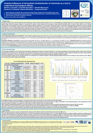
The intracellular distribution of 32 chemicals where measured by validated HPLC methods to provide extracellular (i.e. cell culture supernatant) and three cellular fractions, namely the microsomial,
cytoplasmatic and nuclear fractions. The starting set of chemicals was intentionally diverse to represent a general example: polyphenols of natural origins (6 chemicals), biocides or plant protection
products (9), plasticisers (3) and sex steroid active chemicals (3). The octanol/water (Kow) partitioning constant was retrieved from public literature.
The total intracellular fraction was first compared with Kow. The complete lack of correlation indicates a potential biological-mediated mechanism that is specific for each chemicals; hence, different
from the generally accepted simple passive diffusion.
RESULTS AND DISCUSSION
A straightforward pattern cannot be determined. The total intracellular fraction is above 50% for most chemicals, but VIN, M2 and BPA.
The 6 natural compounds (from API to RESV) are polyphenols with quite similar chemical structures; they all have a higher concentration in the microsomal fraction. In the meanwhile their overall
distribution patterns are different. For example, the nuclear fraction of GEN is definitely lower as compared to QRC.
Other substances preferentially distributed in the microsomal fraction are the reference natural hormone E2 and the three PPPS, with different chemical structures, FEN, GLY and CPF.
The nuclear fraction is high (> 60%) for the reference chemical BIC, and three chemically different PPP, GA, γ-HCH and PERM, with Kow ranging from -4.7 (GA) to 6.6 (PERM).
DEHP has a very high Kow (7.9) while being the only substance equally distributed among medium, cytoplasmic, microsomal and nuclear fractions.
LIST OF SUBSTANCES THAT WERE ANALYSED