SlideShare uma empresa Scribd logo
1 de 608
Baixar para ler offline
Livor mortis - bluish-red discoloration of the
skin and organs
Result of the blood settling or pooling by gravity in
capillaries, which dilate following the cessation of
circulation
Back
Belly
it is absent where there is
pressure which prevents
dilation of capillaries
Livor due to hyopstasis first 8-12Hrs
Livor due to imbibition after 8-12Hrs
(not moved)
Macerated foetus. Note the reddish discoloration of the skin. The epidermis
is detached from the dermis in large laminae.
Courtesy of Kaiser László, MD
Aspiration of gastric content caused obstruction of airways in the patient with deep
coma (lethal condition)
Trachea
Main bronchi
Anemic infarct of the heart: circumscribed yellowish lesion, the
margins are hyperemic
Hemorrhagic infarct of lung: wedge shaped, raised, dark-red
area
34
Hemorrhagic infarct of small bowels
due to arterial occlusion
and there is gas formation
in lumen
Brain infarct: the necrotic area is softened and pale
Internal capsule
Infarcted area
Caudate nucleus
Caseous necrosis
• Immune-mediated distinctive form of coagulative necrosis
in foci of tuberculous infection of the lung
• Grossly, caseous necrosis is white and cheesy
Necrotizing pancreatitis: the pancreas is swollen and displays
several yellowish foci of necrosis
Dry gangrene of the great toe
Obstruction of the CSF flow leads to pressure atrophy of the
brain, with the enlargement of ventricles: hydrocephalus
Hydronephrosis: obstruction of the ureter (arrow) leads to sac-
like dilation of renal pelvis and calyces, and pressure atrophy of
parenchyma
TUMOR
Ischemic
atrophy of the
kidney
because the
supplying
renal artery
was
progressively
narrowed
Hypertrophy of heart, triggered by action of mechanical
stimuli ( workload) and vasoactive substances (e.g.,
angiotensin II). Free wall thickness: above 15 mm
Hypertrophy of the muscles of urinary bladder due to urethral
obstruction
Steatosis: the liver is enlarged, yellow and greasy,
resembles to goose liver
Courtesy of E. Kemény, SZTE Pathology
hypoxia-hepatotoxins
obesity-diabetes
alcohol-protein energy malnutrition
The hepatocytes are vacuolated; representing accumulations
of neutral lipids that have been removed by lipid solvents during
tissue processing
Frozen section, Oil Red O
Jaundice: yellowish discoloration of skin
Dystrophic calcification of aortic valves
(calcifying aortic stenosis) valsalva sinuses are rigid (Ca deposits)
which cause narrowing of aorta
7
Pulmonary edema: the lungs 2 to 3 times exceed their normal
weight; sectioning reveals a foamy mixture of air, edema fluid,
and RBCs
Trachea
Acute LHF
9
Hydrothorax in chronic right-sided HF
it should be almost colorless but blood
during autopsy changed the color
anthracosis
10
Increased hydrostatic pressure in the portal venous circulation
(portal hypertension) by cirrhosis of liver (shown) is an
important cause of ascites
14
Severe lymphedema of arm after mastectomy, surgical
dissection of the axillary lymph nodes and irradiation of the
axillary region because of breast cancer.
Post-mastectomy Lymphedema
Elephantiasis. The patient suffered from morbid obesity,
type 2 diabetes and congestive heart failure.
21
Hepatic congestion: hypoxic/fatty hepatocytes around central
veins are pale yellow, the better oxigenated portal parts are red.
24
Chronic left-sided HF  chronic pulmonary congestion:
brown induration of lungs
Cyanotic induration of the kidney in chronic right-sided heart failure
+Enlarged
Congestive splenomegaly. Weight: up to 500 gm, the organ is
firm, the capsule is thickened, the malphigian corpuscles are
indistinct
Peptic ulcer of the postpyloric duodenum that eroded the pancreaticoduodenal artery
(arrow) and caused lethal bleeding
Classification and nomenclature of hemorrhage
• Surface
- External, internal
• Enclosed within a tissue
Suffusion (2-dimensional)
Hematoma (3-dimensional); may be
- insignificant (bruise)
- lethal (retroperitoneal, subarachnoidal, intracerebral)
Subcutaneous suffusion due to trauma
33
Subarachnoidal
hematoma
rupture of cerebral a.
aneurysm
Basal ganglia hemorrhage as a result of long-standing hypertension
intracerebral
hemmorhage
(HTN crisis)
35
Petechiae: minute, 1-2 mm hemorrhages into skin,
mucous membranes or serosal surfaces, seen in
thrombocytopenia, defective platelet function or
clotting factor deficits
36
Purpura: slightly larger (>3 mm) hemorrhages, may
be associated with similar pathologies, as well as
vasculitis
Purpuras in
small vessel
vasculitis
Sándor Husz, MD,
SZTE Dermatology
39
Hemopericardium because of myocardial rupture (lethal)
blood is in the visceral layer
400 ml can be lethal
Hemocephalus: blood fills the lateral and third ventricles
(the ventricular drainage was unsuccesful)
Draining channel
Iatrogenic (adverse condition in a patient resulting from treatment)
hematoma around the carotid artery (CA) as complication of
catheterization
Internal CA
External CA
Common CA
9
Atherosclerosis of aorta with mural thrombi
Endocarditis. Thrombi on the leaflets
Aneurysms in the aorta filled with thrombi
Left ventricle.
Apical chronic aneurysm
with mural thrombus
(arrow)
17
Auricular thrombus (arrow) in a patient with atrial fibrillation
20
Auricular thrombus: grayish-red, lines of Zahn
Direction of blood flow
Venous thrombi (red or stasis thrombi)
Occur in a stagnating environment, and the thrombi contain more
enmeshed RBCs among sparse fibrin strands;
lAppear as long, red-blue cast of the vein lumen
26
Verrucous vegetations (arrows) on mitral valve composed of
platelets and fibrin. The patient had disseminated cancer.
34
Red thrombi in deep veins
Femoral artery
Saddle embolus (arrow) impacting the bifurcation of the pulmonary
Artery prevented the influx of blood into the lungs (lethal)
Aorta
Pulmonary
vein
Thromboembolism: hemorrhagic infarcts in the terrritory of
middle cerebral artery (hemorrhage due to reperfusion)
Caudate nucleus
Internal capsule
Putamen
55
Ruptured aneurysm of abdominal aorta leading to retroperitoneal
bleading and shock (the probe indicates the rupture)
Decreased perfusion may result in the necrosis of toes
Cardinal signs of bacterial inflammation: calor (warmth), rubor
(redness), tumor (swelling), dolor (pain), and loss of function
Dr. Sándor Husz, SZTE Dermatology
Unilateral bacterial inflammation of rat kidney induced by ligation of ureter
and i.v. injection of bacteria. The animal was sacrified on Day 2. The affected
kidney displayed inflammatory exudation-induced enlargement (“tumor”)
Gross features
Normally, the serous
membrane is smooth and
has a glistening surface.
In serous inflammation, it
becomes reddish and
opaque.
Shown: acute serous
pleurisy
Lethal acute serofibrinous peritonitis: the serosa of bowels is hyperemic;
the bowels adhere to each other with fibrin strands (right: normal serosa)
liver
Edema of larynx induced by
adverse (allergic) reaction
of an antihypertensive drug
administered intravenously in
hypertensive crisis.
Note marked swelling of
laryngeal mucosa which
caused upper airway
obstruction and death
Laryngal edema that caused airway obstruction
Blisters in chickenpox Superinfection with St. aureus:
the blisters are filled with pus
(pustula)
Courtesy of dr. Ildikó Kováts
Acute fibrinous pericarditis: intense hyperemia, fibrin strands
Left: fibrinous pericarditis, right: fibrinous pleurisy (parietal pleura). The thick strands of fibrin
will not not be removed by fibrinolysis, and adhesions develop between serous surfaces
Diaphragm
Thoracic
cage
Lobar pneumonia. Hepatisation: liver-like consistency because alveoli
are packed with fibrin
trachea
Lung: multiple abscesses as complication of pneumonia
Furuncle (boil)
Dr. Sándor Husz, SZTE Dermatology
Folliculitis on the nose; carbuncle on the face
Dr. Sándor Husz, SZTE Dermatology
Cellulitis: sharp, erythematous swelling of the skin
Courtesy of Erika Varga, MD, Department of Dermatology, SZTE
Empyema of thorax
Healing: granulation tissue  secondary calcification: callus of pleura
Purulent mediastinitis
(induced by descending
infection of the neck)
Aorta
Pulm. artery
Pulm.vein
Pericardium
Mediastinum
Mediastinum
Acute purulent peritonitis:
the abdominal cavity was filled
with more than 2000 ml pus
Acute purulent
meningitis
The exudate is
localized in the
subarachnoid space,
the meningeal
vessels are
engorged and
stand out
prominently
Acute purulent
tracheobronchitis
The exudate clogged the
airways and caused
widespread atelectasis
(collapse) of the alveoli
Note: tracheobronchial
toilet is necessary in
patients who cannot
cough actively
Purulent discharge in
Neisseria gonorrhoeae
infection
(sexually-transmitted
disease, termed
gonorrhoea)
Dr. Sándor Husz, SZTE Dermatology
Acute hemorrhagic
cystitis:
the bladder mucosa is
swollen, hyperemic and
displays focal hemorrhages
Pseudomembranous colitis induced by C. difficile: confluent
plaques of yellow fibrin and inflammatory debris adherent to a
reddened mucosa
Pseudomembranous enterocolitis: in severe cases the small
bowels are also affected
Amputated leg because of necrotizing fasciitis
Gas gangrene: necrotizing and gas-producing inflammation of the lower
extemity involving the abdominal wall, perineum and scrotum; the gas
bubbles detach the epidermis from the underlying tissues
Gas gangrene: the gas bubbles accumulated between the epidermis and
the underlying tissues
Acute necrotizing-hemorrhagic pancreatitis
Polyoma virions in the nucleus of an epithelial cell
Influenzavirus-induced
tracheobronchitis
Note intense mucosal
hyperemia
Comment
The influenza A virus subtype
H1N1 infection was lethal in this
young pregnant woman; she was
regrettably not vaccinated
Huge problem
If people refuse vaccination, herd
immunity does not develope
Safety concerns are based on
stories discovered in the media or
received from the acquaintaces
LM structure of chronic abscess
• The central cavity is filled with pus
• The wall of abscess has an inner pyogenic membrane
(granulation tissue rich in neutrophils) and an outer fibroblastic
rim
• Heals with fibrosis
Pus in the
cavity
Pyogenic
membrane
Fibroblastic
rim
Subpleural lung abscess (arrow)
Multiple chronic abscesses in the liver.
The patient suffered from common bile duct obstruction and
purulent cholangitis
Cirrhosis of liver: chronic inflammation of the liver  formation of
fibrous septa, which, in turn, form nodular pseudolobules 
impairment of venous flow through the liver.
Consequence: portal hypertension, hepatic failure
Cirrhosis of liver: nodular appearance of pseudolobules
Chronic glomerulonephritis-induced kidney shrinkage
Chronic inflammation of glomeruli  glomerular scarring
Grossly the kidneys are symmetrically shrunken, the surface is
granular (shown), and the cortex is thinned.
Consequence: chronic renal failure
Honeycomb lung: inflammation or
injury-induced widespread fibrosis in
alveolar septa culminate in grossly
evident lung fibrosis
Consequence: respiratory insufficiency
with hypoxemia and cyanosis
Alveolar septa are widened by deposition of
collagen (trichrome stain: blue).
Chronic peptic ulcer of the stomach
Venous ulceration of the leg due to poor venous drainage from
the lower leg
Decubital ulcer in the sacral region
Healing by
secondary
intention,
leading to
contracture
Courtesy of Prof. Dobozy Attila
Keloid
Raised hyperplastic scar
of the skin
due to accumulation of
great amount of collagen
in the dermis.
Common in Negroid/black
people.
Cosmetic problem.
Shown: keloid after
vaccination
Courtesy of Prof. Dobozy Attila
Laryngal edema causing airway obstruction
Edema of larynx induced by adverse allergic reaction of an anti-
hypertensive drug administered intravenously in hypertensive crisis
Pseudomembranous-ulcerative gastritis induced by Candida
Esophagus
Stomach
Kaposi’s sarcoma.
Tumorous nodules on the leg.
Autopsy case.
Benign tumors in the uterus: expansive growth, well-
defined borders, homogenous cut surface
leiomyoma
Malignant tumor (bronchial carcinoma): irregular infiltrative edges,
foci of necrosis, tissue destruction. Arrows: invasion of vessels
Cerebral metastasis of the bronchial carcinoma shown
Kaposi sarcoma.
Tumorous nodules on the leg.
Autopsy case.
Dermoid cyst of the ovary. Filled with hair, sebaceous
material, and desquamated squames.
Fungating tumor in the lip;
proved carcinoma
histologically
Courtesy of Prof. Sonkodi István, Faculty of Dentistry
Fungating carcinoma of the esophagus
Ulcerated carcinoma in the antrum of stomach:
irregular margins, crater-like raised borders
Annular (napkinring-like) growth in carcinomas of the distal
colon – obstructs the passage of bowel content
Papillomas of the skin
• Two types: basal cell
papilloma, and wart
Basal cell papilloma
(seborrheic keratosis)
• Very common
• In middle aged or older
individuals
• Most frequent on the trunk
• Dark brown lesion
with a granular surface
• Often multiple
Condylomas on the glans and prepuce
Prof. Attila Dobozy, Dermatology
Pedunculated polyp. LM: tubular adenoma; composed of tubular
glands with dysplastic features (elongated nuclei, nuclear
stratification, and architectural atypia)
Stalk
Bowel wall
Head
of the polyp
Resected bowel sample; histologically, the larger pedunculated polyp
proved to be tubular adenoma with malignant transformation
Head of the polyp
Polyp
Stalk
Unilocular serous cystadenoma of the ovary ranging more than 15 cm in
greatest dimension. The outer surface is smooth; the epithelial lining of the inner
surface exhibits small papillary projections.
Courtesy of Bence Nagy, MD, PhD, Department of Pathology, University of Szeged
Mucinous cystadenoma of the ovary: delicate septa form cysts;
mucinous material fills the cysts
Adrenal cortical adenoma: circumscribed, yellowish tumor deriving
from the adrenal cortex. The tumor was hormonally inactive.
Spontaneous hemorrhage of silent adenomas  blockade of
CSF flow  acute increase in liquor pressure  death
Scirrhous carcinoma of stomach (linitis plastica)
:
Signet-ring cc: tube-like thickening of the wall;
LM: the mucin in the cytoplasm pushes the nucleus to te periphery
Mucinous cystadenocarcinoma of the ovari. Foci of necrosis and
hemorrhage indicate malignant tumor. Invasion of ovarian surface →
dissemination in the peritoneal cavity → carcinosis of peritoneum
Special skin cancer:
basal cell cc (basalioma)
• Most frequent among cc-s
• Semimalignant
• Related to chronic sun exposure
• In the elderly; in the face, near
the eyes and nose
• Advanced lesion: ulcerated
(rodent ulcer)
Courtesy of Attila Dobozy, Bőrklinika
Excised breast cancer; infiltrative carcinoma with stellate
appearance; gray-white strands of tumorous tissue radiate into the
surrounding fat
Advanced breast carcinoma: invasion of the skin, with
extensive ulceration
Glioblastoma: continuous spread through corpus callosum
(butterfly tumour)
Renal cell carcinoma: continuous spread involving the cortex,
the medulla and the pelvic fat
Liver metastases of colon carcinoma
Seminal vesicle
Rectum
Carcinoma of the prostate; arrow: extension into the
paravertebral plexus
Vertebral metastases of prostatic carcinoma
Transcelomic spread: carcinosis
of the greater omentum
Greater omentum
Greater omentum
Peritoneally disseminated
ovarian carcinoma –
carcinosis of peritoneum
Greater
omentum
Stomach
Stomach
Liver
Enucleated eye because of retinoblastoma
ABCD of malignant
melanoma
Asymmetrical
lesion with
irregular borders
Bleeding:
spontaneous or
upon minor
trauma
Color: uneven
pigmentation
or change in color
Diameter: >6 mm
Korom Irma, SZTE Dermatology
Vertical growth from the
beginning: nodular melanoma
Gross: deepbrown nodule
LM: anaplastic tumour nests
Highly malignant:
hematogeneous metastases
very early
Korom Irma, SZTE Dermatology
Local effects
Benign tumors:
• Leiomyoma of the uterus: heavy menstrual bleeding, pelvic
pressure/pain, frequent urination, etc.
Uterine cavity
Body of the uterus
Multifocal destruction of vertebrae in multiple myeloma: pain +
pathological fracture
Fungating carcinoma in the lower third of the esophagus
caused progressive stenosis and dysphagia
Trachea
Esophagus Cardia
Left: napkinring-like spread of colonic cc led to stenosis  impaired the passage of
stool; the frequency of passing the stool and the consistency of the stool had changed
Ulcerated carcinoma of the stomach: irregular margins, crater-like raised borders:
iron-deficiency anemia and positive fecal occult blood test
Stenosing cancer of the ileocecal valve: the patient displayed
change in bowel habit and iron-deficiency anemia
Ileum
Coecum
Invasion of pelvis by renal cell carcinoma  painless microhematuria
Urinary bladder: ulcerated carcinoma on the lateral wall caused occult
painless microhematuria and iron deficiency anemia
Trigone
Bronchial cc. The non-tumorous bronchial mucosa (arrow) is
hyperemic, swollen indicating peritumoral bronchitis  caughing
Non-bacterial thrombotic endocarditis. Vegetations along the closure
line of the aortic valve cups. The patient suffered from bronchial
carcinoma
The vegetations
caused embolic
anemic infarction of
the kidney (arrow)
 hematuria
The vegetations caused embolic hemorrhagic infarction of the
small bowel  bloody stool
Sacral decubital ulcer in the bed-ridden patient wearing diaper. The patient
had brain infarction and hemiplegia (complete paralysis of the half of the
body)
Femoral vein thrombosis; pulmonary embolism
vein
artery
Pulm. artery
Embolus
Osteoporosis: severe loss of horizontal trabeculae in vertebral
bodies in the patient who suffered from brain-hemorrhage,
paralysis and prolonged bed rest
5) Ascending urinary tract infections in catheterized
patients: urethritis, cystitis, acute pyelonephritis (shown)
Lobectomy because of
bronchial carcinoma (arrow) in a
heavy smoker
Courtesy of B. Vasas, MD,
Dept. Pathology, Universitiy of Szeged
sq cc
destructed the bronchus
2) Induction of atherosclerosis
Absorbed into the blood, cigarette smoke causes increased
platelet aggregation and dysfunction of the endothelial layer of
aorta and large arteries  development of atheromatous
plaques
Emphysema (alveolar wall destruction, enlarged air spaces) in the non-tumoral areas
of the lobectomy specimen of the heavy smoker
Mucosal defects (erosions) in alcohol-abuse induced gastroduodenitis
Antrum
Duodenum
Hepatic steatosis: the liver is
enlarged (>2000 g), soft, yellow,
and greasy; LM: fat globules in the
hepatocytes; reversible
Pancreas: acinar cell injury (ethanol + heavy, fatty meal) 
acute necrotizing-hemorrhagic pancreatitis: high mortality rate
Micronodular cirrhosis of liver
Pancreas: chronic calcifying pancreatitis: atrophied, fibrotic
pancreas; irregularly dilated ducts obstructed with stones 
pain, malabsorption
Abnormally high deposition of visceral adipose tissue (thickness of subcutaneal
fatty tissue: 10 cm), fatty liver, elevated diaphragm
Obesity-related glomerulopathy: segmental scarring of the tufts
Osteophytes in spondylarthrosis limit movements of the
vertebral columns
Acute myocardial infarction
Most frequent cause of death in diabetes
Cerebral infarct involving the frontoparietal region, the internal capsule and
the striatum (caudate nucleus, putamen)  contralateral hemiparesis
Caudate
nucleus
Putamen
Gangrene of toe
Diabetic retinopathy: new vessels, lipid-cholesterol deposits
Courtesy of Kolozsvári Lajos, SZTE Ophtalmology
Neuropathic diabetic foot ulceration: necrosis + phlegmon of
sole
Courtesy of R. Sipka, MD, Dept. Surgery, University of Szeged
Acute purulent pyelonephritis: several abscesses in the
cortex and medulla
Mucormycosis, with spread to the orbit and CNS
Courtesy of Szabó
Zsuzsanna, MD,
Szent László Kórház
lethal within days after
meninges involved
Myeloma: the tumorous plasma cell nodules destruct the bones;
death in 15% of patients due to consequences of AL-amyloidosis
The amyloid
deposition may not
be evident grossly.
In this case, the
heart weight was
580 gs (normal 350
gs)
LM evaluation
revealed amyloid
deposition
Cystic fibrosis of pancreas: fibrotic areas intermingle with cysts
Lungs of the patient who died of cystic fibrosis: purulent plugs obstruct the bronchi
The plugging of bronchi stem from obstruction and infection of the bronchi
secondary to the viscous mucus secretions
Bronchiectasis, purulent bronchitis and bronchopneumonia in
cystic fibrosis
Atheromatous plaque in the middle cerebral artery: raised
white-yellow lesion in the intima, protruding into the lumen
(formol-fixed brain)
Aorta: the plaques contain a yellow, grumous debris (arrow)
Structure of atheroma on LM
• Intimal lesion
• Central lipid core
• Fibrous ”cap” subendothelially
Coronary occlusion due ulceration of the plaque leading to
thrombosis
Cerebral infarct involving the internal capsule
 contralateral hemiparesis
Abdominal aorta: mural thrombi
Celiac artery Superior
mesenteric artery
Renal artery
24
Ruptured atherosclerotic aneurysm of the infrarenal abdominal aorta; the probe
indicates the communication between the lumen and the extraaortic retroperitoneal
space. The aneurysmal sac is filled with mural thrombus
Thrombosis of superior mesenteric artery: bowel infarction
Ruptured atherosclerotic aneurysm of abdominal aorta, leading to
retroperitoneal bleeding and shock (the probe indicates the rupture)
Iliac arteries
Saccular aneurysms in the thoracic aorta. Note mural thrombi
Degeneration of the tunica media: thinning and dilation of the aortic
root, and intimal tears
Resected ascending aorta because of dissection. By the time of surgery, the
process had not resulted in aortic rupture
Aortic
lumen
Dissecting
column of blood
Tear
Prepared by G. Nyári, MD, Dept. Pathology, Univ. Szeged
Dissection of aorta, hemopericardium
Intimal tears
Aortic valve
Hypertensive kidney disease
• Mild, symmetric shrinkage of kidneys (nephrosclerosis), weight:
120-120 g; the surface is granular
Hypertensive heart disease. Pressure overload-induced left
ventricular hypertrophy  arrhythmias, chronic left-sided heart failure
Vérzések retinopathia hypertonica-ban
Courtesy of Prof. Kolozsvári Lajos
Hypertensive retinopathy. Ophtalmoscopic examination reveals the
thickening of retinal small arteries; microhemorrhages can be present (fundus
hypertonicus)
Microaneurysms undergo rupture in hypertensive crisis 
massive hemorrhage of the basal ganglia
Destruction of the left putamen and internal capsule by fresh
hematoma
Slightly shrunken kidneys; finely granular surface
Heart weight 550 g; enlargement of the left ventricle
Kidneys: hyperplastic arteriolosclerosis
Gross changes
• Early changes: enlarged kidneys, pinpoint petechial
cortical hemorrhages + tiny infarcts
• Later, infarcts are replaced by vascular scars
Brain edema. The gyri are widened and flattened, the sulci are narrowed
Unilateral atrophy of
the kidney
Not shown: the
atherosclerotic
stenosis of the
orifice of the renal
artery
Takayasu arteritis, late phase.
Arrows indicate fibrous narrowing of arterial orifices
Aortic root
Brachioceph. a
Left common carotid a.
Left subclavian a.
Thoracic ao.
Clinical features in cranial GCA
• Fever, fatigue, weight loss; sedimentation rate >50 mm/h
• Palpable nodularity of the tortuous temporal artery
Brain infarcts with secondary hemorrhage in the patient who
died of PAN with kidney, heart, brain and muscle involvement
GPA: cavitation of lung parenchyma, due to necrosis of small
vessels and parenchyma
MPA, lungs: alveolar capillaritis
 bilateral air space
consolidation with relative apical
sparing corresponds to confluent
foci of alveolar hemorrhages
Courtesy of B. Radics, MD, Dept. Pathology, University of Szeged
Vasculitic purpuras
Prof. Husz Sándor, Bôrklinika
Clinical features
• Skin: purpuras, principally
in the lower extremities
• Bowels: abdominal pain,
bleeding
• Joints: arthritis and
arthralgia
• Kidneys: hematuria,
proteinuria and azotemia
Outcome
• Depends on the extent of
crescent formation; overall
prognosis is good
Esophageal varices
• In portal hypertension, the
submucosal veins of distal
esophagus undergo dilation
(portocaval shunt)
• Spontaneous rupture 
hemorrhagic shock 
exsanguination
• Autopsy: 2000-3000 ml-s of
fresh blood fills the stomach
and small bowels
Venous ulceration of the leg (varicose ulcer)
Thrombosis in deep leg veins
Most important consequence  pulmonary embolism
Femoral vein
Femoral artery
Photo: 54-year-old man.
Hemangiomas on his trunk
Cavernous hemangioma of the liver: the vascular channels
produce sponge-like structure
Angiosarcoma of the pulmonary artery; endoluminal spread
lead to occlusion
Morphologic features of concentric hypertrophy of the LV
• small lumen
• markedly increased wall thickness (> 20 mm)
• increased mass (> 500 g)
Septum
Posterior wall
Anterior wall
Lateral wall
Morphology of dilative hypertrophy of the LV: enlarged lumen,
enlarged size, slightly increased wall thickness, increased mass
Septum
Posterior wall
Anterior wall
Lateral wall
Pulmonary edema: lungs >1200 gs, congested and wet,
airways contain bubbly fluid
Brown induration of lungs; atheromatous plaques in pulmonary
arteries indicating sec. pulmonary hypertension
collagen in
alveolar septa
Morphology
RV hypertrophy (thickness › 6
mm) + dilation
and
manifestations of chronic
systemic congestion
Early phase: concentric hypertrophy of LV: wall thickness
> 20 mm; weight > 500 g
Septum
Posterior wall
Anterior wall
Lateral wall
Late phase: LV hypertrophy + dilation: wall thickness < 20 mm;
weight > 550 g
Occlusive thrombus (dissected with the scissor) on
atheromatous plaque (arrow)
MI: yellowish area of necrosis, hyperemic border
66
Hemopericardium due to rupture of the free wall of the LV
rupture
67
Rupture of the interventricular septum
Anteroseptal chronic aneurysm
Apical chronic aneurysm,
with mural thrombus
Septum
Posterior
RV
LV
Anterior
Reperfusion injury: hemorrhage in the septum, and
the anterior and posterior wall; angioplasty 12 hours after the onset of
symptoms
84
Chronic IHD: dilative hypertrophy, apical fibrosis
(arrow), dilated atrium, thrombus in the left auricle
Calcific aortic stenosis in congenitally bicuspid valves
Subendothelial calcific masses within sinuses of Valsalva
Mitral annular calcification: regurgitation + stenosis
Thinned and enlarged leaflets, elongated and attenuated
chordae tendineae, excentric LV hypertrophy
Rheumatic fever: acute verrucous endocarditis,
chordae tendineae are also involved
Dr. Tószegi Anna, SZTE Pathologia, 1972
Mitral stenosis: thickened leaflets, fused at the commissures;
thickened chordae tendinae, dilated LA
Large vegetations on the mitral valve
Nonbacterial thrombotic endocarditis of aortic valve from a patient
with adenocarcinoma of the tail of pancreas.
Histology ruled out infectious endocarditis
Mechanical valve: tilting disk
Bogáts Gábor SZTE Szívsebészet
Bioprothesis
Bogáts Gábor SZTE Szívsebészet
Dilated CM
The heart is
ball-shaped;
there is
pronounced
ventricular
chamber
dilation
The weight of
the heart
was 660 g
DCM in a 2-y-old child
• Extreme cardiomegaly 
lung compression
• Congestive hepatomegaly
HCM: hypertrophy of the septum and the free wall; the volume of the
LV is reduced; subaortic stenosis (arrow) is evident
Note: the thickened left
ventricular wall becomes
stiff, prevents diastolic
filling
Arrhythmogenic CM. Fibrofatty
near-transmural replacement of
ventricular myocardium; the
lesion affected the left ventricle
Courtesy of B. Radics, MD,
Dept Pathol, Univ Szeged
Posterior
wall
Septum
Diffuse mottling in myocarditis
Mural thrombi in myocarditis
Bicuspid pulmonary valve
Aorta
Septum
VSD
Ao valve
Anterior wall
Ventricular septal defect (VSD)
RC
LDA
Atrial septal defect (ASD)
90% secundum type: results
from deficient or fenestrated
fossa ovalis in the central
atrial septum
• RA and RV dilation
• RV hypertrophy
• Dilation of pulmonary artery
J.M. Kissane: Anderson’s Pathology, 1990
Ventricular septal defect
(VSD)
The most common congenital
cardiac anomaly
• Site (90%): membranous
part of the septum
• Dilative hypertrophy of RV
• Small defects may close
spontaneously
J.M. Kissane: Anderson’s Pathology, 1990
Patent ductus
arteriosus (PDA)
The ductus (just distal to
the left subclavian artery)
allows blood flow
between the aorta and
pulmonary artery during
fetal life. Normally closes
within 1 and 2 days of
life.
PDA is associated with
dilation of proximal
pulmonary arteries and
LV
LV and later RV
hypertrophy
J.M. Kissane: Anderson’s Pathology, 1990
Tetralogy of
Fallot (TOF)
• Dextraposed aorta
overriding
• VSD
• Pulmonary stenosis
• RV hypertrophy
J.M. Kissane: Anderson’s Pathology, 1990
Transposition
of the great arteries (TGA)
• Aorta arising from the
RV
• Pulmonary artery
arising from the LV
• A shunt (ASD or VSD
or PDA) for mixing of
blood
J.M. Kissane: Anderson’s Pathology, 1990
Postductal coarctation
(adult-type)
Distal to the obliterated
ductus arteriosus:
• LV hypertrophy
• Hypertension
proximal
and hypotension distal
to the narrowed
segment
• Asymptomatic until adult life
J.M. Kissane: Anderson’s Pathology, 1990
Preductal
coarctation
(infantile-type)
• Hypoplastic aorta
between the left
subclavian artery
and the d. arteriosus
• ASD
• Widely PDA
• Frequently lethal
J.M. Kissane: Anderson’s Pathology, 1990
Virus-induced acute serofibrinous pericarditis
The pericardium is hyperemic, covered by fibrin strands
Carcinosis of pericardium; primary tumor in the lung
Biopsy diagnosis of GN
• Via renal percutaneous biopsy evaluated by light
microscopy (LM), immunofluorescence (IF) and elecron
microscopy (EM)
• Mirrors the inflammatory events occurring in all glomeruli
Severe periorbital edema in a deceased man who had the
nephrotic sy
The kidney in nephrotic syndrome: the pale yellow cortex is due
to lipid accumulation in renal tubules (consequence of lipiduria)
Chronic sclerosing GN: symmetric shrinkage (80-80 g);
granular surface; on section, the cortex is thinned
Bleeding after biopsy
Gross features in advanced disease
• Diabetic nephrosclerosis: symmetrically shrunken kidneys with granular
surface
• Remember: diabetic nephrosclerosis, hypertensive nephrosclerosis, and
chronic glomerulonephritis all lead to symmetrically shrunken kidneys
Acute pyelonephritis: multiple abscesses
Necrotizing papillitis: necrosis of pyramids
Gross:“shock kidney” enlarged, swollen kidney (~ 200 g);
pale, bloodless cortex, dark medulla
The change of “shock kidney”
was photographed from a
deceased patient with
hemorrhagic shock induced
by the rupture of esophageal
varices (arrows)
• Normal ureteral insertion: acts as a valve that prevents
retrograde flow of the urine during micturition.
• Abnormal ureteral insertion: urine refluxes in the ureter and pelvis
during micturition (VUR)
• May be unilateral or bilateral
Kumar et al Pathologic
Basis of Disease, 2005.
Abscesses in the cortex and medulla; the pelvic mucosa is hyperemic
Pelvic mucosa
Confluent abscesses in the cortex of the kidney;
such severe cases result in urosepsis + death
Reflux-induced chronic pyelonephritis leading to kidney shrinkage
Coarse scars overlying blunted calyces in reflux-induced PN
The sharp edges of the stone incised the urothelium of ureter
during the travel  hematuria
Staghorn calculus: remains localized; leads to pyelonephritic
scar
Pyelonephritic scar due to stone in the calyx: obstruction +
infections are keyplayers in scar formation
Hydronephrosis: dilated calyces, atrophied
papillae (arrows), thinned parenchyma
55-y-o man with sepsis induced by intrarenal abscesses. Urgent nephrectomy saved
the life of the patient. The gross evaluation of the specimen revealed congenital
pyeloureteral stenosis (arrow), hydronephrosis, and intrarenal abscesses
Dilated pelvis
Ureter
Gross
Very large cystic kidneys (1000 to 4000 gs); cysts may be 3-4 cm
in diameter
Enourmously enlarged kidneys  elevation of the diaphragm;
pulmonary hypoplasia
The cysts cause spongelike appearance
Oligohydramnios; compression of fetus
By courtesy of L. Kaizer, MD, Dept Pathol, University of Szeged
Renal artery stenosis-induced atrophy of the left kidney.
Note multiple small cysts in the cortex of both kidneys
ESRD with several cysts; renal cell carcinoma in the walls of the
cysts (arrows)
Fresh infarct (arrow): wedge-shaped, yellow, preserved outlines
Embolism-induced vascular scars; thrombus was found in left
auricle during the autopsy of the patient with the history of atrial
fibrillation
End-stage kidneys: severe shrinkage (30 to 50 g each),
medulla and cortex do not separate from each other, and
multiple dialysis-associated cysts
Clear cell carcinoma
• Solitary spherical mass; greatest dimension 6 cm
• Bright yellow
necrosis
Papillary carcinoma
• Large solitary tumorous mass
Chromophobe carcinoma: well-circumscribed, tan-brown
necrosis
Local spread. RCCs invade the parenchyma, the adipose capsule, the pelvis,
the renal sinus, and the renal vein; may spread beyond the Gerota fascia
“Tumor thrombus” in renal vein (arrow), can extend into the
inferior caval vein
Bronchial carcinoma. Distal to the tumor, atelectasis-associated
pneumonia developes
ARDS, gross features: the lungs are heavy (1600-2000 gs),
diffusely firm, red, and boggy
The lungs are solid, airless, and reddish purple
Embolic occlusion of the major pulmonary branches of the
pulmonary artery
Pulmonary atherosclerosis in chronic LSHF
Emphysema: enlargement of alveolar spaces around terminal
bronchioli
Control lung
Emphysema
Pressure atrophy
of the liver where
the diaphragm
compressed it
Bullous E. Large subpleural blebs can develop (greater than 1
cm; usually apical). Risk of ptx!
Cylindrically dilated bronchi, which can be cut up to the pleural surface,
the lumina are filled with pus, the mucosa is hyperemic.
Note foci of associated bronchopneumonia (arrows)
Clubbing of fingers
Trachebronchitis
in H1N1 influenza:
markedly hyperemic mucosa.
Prototype
• Pneumococcal pneumonia
• Leads to the consolidation of an entire lobe (lobar
pneumonia) or patchy consolidation of the lobe
(bronchopneumonia)
JCE Undervood:General and Systematic Pathology,Third edition, 2000.
Lobar pneumonia, red hepatization.
The whole lobe is red, airless, with liver-like consistency.
Lobar pneumonia, gray hepatization. The whole lobe is greyish
and the cut surface is dry.
Lobar pneumonia, fibrinous pleuritis. Greyish-red fibrin on the
pleural surface.
Bronchopneumonia: gray-red patchy lesions slightly elevated from
the cut surface. Bronchi display features of chronic bronchitis
RS virus pneumonia in an infant. The lung is heavy and red.
Bronchial carcinoma: destruction of bronchial wall (arrow), infiltration of
hilar structures
Central tumor: invasion of hilum, pulmonary artery (p), and lung parenchyma;
metastases in hilar lymph nodes (n)
n
n
p
Carcinosis of pleura: grayish-white tumorous invasion of pleural
surface; arrow indicates propagation via lymphatic vessels
Metastases of bronchial carcinoma in vertebrae, suprarenal
glands and calvaria
Brain metastasis of bronchial carcinoma
Post-stenotic bronchiectasis
Chr purulent pneumonia
Large, central carcinoma
Consequences of bronchial obstruction
Adenocarcinoma. Peripheral (subpleural location), simulated pneumonia on X-ray
Courtesy of B. Vasas, MD, Department of Pathology, University of Szeged
Pancoast tumor. Apical tumor, infiltrated the first and second ribs.
rib
rib
METASTATIC TUMORS
Primary sites: tumors drained by the caval system: liver, kidneys,
adrenals, testis, thyroid, nasopharynx
Metastatic involvement
of the lungs: several
tumorous foci in all
lobes
Honeycomb lung. Fibrotic parenchyma surrounding the bronchi retract and
create cobblestone appearance of the pleural surface.
Honeycomb lung. The dense fibrosis causes the destruction of alveolar
architecture and formation of cystic spaces. Bronchi are dilated because of
traction bronchiectasis. Excised pieces of lung fixed in formol.
Traction bronchiectasis
Anthracosis
• Common, mild, asymptomatic, in urban inhabitants, tobacco
smokers
• Morphology: coal dust laden macrophages along
lymphatics and lymph nodes
Silicosis
Gross: small fibrotic nodules, dense scars
Goodpasture sy: anti-GBM autoantibodies crossreact with
alveolar basement membranes  severe lung hemorrhage and symptoms of
crescentic glomerulonephritis
Goodpasture syndrome
Punctuated hemorrhages in the
kidney
Fibrin a félholdban.
Crescentic glomerulonephritis;
fibrin in a crescent
Linear IgG along the GBM
Primary tuberculosis: Ghon complex (the inflammatory
process is asymptomatic)
asysmptomatic)
Subpleural
caseous necrosis
Caseous necrosis in
hilar lymph node
Courtesy of Prof. T. Mikó, Sydney, Australia
Bilateral caseating tuberculotic foci in the upper lobes
Apical lung cavernas in tbc, drained by bronchus
Caseous necrosis affects the the wall of bronchi, drainage of the
caseous debris results in cavity (caverna) formation.
Sputum (infective!!!!)
Miliary tuberculosis in lung. Cut surface of formaldehyde fixed
specimen: numerous small gray-white granulomas in the lung
Bilateral pneumothorax as complication of mechanical ventilation in a patient with
COPD – the lungs were collapsed, the venous return to the heart was impaired
63
Hydrothorax in chronic right-sided HF
Dense fibrous adhesions restrict pulmonary expansion
Empyema of thorax
Precipitation of the exudate
leads to septations of the
pleural space
Carcinosis of pleura: several tumorous nodules on the visceral
pleura. Past medical history: surgically treated breast cancer
Solitary fibrous tumor of the pleura: the cut surface is solid
Pseudomembranous
nasopharyngitis
Agent: Str. pyogenes
(strep throat)
Carcinoma of larynx: fungating tumor destructs the
vocal cords
Thyroid
cartilage
Thyroid
cartilage
The entire circumference of glottic mucosa can be
infiltrated by cancer
Supraglottic tumor: ulcerated carcinoma in the epiglottis
Cleft lip (cheiloschisis)
Unilateral Bilateral
Bilateral cleft lip
Prof. Füzesi Kristóf, SZTE Pediatrics
Terminated pregnancy
because of trisomy 13
(Patau’s sy):
severe clefting of the lip
and palate
Dr László Kaizer, SZTE Pathology
Robbins and Cotran Pathologic
Basis of Disease, 2006
Early complications
•Acute purulent pulpitis (severe toothache)
•Extension of infection throughout the pulp  necrosis of
pulp  loss of tooth
•Extension of infection into apical periodontium: acute apical
abscess  subperiosteal abscess  osteomyelitis 
drainage through the oral mucosa („gumboil”) or to the
adjacent facial skin
Late complications
• Periapical granuloma: necrotic tissue at the apex of the
root canal foramen, surrounded by granulation tissue
infiltrated by lymphocytes and plasma cells.
• Cystic degeneration and epithelialization of the granuloma
 radicular cyst
Recurrent aphthous
stomatitis (canker sores)
Aphtha (arrow)
shallow ulcer on the
inner surface of the lips,
buccal mucosa or the
tongue
surrounded by
hemorrhagic rim
Prof. Sonkodi István, SZTE Faculty of Dentistry
Oral candidiasis (thrush)
White pseudomembranes on
the lingual mucosa
Prof. Sonkodi István, SZTE Faculty of Dentistry
Irritation fibroma
Pedunculated nodule on the apex of the tongue
Prof. Sonkodi István, SZTE Faculty of Dentistry
Peripheral giant cell
granuloma (epulis) in
the gingiva
Prof. Sonkodi István, SZTE Fogászati Klinika
Leukoplakia on the
tongue - histology
revealed in situ cc in
the patient
Prof. Sonkodi István, SZTE Fac.
of Dentistry
Esophageal atresia and
tracheoesophageal fistula
Most common variant (C):
blind upper segment, and
fistula between the lower
segment and the trachea.
Feeding leads to aspiration
pneumonia
Esophageal atresia and
tracheoesophageal fistula
Blind upper segment (yellow
arrow and probe) and fistula
between the lower segment
and the trachea (red arrow)
Congenital
diaphragmatic
hernia.
Stomach, and
bowels and spleen in
the thoracic cavity.
The left lung is
hypoplastic (arrow)
Megaesophagus: dilation of the esophagus proximal to LES,
and also formation of a pulsion diverticulum
Esophageal varices
Tortuously dilated submucosal
veins of the lower esophagus
in response to portal
hypertension
Candidiasis (psuodemembranous esophagitis) in
agranulocytosis
Fungating carcinoma in the lower third of the esophagus
Cardia
Acute hemorrhagic-erosive pangastritis: hyperemic mucosa with
punctate hemorrhages and multiple brownish-black erosions.
Cardia
Acute ulcers in the postpyloric duodenum.
Acute hemorrhagic-erosive pangastritis probably induced by NSAID intake. The 65-y-old
patient with severe coronary atherosclerosis, and arthrosis of hip consumed NSAIDs to releave
pain. Massive gastric bleeding (hematemesis, melena) occurred which led to prolonged
hypotension and, in turn, subendocardial myocardial infarcts. The patient died of acute left-
sided heart failure.
Cardia
CNS trauma-associated acute stress ulcers in the postpyloric duodenum. The ulcers
led to hematemesis, weak pulse, tachycardia, hypotension; the patient ceased.
During the autopsy, 3000 ml blood was found in the intestines.
Large peptic ulcer on the lesser curvature of the stomach;
note round shape and sharp margins
Perforated antral ulcer (probe)
Autoimmune metaplastic atrophic gastritis (AMAG)-associated multiple hyperplastic polyps (P),
polyps with dysplasia (D), and carcinomatous transformation (Cc)
Courtesy of Bence Kővári, MD and Orsolya Oláh, MD, Dept. of Pathol, University of Szeged
P
P
P
D
D
Cc
Advanced gastric carcinoma: large fungating tumorous mass (line);
metastasis in the lymph nodes of greater omentum (M)
M
M
Courtesy of István Németh, MD, Dept. of Pathol, Univ. Szeged
Gastric carcinoma with two ulcer craters (arrow)
Antral gastric carcinoma infiltrating the mucosa and gastric wall
Courtesy of Prof. László Tiszlavicz and Levente Kuthi, MD; Dept. of Pathol, Univ. of Szeged
Linitis plastica (leather bottle stomach): the stomach wall is thicker and more
rigid because of cancerous infiltration
The stomach can’t hold as much and does not move as it should
Krukenberg tumor: bilateral ovarian metastasis of signet-ring cell carcinoma
Courtesy of András Vörös, MD, Dept. Pathol, Univ. Szeged
Perforated duodenal peptic ulcer: duodenal content irritated
the serosa and induced hyperemia
Purulent peritonitis: creamy exudate covers the bowels and
mesentery
Diffuse purulent
peritonitis caused by
perforation of acute ulcer
of the stomach
2000 ml of pus was
found in the abdominal
cavity
Diaphragm
E.Coli
Kleb. pneumo
Strep. pneumo
Carcinosis of omentum; the patient had pancreatic
carcinoma diagnosed during autopsy
Extreme large umbilical hernia
Invagination (intussusception)
The a ag a e ( e e c e ) he d a
loop, like the finger of an inverted glove
Volvulus-induced hemorrhagic infarction of the cecum and
ascending colon. Arrows: not affected colon and ileum
Border of the necrotized and non-necrotized ascending colon
Luminal cause of intestinal obstruction: gallstone ileus.
Proximal to obstruction, the small bowel is dilated
Consequences of intestinal
obstruction
The bowels proximal
to the obstruction
undergo dilation
their wall becomes
thinned
their lumen is filled
with large amount
of fluid and gas
Ileus due to
rectal carcinoma
X-ray of the
abdomen:
distended small
bowel loops
and stomach filled with
fluid and gas.
Note fluid levels.
Courtesy of Morvay Zita,
SZTE Radiology
Consequences of intestinal
obstruction
The bowels proximal
to the obstruction
undergo dilation
their wall becomes
thinned
their lumen is filled
with large amount
of fluid and gas
Elevation of the
diaphragm
+ Strangulation-induced
bowel infarction peritonitis
Thrombosis of superior mesenteric artery (not shown)
hemorrhagic infarction/gangrene of the small bowel +
generalized distension of bowels indicating vascular ileus
cecum
Asc.Colon
Transverse Colon
Small Bowels
Sup. Mes. a. thrombus --> Infarct
in Colon --> vascular ileus and
distention of Colon and SI
Shigella infection
Distal colonic inflammation with shallow ulcers
Colitis
Fulminant Clostridioides difficile colitis: the colon is
markedly distended, confluent pseudomembranes cover
the mucosa
Enteropathy-associated carcinoma
Pseudopolyps and broad-based ulcers in ulcerative colitis
Serpentine fissures and cobblestone-like mucosa in CD
(large bowel)
CD: Serpentine fissures and
cobblestone-like mucosa
CU: broad-based ulcers, pseudopolyps
Gross features that differ between CD and UC
C disease Ulcerative colitis
Bowel region Ileum + colon Rectum > distal colon >
entire colon
Distribution Skip lesions Continuous
Broad-based ulcers and
pseudopolyps
Not
characteristic
Yes
Serpentine fissures and
cobblestone appearance
Yes Not characteristic
Transmural stricture Yes Rare
Wall appearance Thick Thin
Fistulae Yes No
Diverticulosis. The mucosal surface is ridged due to hypertrophy of
the underlying muscle.
Mucosal hyperemia indicates diverticulitis.
Tubular adenomas
About half are found in
the rectosigmoid; may
be single or multiple
Usually <10 mm and
pedunculated
>10 mm: areas of
intramucosal carcinoma
can be present
(invasion of the lamina
propria with no
extension through the
muscularis mucosae
into the submucosa)
Two polyps. The smaller proved to be adenoma; the larger proved to be
adenoma with malignant transformation: stalk-invasive adenocarcinoma and
metastases in mesocolonic lymph nodes were observed
Mesocolonic LN
Villous adenomas
Most often in the
rectum; solitary,
sessile, diameter: up to
10 cm
Composed of villi
(finger-like protrusions
lined with dysplastic
columnar epithelium)
Adenocarcinoma
frequently arises in VA-
s > 4 cm
Villous adenoma with malignant transformation:
adenocarcinoma infiltrates the submucosa
Villous Adenoma
>4cm
Two points of
communication
between Mucosal
Dysplastic glands
and dysplastic
glands in
submucosa
creating the large
Adenocarcinoma
Familial adenomatous polyposis
Cc of the sigmoid bowel: annular (napkinring-like)
growth. Lymph node metastasis in mesocolic adipose
tissue
R. Colon --> Fungating
Into Serosa --> T3?
Carcinoma of the cecum: exophytic, fungous mass is
characteristic
Rectal cc leading to ileus:
extremely dilated colon
proximal to the
obstruction
Ca ci a f he ile cecal al e: he a ie a eak e
induced by iron-deficiency anemia
Ileum
Carcinoma
Neuroendocrine tumor in the ileum close to the
Bauchin valve
T a c L , SZTE Pa a
Mural tumor in the wall of the small intestine causing
subileus. Histology disclosed GIST
Ulcerophlegmonous appendicitis.
The peritoneal surface is covered by fibrinopurulent
exudate. Formol-fixed material
Congenital megacolon; before resection of the
aganglionic segment
P f. F e K f, SZTE Ped a c
Jaundice (icterus)
Yellow discoloration of the skin, sclerae, and mucous membranes due to
increased levels of bilirubin in circulation (> 40 umol/L)
Caused by cholestasis, hemolysis or genetic disease
Chronic hepatitis C virus infection led to cirrhosis of liver:
nodular alteration of the parenchyma
Alcoholic steatosis: the liver is enlarged, soft, yellowish,
the edges are rounded
Autopsy of an obese woman with type 2 diabetes, hypertension, dyslipidemia,
elevated ALT, AST, and bilirubin. Conversion of non-alcoholic steatohepatitis to
cirrhosis was found
Female patient with the medical history of 10 years of
primary sclerosing cholangitis. The normal lobular structure
was replaced by fibrosis.
Hemochromatosis. Fibrous pancreas and cirrhotic liver.
The brownish colour of organs is because of hemosiderin
deposition
Cirrhosis
Fiborus septa convert the normal liver architecture into structurally
abnormal nodules (pseudolobules)
HCV infection usually induces macronodular cirrhosis
5 cm
alcohol - micronodular
Esophageal varices
Dilated veins in submucosa
in the distal esophagus;
can rupture at any time
Focal nodular hyperplasia
In young women (no evidence of a relationship with oral
contraceptive use)
Nodule-forming hyperplastic response of hepatocytes to
focally increased blood flow
Uncapsulated tumorlike lesion with a central stellate scar
Cavernous hemangioma
Red-blue, soft nodules consist of endothelial cell
lined vascular channels and stroma
Blind percutaneous needle biopsy may cause
severe intra-abdominal bleeding
Morphology
Gross: usually in cirrhotic liver
Unifocal
Multifocal
Diffuse
Multifocal hepatocellular carcinoma in cirrhotic liver
Intrahepatic cholangiocarcinoma, multifocal variant
no cirrhosis + history adenocarcinoma = Largeduct iCCA
Metastases in the liver; the patient died of hepatic failure
Several metastatic foci in the liver
Cholesterol stones: from black to yellowish brown;
multiple; faceted surface owing to tight apposition
Gallbladder
Cystic duct
Hepatic duct
Common bile duct
Yellowish-brownish cholesterol stones
Pathogenesis
Crystallization of cholesterol (nucleation) can be
induced by
• Bile supersaturated with cholesterol
• Gallbladder hypomotility and/or defective
gallbladder emptying
• Hypersecretion of gallbladder mucus
Cholesterol can no longer remain dispersed and
nucleates into cholesterol monohydrate crystals
Cholesterol monohydrate crystals
Precipitation of Ca++-salts
Cholesterol stone
Ulcerophlegmonous cholecystitis: hyperemic
mucosa covered with multiple ulcers; the wall is
edematous
Thinned mucosa, thickened gallbladder wall, cholesterol stones
Cholecystoduodenal fistula
Hydrops: the gallbladder is markedly enlarged, the
muscular wall is thinned
Carcinoma developed in chronic calculous
cholecystitis. Note infiltrating growth pattern
Tumorous
Thickening
Of GB wall
Continuous spread to liver
parenchyma
Carcinoma of the gallbladder.
Hematogeneous metastases in the liver.
Liver Metastases with
spontaneous Necrosis
Muliple liver abscesses; the obstruction was managed by
stenting the common bile duct
Green liver in obstructive jaundice
The patient had carcinoma of the head of pancreas; the tumor
infiltrated and obstructed the common bile duct;
liver metastases
Carcinoma of the cystic duct, the hepatoduodenal
ligament and the hepatic duct (probe)
Diffusely infiltrating type
Liver, Nutmeg or
steatotic?
Stimulation
Secretion to the duodenum
Normal site of trypsinogen activation
Acinus
Calcium regulation
• Hypercalcemia
• Alcohol
Trypsin related mutations
• PRSS1+: activation of
trypsinogen
• CTRC+: ineffective
trypsin degradation
• SPINK1+: ineffective
blockade of active
trypsin
Duct
Duct cell secretion
• CFTR
Duct obstruction
• Gallstone
• Duct stones
• Tumor
• Mucus
Genetic and environmental factors that affect acinar cells or ducts
Modified from Muniraj et al. Disease-a-Month 60:530-550, 2014
Classification according to the severity of acute
pancreatitis
Clinically
• mild - morphologically acute interstitial pancreatitis:
interstitial (IS) edema + foci of enzymatic necrosis in the
acini by LM
• moderately severe - morphologically acute necrotizing
pancreatitis: IS edema and gross foci of enzymatic
necrosis
• severe - morphologically acute necrotizing-hemorrhagic
pancreatitis: the entire pancreas is involved; confluent
foci of necrosis and hemorrhage; foci of enzymatic fat
necrosis in the extrapancreatic collections of fat, such
as the mesentery of the bowel and the omentum
Moderately severe - acute necrotizing pancreatitis
IS edema and gross foci of enzymatic necrosis
I idiopathic
G Gallstones
E Ethanol
T Trauma
S Shock
M Mumps
A Autoimmune
S Scorpion Sting
H HyperTG/Ca
E ERCP
D Drugs
Severe - acute necrotizing-hemorrhagic pancreatitis. The
entire pancreas is involved; confluent foci of necrosis and
hemorrhage
Acute necrotizing-hemorrhagic pancreatitis
Severe pancreatitis.
Foci of enzymatic fat necrosis in the mesentery of bowels
Duoddeno-Jejunal
Flexure
Complications in severe pancreatitis
• SIRS (systemic inflammatory response sy)-induced
shock
• Bacterial superinfection of necrotic pancreatic tissue
abscess(es) sepsis
• Disruption of large ducts can result in unilateral
pleural effusion, enlarging peripancreatic fluid
collection, or pancreatic ascites
• > 4 weeks: pseudocyst formation (1 to 15 cm): massive
liquefactive necrosis enclosed by granulation tissue; +
infection of pseudocysts pancreatic abscesses
Pathogenesis
Still not understood; TIGARO classification of risk factors
• Toxic-metabolic: chronic alcohol abuse (mostly in
middle-aged men), hypercalcemia, chronic renal failure,
etc.
• Idiopathic
• Genetic-induced: SPINK1 gene mutation or PRSS1
gene mutation or CFTR gene mutation (no
extrapancreatic manifestations of cystic fibrosis)
• Autoimmune
• Recurrent acute pancreatitis
• Obstruction of pancreatic duct by stone or tumor or
congenital abnormality (pancreas divisum [1 papilla
minor + 1 accesory duct])
Alcoholic chronic pancreatitis: atrophied, markedly fibrotic
pancreas, irregularly dilated ducts obstructed with Ca-
carbonate stones + pseudocysts (not present in this patient)
=Chr. Calcifying Pancreatitis
Carcinoma (cc) in the pancreatic head, infiltrating the papilla of Vater and the
common bile duct (cbd). Distal to the tumor, obstructive pancreatitis developed.
The Wirsungian duct is markedly dilated and tortuous
W
W
cc
cbd
Cc of the pancreas: hard, poor-defined
mass; difficult to distinguish from chronic pancreatitis
Hematogeneous metastases in the liver
Cystadenoma of pancreas
Cystadenocarcinoma of pancreas
Immunopathology of severe SARS-CoV-2 lung disease
Inflammatory lung injury: impaired innate antiviral defense, overactivated innate immune response
Upper airways: evasion of IFN-I response → robust viral replication → infection extends to the lung
Uncurbed
viral
replication
Weak
IFN-I
response
Cytopathic damage to
pneumocytes,
endothelial cells → DAMPs
DAMPs overactivate
alveolar
macrophages
Weak
viral
clearance
Weak CD8+
cytotoxic T cell
response
Influx of activated
monocytes,
neutrophils
Alveolar edema,
hyaline membranes,
alveolar collapse
Progressive diffuse alveolar damage and ARDS + death
Adapted from
Med Res Rev 2020; 1-28
71-y-o male patient with
hypertension, chronic ischemic
heart disease and lethal SARS-
CoV-2 lung disease
Diffuse gray-white consolidation,
admixed with hemorrhage;
thrombi in pulmonary vessels
(arrows). Formol-fixed specimen.
Infected in nursing home 14 days
prior to death. Fracture of femur,
operated urgently 9 days prior to
death.
Fever and progressive ARDS
started 2 days prior to death.
Autopsy performed by B. Radics, MD,
Department of Pathology, University of
Szeged
Timeline of Covid-19
Modified from New Engl J Med 2020;383:2451-60
Onset of
symptoms
Median days
from infection
-5 8 13
-4 -3 -2 -1 0 1 6
5
4
3
2 7 9 10 11 12 14 15 16 17 18
Incubation period
Airways: viral
replication evades
IFN-I response and
extends to alveoli
Fever
Cough
Fatigue
Anorexia
Myalgias
Mild or moderate pneumonia, dyspnea
Severe pneumonia, ARDS, hypoxemia
+ hypercytokinemia, coagulopathy,
shock
19
Plasma
Water
92%
Proteins
7%
Albumin 58%
Globulins 37%
Fibrinogen 4%
Regulatory
proteins 1%
Other solutes
1%
Electrolytes (Na,
K, etc.)
Nutrients (glucose,
amino acids etc.)
Respiratory gases
Waste products
Red cells (erythrocytes)
4-5.5 million/ L
Buffy Coat
Platelets
(thrombocytes)
150.000-400.000/ L
White cells (leukocytes)
4.500-11.000/ L
Neutrophils (54-62%)
Lymphocytes (25-33%)
Monocytes (3-7%)
Eosinophils (1-3%)
Basophils (0-0.75%)
45%
55%
COMPOSITION OF BLOOD
Packed red cell volume: Hematocrit
BONE MARROW
Red marrow: active, blood cell forming
In adults: flat bones (pelvis), vertebrae
Yellow marrow: inactive, fatty tissue → it may turn red
again in cases of blood loss (adaptation), or in myeloid
neoplasias (leukaemias)
BONE MARROW
Top: in AML, the diaphysis is replaced by leukemic red bone marrow.
Bottom: normally, the diaphysis of femur is filled with yellow fatty bone marrow.
red tumorous bone marrow seen in aml and a l l it's a sign of bone
mineral haematopoiesis suppression leading to anemia
thrombocytopenia and sometimes granulocytopenia ( leukemic
leukaemia) JML has many subtypes one of them is PML associated with
translocation 15:17 between PML L Gene and rara play which could be
as viewed as a hey good prognostic sign since atra could be used with
arsenic trioxide to treat it and a special sign of the subtype is the dic due
to the procoagulant release while another subtype with differentiating
monocytic can cause leukaemia cutis
In certain diseases, the HSCs can travel in the blood from the bone marrow in one bone to another bone and
may settle in the liver and spleen (extramedullary hemopoiesis)
Aspiration of the bone marrow yields a bone
marrow cytological smear
Trephine (Jamshidi) biopsy to investigate
the constituents of bone marrow yields
a tissue core of bone marrow
Diseases of blood,
hematopoietic and
lymphoid system
Red blood cell
disorders
White blood cell
disorders
Coagulation
system disorders
Anemia Erythrocytosis
Leukopenia
Reactive
leukocytosis
Neoplasms
Thrombocytopenia
Thrombocytosis
Coagulopathies
Pancytopenia
(anemia, leukopenia,
thrombocytopenia
”Clot or bleed”
Pallor of skin
Pale Skin is a sign of Anemia
there are many causes of anywhere I like to think of them as as deficiency anaemia iron deficiency B12 B9 deficiency then we can talk
about the factory the bone marrow and diseases affecting the bone marrow such as aplastic anaemia tumor infiltration suppression by
irradiation then we can talk about the red blood cells themselves haemoglobin could be affected in thalassemia and sickle cell anaemia
the memory could be defective in spherocytosis and elliptocytosis enzyme deficiencies such as pyruvate kinase gcpd H deficiencies then
we can talk about hemolytic anemias such as autoimmune hemolytic anaemia malaria induced microscopic an geopathic hemolytic
anaemia ( TTP dic( and finally we can talk about blood loss which could be acute in case of traumatic injuries or chronic in case of
gynecology renal or GI pathologies
- Iron deficiency
- Anemia of chronic disease
- Thalassemias
- Sideroblastic anemia
Anemia
Microcytic Normocytic Macrocytic
Reticulocyte
count
Low High
- Megaloblastic anemia
* B12 deficiency
* Folate deficiency
- Non-megaloblastic
*Alcoholic liver disease
*Other liver diseases
Marrow failure
• Aplastic anemia
• Myelofibrosis
• Myelophthisis
• Marrow suppression (drugs,
chemotherapy, radiation)
Chronic renal failure
Anemia of chronic disease
Acute blood loss
Hemolytic anemias
• Sickle cell anemia
• G6PD deficiency
• Hereditary spherocytosis
• Autoimmune hemolitic
anemia
Acute
Massive bleeding from ruptured blood
vessels:
Traumatic wounds (eg.: car accidents)
Ruptured aortic aneurysm
Ruptured esophageal varices
Gastric or duodenal ulcer
Ectopic pregnancy
Etc.
Loss of intravascular volume (>20% loss of
blood) hemorrhagic shock and may lead to
death
If the patient survives hemodilution
(movement of fluid into the vascular space)
normocytic normochromic anemia
Tissue hypoxia EPO RBC production
with reticulocytosis normali ation within 3-4
weeks
Chronic
GI tract (ulcers, polyps, tumors,
IBD, angiodysplasias, hemorrhoids,
etc.)
Gynecological (heavy or frequent
menstrual bleeding, metrorrhagia
abnormal uterine bleeding)
Urological (kidney or bladder
cancer)
IRON DEFICIENCY
ANEMIA
ANEMIA OF BLOOD LOSS: HEMORRHAGE
• Iron in food (2 forms):
– Heme iron (bound to Hb and myoglobin – in meat) – Fe2+ (ferrous)
– Non-heme iron – free iron molecules in Fe3+ (ferric)
• Plant-based foods (spinach, beans, lentils)
IRON METABOLISM
IRON METABOLISM Fe
HCl
HEME IRON
NON-HEME
IRON
Fe3+
Fe2+
Fe2+
Ferri-reductases
Fe2+
Heme-transporter
Fe2+
DMT-1
HCl
Fe2+ Fe2+
Fe3+
Transferrin
(iron transporter)
Duodenal cell Blood
Target tissues
Ferroportin
Hephaestin
Erythropoiesis
Liver
Hepcidine
-
Storage of iron:
intracytoplasmic ferritin
and hemosiderin (eg.: in
macrophages)
Iron loss by
shedding of
epithelial cells
Transferrin-receptor
Stomach
Duodenum
Muscle
Ferritin
Intake: 10-20 mg/day
Gross morphology: yellow fatty marrow at sites of red bone marrow (sternum)
Petechial hemorrhages in severe thrombocytopenia
Myelophthisis: Prostate carcinoma metastases in spine and
femur (bottom: normal trabecular structure after formol fixation)
Lung, pancreas and Breast can also metastasise to bone
Hb Globin
Amino Acids
Heme
Biliverdin
Bilirubin
Heme oxygenase
Biliverdin reductase
Fe2+
A
Bilirubin / albumin complex
MACROPHAGE
I di ec bi i bi c j ga ed bi i bi
Stercobilin
Ha g bi
HEMOLYSIS
RBC c
F ee he g bi
Pink serum
<120 days
INTRA-
VASCULAR
EXTRA-
VASCULAR
CAUSES OF VITAMIN B12 DEFICIENCY
B12
Parietal
cell
Intrinsic
factor (IF)
B12-IF complex
IF receptor
Ileum
Stomach
Blood
Colon
Transcobalamin II
Target tissues
Storage in the Liver
a e B12
Vegeterians
Poor diet
absorption (malabsorption)
- production of IF
- Gastrectomy
- Pernicious anemia
- ileal absorption:
- Ileal resection
- Ileal inflammation: Crohn-
disease
- Consumption by intestinal
bacteria or tapeworms
PERNICIOUS ANEMIA
B12
Parietal
cell
Intrinsic
factor (IF)
B12-IF complex
IF receptor
Ileum
Stomach
Blood
Colon
Transcobalamin II
• Definition:
– A subtype of B12 deficiency
induced megaloblastic
anemia
• Cause:
– Autoimmune disease
against the parietal cells
and intrinsic factor
– IF B12 absorption
– Chronic atrophic corpus
gastritis with intestinal
metaplasia (AMAG)
risk of gastric cancer
Auto-
antibodies
Diseases of blood,
hematopoietic and
lymphoid system
Red blood cell
disorders
White blood cell
disorders
Coagulation
system disorders
Anemia Erythrocytosis
Leukopenia
Reactive
leukocytosis
Neoplasms
Thrombocytopenia
Thrombocytosis
Coagulopathies
Pancytopenia
(anemia, leukopenia,
thrombocytopenia
”Clot or bleed”
CLASSIFICATION BASED ON NORMAL CELL LINES
• Myeloid neoplasms
– Acute myeloid leukaemias (AML)
– Chronic myeloproliferative neoplasms (MPN)
– Myelodysplastic syndromes (MDS)
• Lymphoid neoplasms
– Precursor lymphoid neoplasms (ALL)
– Mature lymphoid neoplasms
• Mature B-cell neoplasms
• Mature T- and NK-cell neoplasms
– Hodgkin lymphoma
• Histiocytic neoplasms
MYELOID NEOPLASMS: CLASSIFICATION
Maturation
arrest + ↑↑
replication
Defective
maturation
Proliferation of
terminally differentiated
myeloid cell lines
Secondary
maturation
arrest + ↑
replication
1) Acute myeloid leukemias
3) Chronic myeloproliferative neoplasms
3) Myelodysplastic syndromes
New
oncogenic
mutations
Grossly, the sites of yellow bone marrow are infiltrated by tumorous red bone
marrow.
Top: in AML, the diaphysis is replaced by leukemic red bone marrow.
Bottom: normally, the diaphysis of femur is filled with yellow fatty bone
marrow.
CML Polycythemia vera Essential thrombocytosis Primary myelofibrosis
Clinical features
Peak age 50-60 yrs 40-60 yrs 70 yrs 70 yrs
Splenomegaly Yes (Massive) Yes 30% (slight) Yes (Massive)
Bone marrow
Panhyperplasia
(predominantly
granulopoiesis)
Panhyperplasia
(predominantly
erythropoiesis)
Isolated megakarycytic
hyperplasia with large
megakaryocytes in clusters
Panhyperplasia with
fibrosis
Peripheral blood
Erythrocytes Mild anemia Erythrocytosis
(polycythemia)
Mild anemia Mild anemia
Granulocytes Markedly increased Norm.-mildly increased Normal (slightly increased) Normal to moderately
increased
Platalets Normal to moderately
increased
Normal to moderately
increased
Markedly increased (giant
thrombocytes)
Inreased to decreased
Genetics Philadelphia
chromosome (BCR/ABL
rearrangement)
JAK2 activating mutation JAK2 activating mutation;
MPL activating mutation
JAK2 activating mutation
MPL activating mutation
Myelofibrosis (right): osteosclerosis of the marrow spaces of
sternum. Left: normal sternum
Myelofibrosis
Note extreme splenomegaly
induced by extramedullary
hemopoiesis (myeloid metaplasia).
The arrow indicates spontaneous
infarction of the spleen
parenchyma.
The patient died of
consequences of transformation
to acute myeloid leukemia.
Liver
Spleen
Stomach
Non-Hodgkin lymphoma: enlarged lymph nodes, the cut
surface is homogeneous, grayish-white
lymphoma that could be Hodgkin's or non-Hodgkin's hodgkin's lymphoma is characterized by contiguous surprised
lymphadenopathy that is usually the cervical region it is characterised by symptoms such as fever night sweats and weight loss also
characterized by presence of certain cells such as reed-sternberg cells hodgkin's cells and lacunar cells it has Ford main subtypes
for the classical hodgkin's lymphoma the nodular sclerosing mixed hypercellularity lymphocyte rich and deleted while then non
Hodgkin lymphoma could be further classified based on whether the tumor is aggressive2 or indolent
Marginal zone lymphoma in the stomach and the regional
lymph nodes (formol-fixed specimen)
Pancreas
Stomach
this is a GI lymphoma most common GI landform what is the diffuse large b-cell lymphoma
and then the marginal zone lymphoma and then the celiac disease associated t-cell
lymphoma they are associated with chronic inflammation aka helicobacter pylori infection or
celiac disease they are highly aggressive
Marked enlargement of the hilar and paratracheal lymph nodes;
the disease was diagnosed as DLBCL during autopsy
Larynx
Trachea
Tongue
Superior vena cava sy caused by an agressive tumor;
autopsy revealed enlarged paratracheal, hilar and mediastinal
lymph nodes; T-cell lymphoma was confirmed
Erosions of cancellous bone of calvarium in myeloma
Erosions of cancellous bone of vertebrae in myeloma
myelomas are tumors arising from mature plasma cells that produce immunoglobulin light chain bence-jones protein that leads to renal failure
and gastric property and bence Jones proteinuria it is also associated with hypercalcemia due to the stimulation of osteoclast through cytokines
you can also cause anaemia and Bone defects presenting as pathological fractures the disease starts as monoclonal gammopathy of uncertain
significance asymptomatic myeloma then symptomatic over to myeloma it is characterized by by m-protein Spike and the produced
immunoglobulins could act as autoantibodies and generally they are dysfunctional so we will have hypogammaglobulinemia
Toxoplasma-lymphadenitis
Ingestion of oocysts from cat feces; the protozoon
disseminates in the human body; the inflammatory
response, including lymphadenitis, destructs the
parasites).
Markedl enlarged pleen ( 1000 g) in ad l
Myelofibrosis
CML
CLL
Myelofibrosis.
Note extreme splenomegaly
induced by extramedullary
hemopoiesis (myeloid metaplasia).
The arrow indicates spontaneous
infarction of the spleen
parenchyma.
The patient died of
consequences of transformation
to acute myeloid leukemia.
Liver
Spleen
Stomach
Invasive thymoma:
The surgical resection margin (blue) is infiltrated by
the tumor
litterer siwe disease affect children, filtration of skin spleen liver bone marrow can be lethal
third disease is is hand-schuler-christain having a triad of exopthalmus diabetes insipidus
calvarial bone defects
P of. Ba P l, Ne o ge
Diffuse brain
edema: ventricular
spaces are
severely
narrowed
1. Subfalcine herniation
Unilateral mass lesion
forces the ipsilateral
cingulate gyrus to be
compressed
underneath the falx
cerebri
focal necrosis and
hemorrhage in the
herniated tissue
+
compression of the
anterior cerebral artery
Kumar, Cotran, Robbins: Basic
Pathology 2003
Subfalcine
herniation
2. Transtentorial herniation
Transtentorial
herniation
• Expansion of the
hemisphere the uncal
gyrus of hippocampus is
herniated underneath the
free edge of the cerebellar
tentorium
2. Transtentorial herniation
Transtentorial
herniation
•The ipsilateral oculomotor
nerve undergoes
compression: ipsilateral
fixed pupil
• Posterior cerebral artery
compression occipital
infarction, cortical
blindness
• Cerebral peduncle
compression upper
motor neuron signs
• Brainstem compression
cardiorespiratory failure,
death
3. Tonsillar herniation
Tonsillar
herniation
Transtentorial
herniation
The cerebellar tonsils are
forced into the foramen
magnum and compress
the respiratory and
cardiac centers within the
medulla
cardiorespiratory failure,
death
Compression of cerebellar tonsils and medulla
oblongata by the foramen magnum
Green=Deep lesion
Red= Necrotic features
Striped hemorrhages in the pons in response to tearing
of small vessels(Duret hemorrhages)
Secondary hemorrhages to a space occupying leion
Can be seen in Tonsillar herniation
Hydrocephalus
Preparation of late
P of D . Gell Albe
SZTE Anatomy Department
Hydrocephalus: severe enlargement of the ventricles,
atrophy of the brain
Usually Atrophy of the brain leads to
ventricular enlargement
But in this case the enlargement of
the ventricles is huge so maybe the
ventricular enlargment (Primary
Hydrocephalus) preceeded and
cause pressure atrophy of the brain
Sec. (compensatory) hydrocephalus
The increase in
CSF volume
occurs following
brain atrophy:
HC ex vacuo
The intracranial
pressure is
normal
Fracture contusion with necrosis of GM and WM,
hemorrhage and brain oedema
Coup
Contre-coup
The branches of middle meningeal artery run between the dura
mater and the skull
Epidural hematoma
• Temporal bone fracture rupture of middle
meningeal artery accumulation of arterial blood
between the dura and the skull: EH
• Lucid interval followed by a rapid in intracranial
pressure
P of. Ba P l, Ne o ge
The bridging veins between the dura mater and the arachnoid membrane
The middle cerebral artery is the most frequent site
of occlusion in association with atherosclerosis
Thromboembolism: several infarcts with secondary reperfusion
hemorrhage in the territory of the middle cerebral artery
Caudate nucleus
Internal capsule
Putamen of
lentiform nucleus
Supplied by Ant.
cerebral a.
Midline shift is also present Cavity of Septum pallucidum
"Cavum Septi Pellucidi"
It is normaly present in fetuses
and closes on its own, finding it
in adults is not a pathologcal
finding per se, but rather an
antomical variation, sometimes
could be considered as a
marker of neural
maldevelopment.
Cerebral infarction: wedge-shaped softening of the parietal
lobe and the internal capsule
Caudate nucleus
Thalamus
infarction
Globus pallidus
infarction
Whitesh yellowish
necrotic area
with irregular
border and
surrounded by
edema
Blurring of WM and
GM on left side
compared to the well
demarcated right side
Caudate is infarcte
GP is spared
Hypertensive hemorrhage in the left hemisphere, lacunar state
in the right basal ganglia
Formol-fixed brain
Huge hemorrhagic
lesion "Apoplexy"
Hypertensive patient with psychomotor slowness:
bilateral lacunar infarcts in the deep WM focal myelin loss
From psychomotor
slowness it could
progress so vascular
dementia
Fresh hematoma distructs the putamen, the WM of parietal
lobe, and the internal capsule Midline shift
Hemocephalus in the fourth ventricle - lethal
Extension to subarachnoidal space
--> Subarachnoidal hemorrhage
Substantia Nigra could be
seen as a thin dark line
Maybe some signs of
Duret Hemorrhage
Multiple hemorrhages in blastic crisis
Berry aneurysm of the circle of Willis
Subarachnoid hemorrhage due to ruptured berry
aneurysm
Glioblastoma. Distortion of the temporal WM and thalamus,
note foci of necrosis, cystic change and hemorrhage
Heterogenous appearance
"Glioblastoma Multiforme"
Glioblastoma. Butterfly pattern of spread through the
corpus callosum
Cerebellar metastasis of breast carcinoma
(autopsy disclosed the primary tumour)
Well circumscribed
Lung cancer and melanoma are most likely to present with multiple metastasis, whereas breast,
colon, and renal cancers are more likely to present with a single metastasis
primary brain tumors could be pilocytic astrocytoma diffuse astrocytoma anaplastic
astrocytoma glioblastoma oligodendrogliomas ependymoma medulla blastoma
meningioma schwanoma and neuroblastoma
Parasagittal meningioma
Falx cerebri
Found incidentaly
Hard to excise since it is near the Falx cerebri
Tendency to recur
Gray-tan, firm plaques in the WM of brain and spinal cord; commonly beside
the lateral ventricles; optic nerve is frequently involved
Multiple Sclerosis "MS"
The plaques are located in the white matter: well-circumscribed,
glassy, gray-tan, irregularly shaped lesions
Multiple Sclerosis "MS"
Alzheimer disease. Cortical atrophy, characterized by
narrow gyri and widened sulci (meninges had been removed)
The medial structures of the temporal
lobe display marked atrophy.
Note compensatory ventricular
enlargement – HC ex vacuo
Sup. and Middle Temporal Gyrus
Atrophied
Cingulate Gyrus
Relatively preserved
Middle Frontal
Gyrus Atrophied
Gross: depigmentation of the substantia nigra and
locus coeruleus
Acute purulent meningitis of the brain and the
spinal cord
Engorged veins
Multiplex brain abscess, pyocephalus internus; the patient was hypertensive, the
reason why lacunar state was also observed
Pyocephalus internus
Lacunar infarcts
Abscess
Chronic abscess of the spinal cord
Anencephaly - lethal
• The calvaria and the
hemispheres are
absent
• The base of the skull is
covered by a mass of
vascular granulation
tissue (area
cerebrovasculosa)
D . Kai e L , SZTE Pa h g
Encephalocele
Malformed CNS
tissue extends
through a defect in
the cranium
Occurs in the
occipital region or
in the posterior
fossa
Lethal
D . Kai e L , SZTE Pa h g
Neuro - Fejlődési rendellenesség - Spina bifida
Myelomeningocele and rachischisis
D . Kai e L , SZTE Pa h g
Holoprosencephaly: incomplete separation of
hemispheres
D . Kai e L , SZTE Pa h g
Agenesis of corpus callosum
D . Kai e L , SZTE Pa h g
Phyllodes tumor
BC Screening
Before the advent of
breast cancer
screening programs
breast cancer was
mainly detected as a
symptomatic disease.
In the era of BC
screening many cases
are detected in an
asymptomatic (non-
palpable) stage; many
in the non-invasive or
in situ ha e
Carcinoma of cervix
Specimen removal termed:
Hysterectomy and salpingo-oopherectomy
Carcinoma of cervix
Carcinoma of cervix
Cervical cancer in advanced stage
causing hydronephrosis
Leiomyoma
Uterine Cavity
Solitary circumscribed leiomyoma
If located submucosaly they cause bleeding
If intrnurally then it can block/narrow lumen of fallopian tubes
Rarely they rise subserosaly bulging out of the contours of uterus
Serous cystadenocarcinoma
Cysts are visible on the cut surface
"Next image"
Serous cystadenocarcinoma Cysts with Papillary ingrowth pattern
Mucinous cystadenoma, multicystic
Gelatinous substance
remains in the cyst
after the cut Mucin flowed out
DERMOID CYST
filled with hair,
sebaceuous material
DERMOID CYST
Urolithiasis of the bladder
Note inflammatory hyperemia of
trigone
Ureter
Ureter
Autopsy case of multifocal, cauliflower-like exophytic bladder tumor (the 78-y-old patient had
iron deficiency anemia). Histologic evaluation revelealed nonmuscle-invasive papillary UCC.
Lateral wall
Posterior wall
Trigone
Ureter
Muscle invasive UCC: the layers of bladder (mucosa, submucosa,
muscularis propria) are invaded by the cancer
Detrusor muscle
Serosa
Mucosa
Slices of radical cystectomy specimen fixed in formol. Muscle-invasive UCC; the tumor has
invaded the perivesical fatty tissue (arrow)
Courtesy of F. Sükösd, SZTE Pathology
54-year-old, smoking male evaluated because of weakness.
Iron deficient anemia and microhematuria were found; abdominal ultrasound scans revealed
a tumorous mass in the renal pelvis. The symptoms were caused by papillary UCC (arrow),
evidenced by the pathological examination of the nephrectomy specimen
Courtesy of L. Kuthi, SZTE Pathology
UCC of the ureter: the exophytic tumor clogged the lumen and caused
hydronephrosis (next slide)
The tumor caused dilation of the ureter and hydronephrosis
Control of pregnancy by ultrasound
examination of the fetus.
Oligohydramnios, abdominal cyst?,
bilateral hydronephrosis were observed, and
the pregnancy was interrupted.
The autopsy identified the posterior urethral
valve and the extreme dilation of the bladder
as the consequence of urinary tract
obstruction developed in the fetus.
Courtesy of L. Kaiser, SZTE Pathology
Gonorrhoea
• 2-7 days after exposure: acute purulent
urethritis
• Complications: purulent prostatitis,
seminal vesiculitis, epididymitis
Late consequences:
• Urethral stricture  urinary tract
obstruction
• Fibrosis of the prostate
• Fibrosis of the epididymis; if bilateral:
obstructive azoospermia
Dr. Husz Sándor, Dermatology
Venereal ulceration of the glans penis
• Genital herpes: HSV2, HSV1 (increasing incidence due to practice of oral
sex); painful vesicles  ulcer + inguinal lymphadenitis
• Firm chancre: Treponema pallidum; painless firm ulcer + painless inguinal
lymphadenitis; heals with a subtle scar; 2 months later: secondary syphilis:
gen. lymph node enlargement, mucocutaneous lesions
• Soft chancre (chancroid): Hemophilus ducreyi; common in Africa and
Southeast Asia; painful soft ulcer + painful inguinal lymphadenitis with
central abscesses
Firm (luetic) chancre:
painless ulcer
at the site of
initial
inoculation
Dr. Husz Sándor, SZTE Dermatology
Phimosis
Abnormally small orifice in the foreskin; does not permit the retraction of the
foreskin over the glans penis
Füzesi Kristóf, SZTE, Pediatrics
Condyloma acuminatum (venereal wart). Cauliflowerlike lesions involving the
coronal sulcus, the glans, and inner prepuce
Dr. Husz Sándor, Dermatology
Penile intraepithelial neoplasia.
Plaques on the glans, prepuce and orifice.
Dr. Husz Sándor, Dermatology
Carcinoma of the penis
Dr. Husz Sándor, Dermatology
Partial penile
amputation
because of
invasive SCC of
preputium, coronal
sulcus and glans.
Formol-fixed
specimen.
Courtesy of L. Kuthi, SZTE Pathology
62-y-old man with type 2 diabetes who
suffered from a large abscess of
prostate (arrow) and purulent cystitis
and died of urosepsis.
E. coli grew from the exudate.
L. Kuthi, Department of Pathology
Morphology
Gross
• Nodular enlargement
(60-100 g; normal: 20 g),
• The nodules have no
capsule
Frequent: the hyperplastic nodule elevates the trigone and compresses the internal urethral
meatus. Note trabecular hypertrophy and acute cystitis
Obstruction of urinary flow caused by hyperplastic nodule
Nodular hyperplasia of
prostate, obstruction of
urinary flow,
bilateral ureterectasis,
pyelectasis
and hydronephrosis.
The patient had renal
failure
Courtesy of L. Kuthi, SZTE Pathology
Prostate
Bladder
Infection of residual urine in the bladder: acute cystitis, ascending pyelonephritis
Morphology
Gross
• PCCs arise
multifocally in the
peripheral posterior
zone of the prostate,
facilitating palpation
during rectal digital
examination
• Appear as multifocal
firm, grayish-
yellowish masses
(arrow)
Urinary bladder
Carcinoma invading the periprostatic tissues; the prostatic urethra is narrowed
(arrow).
Continuous spread: involvement of seminal vesicles.
Lymphatic metastases (arrow) along the iliac arteries
Rectum
Seminal
vesicle
Prostatic carcinoma: hematogeneous metastases in the spine
(retrograde venous spread)
Twisting of the spermatic cord  hemorrhagic necrosis of the
testis; orchiectomy had to be performed
Füzesi Kristóf, SZTE, Pediatrics
Varicocele (marked) and small seminoma were verified during the evaluation
of 26-y-old man with oligospermia
Seminoma invading the paratesticular structures:
homogeneous, lobulated cut surface
Non-seminomatous GCT
involving almost the entire testis,
invading the extratesticular structures,
and spread along the spermatic cord.
The heterogeneous cut surface
indicates extensive necrosis within
the tumor; the feature of NSGCTs
Non-seminomatous mixed GCT
(this case: seminoma + embryonal cc + teratoma)
T
E
S
Phyllodes tumor
BC Screening
• Before the advent of
breast cancer
screening programs
breast cancer was
mainly detected as a
symptomatic disease.
• In the era of BC
screening many cases
are detected in an
asymptomatic (non-
palpable) stage; many
in the non-invasive or
„in situ” phase
Malar rash
Erythema affecting the
facial butterfly (malar)
area (bridge of the nose
and cheeks);
upper eyelids and
nasolabial folds are
not involved
Husz Sándor, Dermatology
Photosensitivity
Dermatitis at sites
exposed to sunlight
Pokorny Gyula, Rheumatology
Angoinvasive aspergillosis
Angioinvasive aspergillosis
Confluent foci of lethal
pulmonary hemorrhage in
SLE, not verified clinically
23-year-old female. Coughs
for 2 weeks, dyspnoe,
weakness and pain in her
knees and shoulders. Low
grade fever.
Chest X-ray: bilateral patchy
opacities; left-sided pleural
effusion.
Proteinuria 2+, urinary
sediment: 25 RBCs,
se-creatinin 180 umol/L;
Hematocrit: 26
5 manifestations were present
simultaneously
Coughs, dyspnoe, bilateral
patchy opacities  pulmonary
hemorrhage
Weakness and pain in her
knees and shoulders 
polyarthritis
Low grade fever  active
disease
Left-sided pleural effusion 
pleurisy
Proteinuria 2+, urinary
sediment: 25 RBCs,
se-creatinine 180 umol/L 
lupus nephritis
Hematocrit: 26  anemia
Radiographic hallmark of RA: erosions in joints
Pokorny Gyula, Rheumatology
Normal
joints
Pokorny Gyula, Rheumatology
Fusiform swelling of the joints
Progressive pulmonary fibrosis
Cobblestoned pleural surface owing to the retraction of fibrosis
along the interlobular septa
Dermal fibrous thickening: prevents movements of fingers
Prof. Pokorny Gyula, Rheumatology
Scleroderma
Skin thickinenin of both hands extending
proximal to metacaropphalengeal joints
Prof. Pokorny Gyula, Rheumatology
Thickening and tightness of the
skin  mask-like face
c c
Raynaud’s phenomenon due to small vessel obliteration.
Gangraene of toes  autoamputation
Ondrik Zoltán, SZTE, Internal Medicine
Clinical syndromes of pituitary disease
Anterior pituitary
syndromes
Posterior pituitary
syndromes
- Nonfunctional adenomas / carcinomas
- Metastases
- Surgery, irradiation
- Inflammation
- Ischemic necrosis
- Pituitary haemorrhage (apoplexy)
- Hypothalamic diseases (rare)
Hyperpituitarism
Hypopituitarism
Local mass
effects
- Functional adenomas
- Functional carcinomas (very
rare)
- Hypothalamic diseases (rare)
Diabetes insipidus
ADH deficiency
Syndrome of
inappropriate ADH
secretion (SIADH)
ADH excess
↑ secretion of
trophic hormones
↓ secretion of
trophic hormones
Gross: Nonfunctional adenoma bulges from the sella (autopsy case)
Macroadenoma
more than 1 cm
Downward extension to inferior sella turcica
Cross section of the adenoma
Cavernous sinus
Carotid
Carotid Tumor
Tumor
Oculomotor nerve
Carotid
Carotid
Grayish tan with cystic degeneration
Necrosis and Hg can be seen
Irrguular shape
Infiltrate or compress neighbouring structures
patho(gross).pdf
patho(gross).pdf
patho(gross).pdf
patho(gross).pdf
patho(gross).pdf
patho(gross).pdf
patho(gross).pdf
patho(gross).pdf
patho(gross).pdf
patho(gross).pdf
patho(gross).pdf
patho(gross).pdf
patho(gross).pdf
patho(gross).pdf
patho(gross).pdf
patho(gross).pdf
patho(gross).pdf
patho(gross).pdf
patho(gross).pdf
patho(gross).pdf
patho(gross).pdf
patho(gross).pdf
patho(gross).pdf
patho(gross).pdf
patho(gross).pdf
patho(gross).pdf
patho(gross).pdf
patho(gross).pdf
patho(gross).pdf
patho(gross).pdf
patho(gross).pdf
patho(gross).pdf
patho(gross).pdf
patho(gross).pdf
patho(gross).pdf
patho(gross).pdf
patho(gross).pdf
patho(gross).pdf

Mais conteúdo relacionado

Semelhante a patho(gross).pdf

Hemodynamics - Hyperemia
Hemodynamics - HyperemiaHemodynamics - Hyperemia
Hemodynamics - HyperemiaJyothi Reshma S
 
HEMODYNAMIC DISORDERS.pptx HAWASSA UNIVERSITY
HEMODYNAMIC DISORDERS.pptx HAWASSA UNIVERSITYHEMODYNAMIC DISORDERS.pptx HAWASSA UNIVERSITY
HEMODYNAMIC DISORDERS.pptx HAWASSA UNIVERSITYEYOSIASABIY
 
Hemodynamic disorders- exposicion de patologia
Hemodynamic disorders- exposicion de patologiaHemodynamic disorders- exposicion de patologia
Hemodynamic disorders- exposicion de patologiastephany vallecia
 
Thrombosis, embolism and infarction
Thrombosis, embolism and infarctionThrombosis, embolism and infarction
Thrombosis, embolism and infarctionDr. Varughese George
 
Circulatory disturbances
Circulatory disturbances Circulatory disturbances
Circulatory disturbances Gaurav Pawar
 
Pathology+slides
Pathology+slidesPathology+slides
Pathology+slidesshabeel pn
 
Hemodynamic disturbance
Hemodynamic disturbanceHemodynamic disturbance
Hemodynamic disturbanceNguyen Khue
 
thrombosisembolismandinfarction-180117180555.pptx
thrombosisembolismandinfarction-180117180555.pptxthrombosisembolismandinfarction-180117180555.pptx
thrombosisembolismandinfarction-180117180555.pptxmalti19
 
thrombosisembolismandinfarction-180117180555.pptx
thrombosisembolismandinfarction-180117180555.pptxthrombosisembolismandinfarction-180117180555.pptx
thrombosisembolismandinfarction-180117180555.pptxmalti19
 
Circulatory disorders
Circulatory disordersCirculatory disorders
Circulatory disordersRASHA
 
Edema congestion 15th.pdf
Edema congestion 15th.pdfEdema congestion 15th.pdf
Edema congestion 15th.pdfchamithdilshan3
 
Fluid and Hemodynamic Derrangements
Fluid and Hemodynamic DerrangementsFluid and Hemodynamic Derrangements
Fluid and Hemodynamic DerrangementsPam Fabie
 
MD61 Group2 - Chronic Venous Insufficiency (Presentation).pdf
MD61 Group2 - Chronic Venous Insufficiency (Presentation).pdfMD61 Group2 - Chronic Venous Insufficiency (Presentation).pdf
MD61 Group2 - Chronic Venous Insufficiency (Presentation).pdfSiyeanHel2
 

Semelhante a patho(gross).pdf (20)

Hemodynamic Disorders
Hemodynamic DisordersHemodynamic Disorders
Hemodynamic Disorders
 
Circulatory disturbances
Circulatory disturbancesCirculatory disturbances
Circulatory disturbances
 
Hemodynamics - Hyperemia
Hemodynamics - HyperemiaHemodynamics - Hyperemia
Hemodynamics - Hyperemia
 
HEMODYNAMIC DISORDERS.pptx HAWASSA UNIVERSITY
HEMODYNAMIC DISORDERS.pptx HAWASSA UNIVERSITYHEMODYNAMIC DISORDERS.pptx HAWASSA UNIVERSITY
HEMODYNAMIC DISORDERS.pptx HAWASSA UNIVERSITY
 
Hemodynamic disorders- exposicion de patologia
Hemodynamic disorders- exposicion de patologiaHemodynamic disorders- exposicion de patologia
Hemodynamic disorders- exposicion de patologia
 
Thrombosis, embolism and infarction
Thrombosis, embolism and infarctionThrombosis, embolism and infarction
Thrombosis, embolism and infarction
 
Hemodynamic disorders
Hemodynamic disorders Hemodynamic disorders
Hemodynamic disorders
 
Circulatory disturbances
Circulatory disturbances Circulatory disturbances
Circulatory disturbances
 
1 edema
1 edema1 edema
1 edema
 
Pathology+slides
Pathology+slidesPathology+slides
Pathology+slides
 
Hemodynamic disorders
Hemodynamic disorders Hemodynamic disorders
Hemodynamic disorders
 
Hemodynamic disturbance
Hemodynamic disturbanceHemodynamic disturbance
Hemodynamic disturbance
 
thrombosisembolismandinfarction-180117180555.pptx
thrombosisembolismandinfarction-180117180555.pptxthrombosisembolismandinfarction-180117180555.pptx
thrombosisembolismandinfarction-180117180555.pptx
 
thrombosisembolismandinfarction-180117180555.pptx
thrombosisembolismandinfarction-180117180555.pptxthrombosisembolismandinfarction-180117180555.pptx
thrombosisembolismandinfarction-180117180555.pptx
 
Circulatory disorders
Circulatory disordersCirculatory disorders
Circulatory disorders
 
Haemorrhage and management
Haemorrhage and managementHaemorrhage and management
Haemorrhage and management
 
Haemodynamic disorders.
Haemodynamic disorders.Haemodynamic disorders.
Haemodynamic disorders.
 
Edema congestion 15th.pdf
Edema congestion 15th.pdfEdema congestion 15th.pdf
Edema congestion 15th.pdf
 
Fluid and Hemodynamic Derrangements
Fluid and Hemodynamic DerrangementsFluid and Hemodynamic Derrangements
Fluid and Hemodynamic Derrangements
 
MD61 Group2 - Chronic Venous Insufficiency (Presentation).pdf
MD61 Group2 - Chronic Venous Insufficiency (Presentation).pdfMD61 Group2 - Chronic Venous Insufficiency (Presentation).pdf
MD61 Group2 - Chronic Venous Insufficiency (Presentation).pdf
 

Mais de bnvj

conn_tissue_fibres_types_em1_VV_2018.ppt
conn_tissue_fibres_types_em1_VV_2018.pptconn_tissue_fibres_types_em1_VV_2018.ppt
conn_tissue_fibres_types_em1_VV_2018.pptbnvj
 
13852815.ppt
13852815.ppt13852815.ppt
13852815.pptbnvj
 
Lecture_Week_3.ppt
Lecture_Week_3.pptLecture_Week_3.ppt
Lecture_Week_3.pptbnvj
 
היסטולוגיה!.ppt
היסטולוגיה!.pptהיסטולוגיה!.ppt
היסטולוגיה!.pptbnvj
 
essential-phs.ppt
essential-phs.pptessential-phs.ppt
essential-phs.pptbnvj
 
webinar 1 (2).pptx
webinar 1 (2).pptxwebinar 1 (2).pptx
webinar 1 (2).pptxbnvj
 
phw1wyuuswgwao8gzhfj-signature-091447bade647bc70325d98b994f7c3a3901d946a34a45...
phw1wyuuswgwao8gzhfj-signature-091447bade647bc70325d98b994f7c3a3901d946a34a45...phw1wyuuswgwao8gzhfj-signature-091447bade647bc70325d98b994f7c3a3901d946a34a45...
phw1wyuuswgwao8gzhfj-signature-091447bade647bc70325d98b994f7c3a3901d946a34a45...bnvj
 
Time Schedule.pdf
Time Schedule.pdfTime Schedule.pdf
Time Schedule.pdfbnvj
 
7xkvfxwiqdaxzqaaxydl-signature-091447bade647bc70325d98b994f7c3a3901d946a34a45...
7xkvfxwiqdaxzqaaxydl-signature-091447bade647bc70325d98b994f7c3a3901d946a34a45...7xkvfxwiqdaxzqaaxydl-signature-091447bade647bc70325d98b994f7c3a3901d946a34a45...
7xkvfxwiqdaxzqaaxydl-signature-091447bade647bc70325d98b994f7c3a3901d946a34a45...bnvj
 
U1 The_function_of_meaning_and_pu.PDF
U1 The_function_of_meaning_and_pu.PDFU1 The_function_of_meaning_and_pu.PDF
U1 The_function_of_meaning_and_pu.PDFbnvj
 
9789241514088-eng.pdf
9789241514088-eng.pdf9789241514088-eng.pdf
9789241514088-eng.pdfbnvj
 

Mais de bnvj (11)

conn_tissue_fibres_types_em1_VV_2018.ppt
conn_tissue_fibres_types_em1_VV_2018.pptconn_tissue_fibres_types_em1_VV_2018.ppt
conn_tissue_fibres_types_em1_VV_2018.ppt
 
13852815.ppt
13852815.ppt13852815.ppt
13852815.ppt
 
Lecture_Week_3.ppt
Lecture_Week_3.pptLecture_Week_3.ppt
Lecture_Week_3.ppt
 
היסטולוגיה!.ppt
היסטולוגיה!.pptהיסטולוגיה!.ppt
היסטולוגיה!.ppt
 
essential-phs.ppt
essential-phs.pptessential-phs.ppt
essential-phs.ppt
 
webinar 1 (2).pptx
webinar 1 (2).pptxwebinar 1 (2).pptx
webinar 1 (2).pptx
 
phw1wyuuswgwao8gzhfj-signature-091447bade647bc70325d98b994f7c3a3901d946a34a45...
phw1wyuuswgwao8gzhfj-signature-091447bade647bc70325d98b994f7c3a3901d946a34a45...phw1wyuuswgwao8gzhfj-signature-091447bade647bc70325d98b994f7c3a3901d946a34a45...
phw1wyuuswgwao8gzhfj-signature-091447bade647bc70325d98b994f7c3a3901d946a34a45...
 
Time Schedule.pdf
Time Schedule.pdfTime Schedule.pdf
Time Schedule.pdf
 
7xkvfxwiqdaxzqaaxydl-signature-091447bade647bc70325d98b994f7c3a3901d946a34a45...
7xkvfxwiqdaxzqaaxydl-signature-091447bade647bc70325d98b994f7c3a3901d946a34a45...7xkvfxwiqdaxzqaaxydl-signature-091447bade647bc70325d98b994f7c3a3901d946a34a45...
7xkvfxwiqdaxzqaaxydl-signature-091447bade647bc70325d98b994f7c3a3901d946a34a45...
 
U1 The_function_of_meaning_and_pu.PDF
U1 The_function_of_meaning_and_pu.PDFU1 The_function_of_meaning_and_pu.PDF
U1 The_function_of_meaning_and_pu.PDF
 
9789241514088-eng.pdf
9789241514088-eng.pdf9789241514088-eng.pdf
9789241514088-eng.pdf
 

Último

Bhubaneswar🌹Call Girls Bhubaneswar ❤Komal 9777949614 💟 Full Trusted CALL GIRL...
Bhubaneswar🌹Call Girls Bhubaneswar ❤Komal 9777949614 💟 Full Trusted CALL GIRL...Bhubaneswar🌹Call Girls Bhubaneswar ❤Komal 9777949614 💟 Full Trusted CALL GIRL...
Bhubaneswar🌹Call Girls Bhubaneswar ❤Komal 9777949614 💟 Full Trusted CALL GIRL...Call Girls Mumbai
 
Thermal Engineering-R & A / C - unit - V
Thermal Engineering-R & A / C - unit - VThermal Engineering-R & A / C - unit - V
Thermal Engineering-R & A / C - unit - VDineshKumar4165
 
DeepFakes presentation : brief idea of DeepFakes
DeepFakes presentation : brief idea of DeepFakesDeepFakes presentation : brief idea of DeepFakes
DeepFakes presentation : brief idea of DeepFakesMayuraD1
 
HOA1&2 - Module 3 - PREHISTORCI ARCHITECTURE OF KERALA.pptx
HOA1&2 - Module 3 - PREHISTORCI ARCHITECTURE OF KERALA.pptxHOA1&2 - Module 3 - PREHISTORCI ARCHITECTURE OF KERALA.pptx
HOA1&2 - Module 3 - PREHISTORCI ARCHITECTURE OF KERALA.pptxSCMS School of Architecture
 
kiln thermal load.pptx kiln tgermal load
kiln thermal load.pptx kiln tgermal loadkiln thermal load.pptx kiln tgermal load
kiln thermal load.pptx kiln tgermal loadhamedmustafa094
 
A Study of Urban Area Plan for Pabna Municipality
A Study of Urban Area Plan for Pabna MunicipalityA Study of Urban Area Plan for Pabna Municipality
A Study of Urban Area Plan for Pabna MunicipalityMorshed Ahmed Rahath
 
Learn the concepts of Thermodynamics on Magic Marks
Learn the concepts of Thermodynamics on Magic MarksLearn the concepts of Thermodynamics on Magic Marks
Learn the concepts of Thermodynamics on Magic MarksMagic Marks
 
Introduction to Serverless with AWS Lambda
Introduction to Serverless with AWS LambdaIntroduction to Serverless with AWS Lambda
Introduction to Serverless with AWS LambdaOmar Fathy
 
School management system project Report.pdf
School management system project Report.pdfSchool management system project Report.pdf
School management system project Report.pdfKamal Acharya
 
Employee leave management system project.
Employee leave management system project.Employee leave management system project.
Employee leave management system project.Kamal Acharya
 
Design For Accessibility: Getting it right from the start
Design For Accessibility: Getting it right from the startDesign For Accessibility: Getting it right from the start
Design For Accessibility: Getting it right from the startQuintin Balsdon
 
Thermal Engineering Unit - I & II . ppt
Thermal Engineering  Unit - I & II . pptThermal Engineering  Unit - I & II . ppt
Thermal Engineering Unit - I & II . pptDineshKumar4165
 
Minimum and Maximum Modes of microprocessor 8086
Minimum and Maximum Modes of microprocessor 8086Minimum and Maximum Modes of microprocessor 8086
Minimum and Maximum Modes of microprocessor 8086anil_gaur
 
Work-Permit-Receiver-in-Saudi-Aramco.pptx
Work-Permit-Receiver-in-Saudi-Aramco.pptxWork-Permit-Receiver-in-Saudi-Aramco.pptx
Work-Permit-Receiver-in-Saudi-Aramco.pptxJuliansyahHarahap1
 
COST-EFFETIVE and Energy Efficient BUILDINGS ptx
COST-EFFETIVE  and Energy Efficient BUILDINGS ptxCOST-EFFETIVE  and Energy Efficient BUILDINGS ptx
COST-EFFETIVE and Energy Efficient BUILDINGS ptxJIT KUMAR GUPTA
 
Online food ordering system project report.pdf
Online food ordering system project report.pdfOnline food ordering system project report.pdf
Online food ordering system project report.pdfKamal Acharya
 
+97470301568>> buy weed in qatar,buy thc oil qatar,buy weed and vape oil in d...
+97470301568>> buy weed in qatar,buy thc oil qatar,buy weed and vape oil in d...+97470301568>> buy weed in qatar,buy thc oil qatar,buy weed and vape oil in d...
+97470301568>> buy weed in qatar,buy thc oil qatar,buy weed and vape oil in d...Health
 

Último (20)

Bhubaneswar🌹Call Girls Bhubaneswar ❤Komal 9777949614 💟 Full Trusted CALL GIRL...
Bhubaneswar🌹Call Girls Bhubaneswar ❤Komal 9777949614 💟 Full Trusted CALL GIRL...Bhubaneswar🌹Call Girls Bhubaneswar ❤Komal 9777949614 💟 Full Trusted CALL GIRL...
Bhubaneswar🌹Call Girls Bhubaneswar ❤Komal 9777949614 💟 Full Trusted CALL GIRL...
 
Thermal Engineering-R & A / C - unit - V
Thermal Engineering-R & A / C - unit - VThermal Engineering-R & A / C - unit - V
Thermal Engineering-R & A / C - unit - V
 
DeepFakes presentation : brief idea of DeepFakes
DeepFakes presentation : brief idea of DeepFakesDeepFakes presentation : brief idea of DeepFakes
DeepFakes presentation : brief idea of DeepFakes
 
HOA1&2 - Module 3 - PREHISTORCI ARCHITECTURE OF KERALA.pptx
HOA1&2 - Module 3 - PREHISTORCI ARCHITECTURE OF KERALA.pptxHOA1&2 - Module 3 - PREHISTORCI ARCHITECTURE OF KERALA.pptx
HOA1&2 - Module 3 - PREHISTORCI ARCHITECTURE OF KERALA.pptx
 
kiln thermal load.pptx kiln tgermal load
kiln thermal load.pptx kiln tgermal loadkiln thermal load.pptx kiln tgermal load
kiln thermal load.pptx kiln tgermal load
 
A Study of Urban Area Plan for Pabna Municipality
A Study of Urban Area Plan for Pabna MunicipalityA Study of Urban Area Plan for Pabna Municipality
A Study of Urban Area Plan for Pabna Municipality
 
Learn the concepts of Thermodynamics on Magic Marks
Learn the concepts of Thermodynamics on Magic MarksLearn the concepts of Thermodynamics on Magic Marks
Learn the concepts of Thermodynamics on Magic Marks
 
Introduction to Serverless with AWS Lambda
Introduction to Serverless with AWS LambdaIntroduction to Serverless with AWS Lambda
Introduction to Serverless with AWS Lambda
 
School management system project Report.pdf
School management system project Report.pdfSchool management system project Report.pdf
School management system project Report.pdf
 
Cara Menggugurkan Sperma Yang Masuk Rahim Biyar Tidak Hamil
Cara Menggugurkan Sperma Yang Masuk Rahim Biyar Tidak HamilCara Menggugurkan Sperma Yang Masuk Rahim Biyar Tidak Hamil
Cara Menggugurkan Sperma Yang Masuk Rahim Biyar Tidak Hamil
 
Call Girls in South Ex (delhi) call me [🔝9953056974🔝] escort service 24X7
Call Girls in South Ex (delhi) call me [🔝9953056974🔝] escort service 24X7Call Girls in South Ex (delhi) call me [🔝9953056974🔝] escort service 24X7
Call Girls in South Ex (delhi) call me [🔝9953056974🔝] escort service 24X7
 
Employee leave management system project.
Employee leave management system project.Employee leave management system project.
Employee leave management system project.
 
Design For Accessibility: Getting it right from the start
Design For Accessibility: Getting it right from the startDesign For Accessibility: Getting it right from the start
Design For Accessibility: Getting it right from the start
 
Thermal Engineering Unit - I & II . ppt
Thermal Engineering  Unit - I & II . pptThermal Engineering  Unit - I & II . ppt
Thermal Engineering Unit - I & II . ppt
 
Minimum and Maximum Modes of microprocessor 8086
Minimum and Maximum Modes of microprocessor 8086Minimum and Maximum Modes of microprocessor 8086
Minimum and Maximum Modes of microprocessor 8086
 
Work-Permit-Receiver-in-Saudi-Aramco.pptx
Work-Permit-Receiver-in-Saudi-Aramco.pptxWork-Permit-Receiver-in-Saudi-Aramco.pptx
Work-Permit-Receiver-in-Saudi-Aramco.pptx
 
COST-EFFETIVE and Energy Efficient BUILDINGS ptx
COST-EFFETIVE  and Energy Efficient BUILDINGS ptxCOST-EFFETIVE  and Energy Efficient BUILDINGS ptx
COST-EFFETIVE and Energy Efficient BUILDINGS ptx
 
Online food ordering system project report.pdf
Online food ordering system project report.pdfOnline food ordering system project report.pdf
Online food ordering system project report.pdf
 
FEA Based Level 3 Assessment of Deformed Tanks with Fluid Induced Loads
FEA Based Level 3 Assessment of Deformed Tanks with Fluid Induced LoadsFEA Based Level 3 Assessment of Deformed Tanks with Fluid Induced Loads
FEA Based Level 3 Assessment of Deformed Tanks with Fluid Induced Loads
 
+97470301568>> buy weed in qatar,buy thc oil qatar,buy weed and vape oil in d...
+97470301568>> buy weed in qatar,buy thc oil qatar,buy weed and vape oil in d...+97470301568>> buy weed in qatar,buy thc oil qatar,buy weed and vape oil in d...
+97470301568>> buy weed in qatar,buy thc oil qatar,buy weed and vape oil in d...
 

patho(gross).pdf

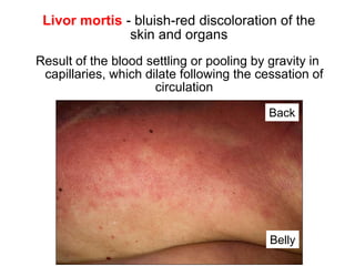
  • 1. Livor mortis - bluish-red discoloration of the skin and organs Result of the blood settling or pooling by gravity in capillaries, which dilate following the cessation of circulation Back Belly it is absent where there is pressure which prevents dilation of capillaries Livor due to hyopstasis first 8-12Hrs Livor due to imbibition after 8-12Hrs (not moved)
  • 2. Macerated foetus. Note the reddish discoloration of the skin. The epidermis is detached from the dermis in large laminae. Courtesy of Kaiser László, MD
  • 3. Aspiration of gastric content caused obstruction of airways in the patient with deep coma (lethal condition) Trachea Main bronchi
  • 4. Anemic infarct of the heart: circumscribed yellowish lesion, the margins are hyperemic
  • 5. Hemorrhagic infarct of lung: wedge shaped, raised, dark-red area
  • 6. 34 Hemorrhagic infarct of small bowels due to arterial occlusion and there is gas formation in lumen
  • 7. Brain infarct: the necrotic area is softened and pale Internal capsule Infarcted area Caudate nucleus
  • 8. Caseous necrosis • Immune-mediated distinctive form of coagulative necrosis in foci of tuberculous infection of the lung • Grossly, caseous necrosis is white and cheesy
  • 9. Necrotizing pancreatitis: the pancreas is swollen and displays several yellowish foci of necrosis
  • 10. Dry gangrene of the great toe
  • 11. Obstruction of the CSF flow leads to pressure atrophy of the brain, with the enlargement of ventricles: hydrocephalus
  • 12. Hydronephrosis: obstruction of the ureter (arrow) leads to sac- like dilation of renal pelvis and calyces, and pressure atrophy of parenchyma TUMOR
  • 13. Ischemic atrophy of the kidney because the supplying renal artery was progressively narrowed
  • 14. Hypertrophy of heart, triggered by action of mechanical stimuli ( workload) and vasoactive substances (e.g., angiotensin II). Free wall thickness: above 15 mm
  • 15. Hypertrophy of the muscles of urinary bladder due to urethral obstruction
  • 16. Steatosis: the liver is enlarged, yellow and greasy, resembles to goose liver Courtesy of E. Kemény, SZTE Pathology hypoxia-hepatotoxins obesity-diabetes alcohol-protein energy malnutrition
  • 17. The hepatocytes are vacuolated; representing accumulations of neutral lipids that have been removed by lipid solvents during tissue processing Frozen section, Oil Red O
  • 19. Dystrophic calcification of aortic valves (calcifying aortic stenosis) valsalva sinuses are rigid (Ca deposits) which cause narrowing of aorta
  • 20. 7 Pulmonary edema: the lungs 2 to 3 times exceed their normal weight; sectioning reveals a foamy mixture of air, edema fluid, and RBCs Trachea Acute LHF
  • 21. 9 Hydrothorax in chronic right-sided HF it should be almost colorless but blood during autopsy changed the color anthracosis
  • 22. 10 Increased hydrostatic pressure in the portal venous circulation (portal hypertension) by cirrhosis of liver (shown) is an important cause of ascites
  • 23. 14 Severe lymphedema of arm after mastectomy, surgical dissection of the axillary lymph nodes and irradiation of the axillary region because of breast cancer. Post-mastectomy Lymphedema
  • 24. Elephantiasis. The patient suffered from morbid obesity, type 2 diabetes and congestive heart failure.
  • 25. 21 Hepatic congestion: hypoxic/fatty hepatocytes around central veins are pale yellow, the better oxigenated portal parts are red.
  • 26. 24 Chronic left-sided HF  chronic pulmonary congestion: brown induration of lungs
  • 27. Cyanotic induration of the kidney in chronic right-sided heart failure +Enlarged
  • 28. Congestive splenomegaly. Weight: up to 500 gm, the organ is firm, the capsule is thickened, the malphigian corpuscles are indistinct
  • 29. Peptic ulcer of the postpyloric duodenum that eroded the pancreaticoduodenal artery (arrow) and caused lethal bleeding
  • 30. Classification and nomenclature of hemorrhage • Surface - External, internal • Enclosed within a tissue Suffusion (2-dimensional) Hematoma (3-dimensional); may be - insignificant (bruise) - lethal (retroperitoneal, subarachnoidal, intracerebral) Subcutaneous suffusion due to trauma
  • 32. Basal ganglia hemorrhage as a result of long-standing hypertension intracerebral hemmorhage (HTN crisis)
  • 33. 35 Petechiae: minute, 1-2 mm hemorrhages into skin, mucous membranes or serosal surfaces, seen in thrombocytopenia, defective platelet function or clotting factor deficits
  • 34. 36 Purpura: slightly larger (>3 mm) hemorrhages, may be associated with similar pathologies, as well as vasculitis Purpuras in small vessel vasculitis Sándor Husz, MD, SZTE Dermatology
  • 35. 39 Hemopericardium because of myocardial rupture (lethal) blood is in the visceral layer 400 ml can be lethal
  • 36. Hemocephalus: blood fills the lateral and third ventricles (the ventricular drainage was unsuccesful) Draining channel
  • 37. Iatrogenic (adverse condition in a patient resulting from treatment) hematoma around the carotid artery (CA) as complication of catheterization Internal CA External CA Common CA
  • 38. 9 Atherosclerosis of aorta with mural thrombi
  • 39. Endocarditis. Thrombi on the leaflets
  • 40. Aneurysms in the aorta filled with thrombi
  • 41. Left ventricle. Apical chronic aneurysm with mural thrombus (arrow)
  • 42. 17 Auricular thrombus (arrow) in a patient with atrial fibrillation
  • 44. Direction of blood flow Venous thrombi (red or stasis thrombi) Occur in a stagnating environment, and the thrombi contain more enmeshed RBCs among sparse fibrin strands; lAppear as long, red-blue cast of the vein lumen
  • 45. 26 Verrucous vegetations (arrows) on mitral valve composed of platelets and fibrin. The patient had disseminated cancer.
  • 46. 34 Red thrombi in deep veins Femoral artery
  • 47. Saddle embolus (arrow) impacting the bifurcation of the pulmonary Artery prevented the influx of blood into the lungs (lethal) Aorta Pulmonary vein
  • 48. Thromboembolism: hemorrhagic infarcts in the terrritory of middle cerebral artery (hemorrhage due to reperfusion) Caudate nucleus Internal capsule Putamen
  • 49. 55 Ruptured aneurysm of abdominal aorta leading to retroperitoneal bleading and shock (the probe indicates the rupture)
  • 50. Decreased perfusion may result in the necrosis of toes
  • 51. Cardinal signs of bacterial inflammation: calor (warmth), rubor (redness), tumor (swelling), dolor (pain), and loss of function Dr. Sándor Husz, SZTE Dermatology
  • 52. Unilateral bacterial inflammation of rat kidney induced by ligation of ureter and i.v. injection of bacteria. The animal was sacrified on Day 2. The affected kidney displayed inflammatory exudation-induced enlargement (“tumor”)
  • 53. Gross features Normally, the serous membrane is smooth and has a glistening surface. In serous inflammation, it becomes reddish and opaque. Shown: acute serous pleurisy
  • 54. Lethal acute serofibrinous peritonitis: the serosa of bowels is hyperemic; the bowels adhere to each other with fibrin strands (right: normal serosa) liver
  • 55. Edema of larynx induced by adverse (allergic) reaction of an antihypertensive drug administered intravenously in hypertensive crisis. Note marked swelling of laryngeal mucosa which caused upper airway obstruction and death
  • 56. Laryngal edema that caused airway obstruction
  • 57. Blisters in chickenpox Superinfection with St. aureus: the blisters are filled with pus (pustula) Courtesy of dr. Ildikó Kováts
  • 58. Acute fibrinous pericarditis: intense hyperemia, fibrin strands
  • 59. Left: fibrinous pericarditis, right: fibrinous pleurisy (parietal pleura). The thick strands of fibrin will not not be removed by fibrinolysis, and adhesions develop between serous surfaces Diaphragm Thoracic cage
  • 60. Lobar pneumonia. Hepatisation: liver-like consistency because alveoli are packed with fibrin trachea
  • 61. Lung: multiple abscesses as complication of pneumonia
  • 62. Furuncle (boil) Dr. Sándor Husz, SZTE Dermatology
  • 63. Folliculitis on the nose; carbuncle on the face Dr. Sándor Husz, SZTE Dermatology
  • 64. Cellulitis: sharp, erythematous swelling of the skin Courtesy of Erika Varga, MD, Department of Dermatology, SZTE
  • 65. Empyema of thorax Healing: granulation tissue  secondary calcification: callus of pleura
  • 66. Purulent mediastinitis (induced by descending infection of the neck) Aorta Pulm. artery Pulm.vein Pericardium Mediastinum Mediastinum
  • 67. Acute purulent peritonitis: the abdominal cavity was filled with more than 2000 ml pus
  • 68. Acute purulent meningitis The exudate is localized in the subarachnoid space, the meningeal vessels are engorged and stand out prominently
  • 69. Acute purulent tracheobronchitis The exudate clogged the airways and caused widespread atelectasis (collapse) of the alveoli Note: tracheobronchial toilet is necessary in patients who cannot cough actively
  • 70. Purulent discharge in Neisseria gonorrhoeae infection (sexually-transmitted disease, termed gonorrhoea) Dr. Sándor Husz, SZTE Dermatology
  • 71. Acute hemorrhagic cystitis: the bladder mucosa is swollen, hyperemic and displays focal hemorrhages
  • 72. Pseudomembranous colitis induced by C. difficile: confluent plaques of yellow fibrin and inflammatory debris adherent to a reddened mucosa
  • 73. Pseudomembranous enterocolitis: in severe cases the small bowels are also affected
  • 74. Amputated leg because of necrotizing fasciitis
  • 75. Gas gangrene: necrotizing and gas-producing inflammation of the lower extemity involving the abdominal wall, perineum and scrotum; the gas bubbles detach the epidermis from the underlying tissues
  • 76. Gas gangrene: the gas bubbles accumulated between the epidermis and the underlying tissues
  • 78. Polyoma virions in the nucleus of an epithelial cell
  • 80. Comment The influenza A virus subtype H1N1 infection was lethal in this young pregnant woman; she was regrettably not vaccinated Huge problem If people refuse vaccination, herd immunity does not develope Safety concerns are based on stories discovered in the media or received from the acquaintaces
  • 81. LM structure of chronic abscess • The central cavity is filled with pus • The wall of abscess has an inner pyogenic membrane (granulation tissue rich in neutrophils) and an outer fibroblastic rim • Heals with fibrosis Pus in the cavity Pyogenic membrane Fibroblastic rim Subpleural lung abscess (arrow)
  • 82. Multiple chronic abscesses in the liver. The patient suffered from common bile duct obstruction and purulent cholangitis
  • 83. Cirrhosis of liver: chronic inflammation of the liver  formation of fibrous septa, which, in turn, form nodular pseudolobules  impairment of venous flow through the liver. Consequence: portal hypertension, hepatic failure
  • 84. Cirrhosis of liver: nodular appearance of pseudolobules
  • 85. Chronic glomerulonephritis-induced kidney shrinkage Chronic inflammation of glomeruli  glomerular scarring Grossly the kidneys are symmetrically shrunken, the surface is granular (shown), and the cortex is thinned. Consequence: chronic renal failure
  • 86. Honeycomb lung: inflammation or injury-induced widespread fibrosis in alveolar septa culminate in grossly evident lung fibrosis Consequence: respiratory insufficiency with hypoxemia and cyanosis Alveolar septa are widened by deposition of collagen (trichrome stain: blue).
  • 87. Chronic peptic ulcer of the stomach
  • 88. Venous ulceration of the leg due to poor venous drainage from the lower leg
  • 89. Decubital ulcer in the sacral region
  • 91. Keloid Raised hyperplastic scar of the skin due to accumulation of great amount of collagen in the dermis. Common in Negroid/black people. Cosmetic problem. Shown: keloid after vaccination Courtesy of Prof. Dobozy Attila
  • 92. Laryngal edema causing airway obstruction Edema of larynx induced by adverse allergic reaction of an anti- hypertensive drug administered intravenously in hypertensive crisis
  • 93. Pseudomembranous-ulcerative gastritis induced by Candida Esophagus Stomach
  • 94. Kaposi’s sarcoma. Tumorous nodules on the leg. Autopsy case.
  • 95. Benign tumors in the uterus: expansive growth, well- defined borders, homogenous cut surface leiomyoma
  • 96. Malignant tumor (bronchial carcinoma): irregular infiltrative edges, foci of necrosis, tissue destruction. Arrows: invasion of vessels
  • 97. Cerebral metastasis of the bronchial carcinoma shown
  • 98. Kaposi sarcoma. Tumorous nodules on the leg. Autopsy case.
  • 99. Dermoid cyst of the ovary. Filled with hair, sebaceous material, and desquamated squames.
  • 100. Fungating tumor in the lip; proved carcinoma histologically Courtesy of Prof. Sonkodi István, Faculty of Dentistry
  • 101. Fungating carcinoma of the esophagus
  • 102. Ulcerated carcinoma in the antrum of stomach: irregular margins, crater-like raised borders
  • 103. Annular (napkinring-like) growth in carcinomas of the distal colon – obstructs the passage of bowel content
  • 104. Papillomas of the skin • Two types: basal cell papilloma, and wart Basal cell papilloma (seborrheic keratosis) • Very common • In middle aged or older individuals • Most frequent on the trunk • Dark brown lesion with a granular surface • Often multiple
  • 105. Condylomas on the glans and prepuce Prof. Attila Dobozy, Dermatology
  • 106. Pedunculated polyp. LM: tubular adenoma; composed of tubular glands with dysplastic features (elongated nuclei, nuclear stratification, and architectural atypia) Stalk Bowel wall Head of the polyp
  • 107. Resected bowel sample; histologically, the larger pedunculated polyp proved to be tubular adenoma with malignant transformation Head of the polyp Polyp Stalk
  • 108. Unilocular serous cystadenoma of the ovary ranging more than 15 cm in greatest dimension. The outer surface is smooth; the epithelial lining of the inner surface exhibits small papillary projections. Courtesy of Bence Nagy, MD, PhD, Department of Pathology, University of Szeged
  • 109. Mucinous cystadenoma of the ovary: delicate septa form cysts; mucinous material fills the cysts
  • 110. Adrenal cortical adenoma: circumscribed, yellowish tumor deriving from the adrenal cortex. The tumor was hormonally inactive.
  • 111. Spontaneous hemorrhage of silent adenomas  blockade of CSF flow  acute increase in liquor pressure  death
  • 112. Scirrhous carcinoma of stomach (linitis plastica) :
  • 113. Signet-ring cc: tube-like thickening of the wall; LM: the mucin in the cytoplasm pushes the nucleus to te periphery
  • 114. Mucinous cystadenocarcinoma of the ovari. Foci of necrosis and hemorrhage indicate malignant tumor. Invasion of ovarian surface → dissemination in the peritoneal cavity → carcinosis of peritoneum
  • 115. Special skin cancer: basal cell cc (basalioma) • Most frequent among cc-s • Semimalignant • Related to chronic sun exposure • In the elderly; in the face, near the eyes and nose • Advanced lesion: ulcerated (rodent ulcer) Courtesy of Attila Dobozy, Bőrklinika
  • 116. Excised breast cancer; infiltrative carcinoma with stellate appearance; gray-white strands of tumorous tissue radiate into the surrounding fat
  • 117. Advanced breast carcinoma: invasion of the skin, with extensive ulceration
  • 118. Glioblastoma: continuous spread through corpus callosum (butterfly tumour)
  • 119. Renal cell carcinoma: continuous spread involving the cortex, the medulla and the pelvic fat
  • 120. Liver metastases of colon carcinoma
  • 121. Seminal vesicle Rectum Carcinoma of the prostate; arrow: extension into the paravertebral plexus
  • 122. Vertebral metastases of prostatic carcinoma
  • 123. Transcelomic spread: carcinosis of the greater omentum Greater omentum Greater omentum
  • 124. Peritoneally disseminated ovarian carcinoma – carcinosis of peritoneum Greater omentum Stomach Stomach Liver
  • 125. Enucleated eye because of retinoblastoma
  • 126. ABCD of malignant melanoma Asymmetrical lesion with irregular borders Bleeding: spontaneous or upon minor trauma Color: uneven pigmentation or change in color Diameter: >6 mm Korom Irma, SZTE Dermatology
  • 127. Vertical growth from the beginning: nodular melanoma Gross: deepbrown nodule LM: anaplastic tumour nests Highly malignant: hematogeneous metastases very early Korom Irma, SZTE Dermatology
  • 128. Local effects Benign tumors: • Leiomyoma of the uterus: heavy menstrual bleeding, pelvic pressure/pain, frequent urination, etc. Uterine cavity Body of the uterus
  • 129. Multifocal destruction of vertebrae in multiple myeloma: pain + pathological fracture
  • 130. Fungating carcinoma in the lower third of the esophagus caused progressive stenosis and dysphagia Trachea Esophagus Cardia
  • 131. Left: napkinring-like spread of colonic cc led to stenosis  impaired the passage of stool; the frequency of passing the stool and the consistency of the stool had changed
  • 132. Ulcerated carcinoma of the stomach: irregular margins, crater-like raised borders: iron-deficiency anemia and positive fecal occult blood test
  • 133. Stenosing cancer of the ileocecal valve: the patient displayed change in bowel habit and iron-deficiency anemia Ileum Coecum
  • 134. Invasion of pelvis by renal cell carcinoma  painless microhematuria
  • 135. Urinary bladder: ulcerated carcinoma on the lateral wall caused occult painless microhematuria and iron deficiency anemia Trigone
  • 136. Bronchial cc. The non-tumorous bronchial mucosa (arrow) is hyperemic, swollen indicating peritumoral bronchitis  caughing
  • 137. Non-bacterial thrombotic endocarditis. Vegetations along the closure line of the aortic valve cups. The patient suffered from bronchial carcinoma
  • 138. The vegetations caused embolic anemic infarction of the kidney (arrow)  hematuria
  • 139. The vegetations caused embolic hemorrhagic infarction of the small bowel  bloody stool
  • 140. Sacral decubital ulcer in the bed-ridden patient wearing diaper. The patient had brain infarction and hemiplegia (complete paralysis of the half of the body)
  • 141. Femoral vein thrombosis; pulmonary embolism vein artery Pulm. artery Embolus
  • 142. Osteoporosis: severe loss of horizontal trabeculae in vertebral bodies in the patient who suffered from brain-hemorrhage, paralysis and prolonged bed rest
  • 143. 5) Ascending urinary tract infections in catheterized patients: urethritis, cystitis, acute pyelonephritis (shown)
  • 144. Lobectomy because of bronchial carcinoma (arrow) in a heavy smoker Courtesy of B. Vasas, MD, Dept. Pathology, Universitiy of Szeged sq cc destructed the bronchus
  • 145. 2) Induction of atherosclerosis Absorbed into the blood, cigarette smoke causes increased platelet aggregation and dysfunction of the endothelial layer of aorta and large arteries  development of atheromatous plaques
  • 146. Emphysema (alveolar wall destruction, enlarged air spaces) in the non-tumoral areas of the lobectomy specimen of the heavy smoker
  • 147. Mucosal defects (erosions) in alcohol-abuse induced gastroduodenitis Antrum Duodenum
  • 148. Hepatic steatosis: the liver is enlarged (>2000 g), soft, yellow, and greasy; LM: fat globules in the hepatocytes; reversible
  • 149. Pancreas: acinar cell injury (ethanol + heavy, fatty meal)  acute necrotizing-hemorrhagic pancreatitis: high mortality rate
  • 151. Pancreas: chronic calcifying pancreatitis: atrophied, fibrotic pancreas; irregularly dilated ducts obstructed with stones  pain, malabsorption
  • 152. Abnormally high deposition of visceral adipose tissue (thickness of subcutaneal fatty tissue: 10 cm), fatty liver, elevated diaphragm
  • 154. Osteophytes in spondylarthrosis limit movements of the vertebral columns
  • 155. Acute myocardial infarction Most frequent cause of death in diabetes
  • 156. Cerebral infarct involving the frontoparietal region, the internal capsule and the striatum (caudate nucleus, putamen)  contralateral hemiparesis Caudate nucleus Putamen
  • 158. Diabetic retinopathy: new vessels, lipid-cholesterol deposits Courtesy of Kolozsvári Lajos, SZTE Ophtalmology
  • 159. Neuropathic diabetic foot ulceration: necrosis + phlegmon of sole Courtesy of R. Sipka, MD, Dept. Surgery, University of Szeged
  • 160. Acute purulent pyelonephritis: several abscesses in the cortex and medulla
  • 161. Mucormycosis, with spread to the orbit and CNS Courtesy of Szabó Zsuzsanna, MD, Szent László Kórház lethal within days after meninges involved
  • 162. Myeloma: the tumorous plasma cell nodules destruct the bones; death in 15% of patients due to consequences of AL-amyloidosis
  • 163. The amyloid deposition may not be evident grossly. In this case, the heart weight was 580 gs (normal 350 gs) LM evaluation revealed amyloid deposition
  • 164. Cystic fibrosis of pancreas: fibrotic areas intermingle with cysts
  • 165. Lungs of the patient who died of cystic fibrosis: purulent plugs obstruct the bronchi
  • 166. The plugging of bronchi stem from obstruction and infection of the bronchi secondary to the viscous mucus secretions
  • 167. Bronchiectasis, purulent bronchitis and bronchopneumonia in cystic fibrosis
  • 168. Atheromatous plaque in the middle cerebral artery: raised white-yellow lesion in the intima, protruding into the lumen (formol-fixed brain)
  • 169. Aorta: the plaques contain a yellow, grumous debris (arrow)
  • 170. Structure of atheroma on LM • Intimal lesion • Central lipid core • Fibrous ”cap” subendothelially
  • 171. Coronary occlusion due ulceration of the plaque leading to thrombosis
  • 172. Cerebral infarct involving the internal capsule  contralateral hemiparesis
  • 173. Abdominal aorta: mural thrombi Celiac artery Superior mesenteric artery Renal artery
  • 174. 24 Ruptured atherosclerotic aneurysm of the infrarenal abdominal aorta; the probe indicates the communication between the lumen and the extraaortic retroperitoneal space. The aneurysmal sac is filled with mural thrombus
  • 175. Thrombosis of superior mesenteric artery: bowel infarction
  • 176. Ruptured atherosclerotic aneurysm of abdominal aorta, leading to retroperitoneal bleeding and shock (the probe indicates the rupture) Iliac arteries
  • 177. Saccular aneurysms in the thoracic aorta. Note mural thrombi
  • 178. Degeneration of the tunica media: thinning and dilation of the aortic root, and intimal tears
  • 179. Resected ascending aorta because of dissection. By the time of surgery, the process had not resulted in aortic rupture Aortic lumen Dissecting column of blood Tear Prepared by G. Nyári, MD, Dept. Pathology, Univ. Szeged
  • 180. Dissection of aorta, hemopericardium Intimal tears Aortic valve
  • 181. Hypertensive kidney disease • Mild, symmetric shrinkage of kidneys (nephrosclerosis), weight: 120-120 g; the surface is granular
  • 182. Hypertensive heart disease. Pressure overload-induced left ventricular hypertrophy  arrhythmias, chronic left-sided heart failure
  • 183. Vérzések retinopathia hypertonica-ban Courtesy of Prof. Kolozsvári Lajos Hypertensive retinopathy. Ophtalmoscopic examination reveals the thickening of retinal small arteries; microhemorrhages can be present (fundus hypertonicus)
  • 184. Microaneurysms undergo rupture in hypertensive crisis  massive hemorrhage of the basal ganglia
  • 185. Destruction of the left putamen and internal capsule by fresh hematoma
  • 186. Slightly shrunken kidneys; finely granular surface
  • 187. Heart weight 550 g; enlargement of the left ventricle
  • 188. Kidneys: hyperplastic arteriolosclerosis Gross changes • Early changes: enlarged kidneys, pinpoint petechial cortical hemorrhages + tiny infarcts • Later, infarcts are replaced by vascular scars
  • 189. Brain edema. The gyri are widened and flattened, the sulci are narrowed
  • 190. Unilateral atrophy of the kidney Not shown: the atherosclerotic stenosis of the orifice of the renal artery
  • 191. Takayasu arteritis, late phase. Arrows indicate fibrous narrowing of arterial orifices Aortic root Brachioceph. a Left common carotid a. Left subclavian a. Thoracic ao.
  • 192. Clinical features in cranial GCA • Fever, fatigue, weight loss; sedimentation rate >50 mm/h • Palpable nodularity of the tortuous temporal artery
  • 193. Brain infarcts with secondary hemorrhage in the patient who died of PAN with kidney, heart, brain and muscle involvement
  • 194. GPA: cavitation of lung parenchyma, due to necrosis of small vessels and parenchyma
  • 195. MPA, lungs: alveolar capillaritis  bilateral air space consolidation with relative apical sparing corresponds to confluent foci of alveolar hemorrhages Courtesy of B. Radics, MD, Dept. Pathology, University of Szeged
  • 196. Vasculitic purpuras Prof. Husz Sándor, Bôrklinika
  • 197. Clinical features • Skin: purpuras, principally in the lower extremities • Bowels: abdominal pain, bleeding • Joints: arthritis and arthralgia • Kidneys: hematuria, proteinuria and azotemia Outcome • Depends on the extent of crescent formation; overall prognosis is good
  • 198. Esophageal varices • In portal hypertension, the submucosal veins of distal esophagus undergo dilation (portocaval shunt) • Spontaneous rupture  hemorrhagic shock  exsanguination • Autopsy: 2000-3000 ml-s of fresh blood fills the stomach and small bowels
  • 199. Venous ulceration of the leg (varicose ulcer)
  • 200. Thrombosis in deep leg veins Most important consequence  pulmonary embolism Femoral vein Femoral artery
  • 202. Cavernous hemangioma of the liver: the vascular channels produce sponge-like structure
  • 203. Angiosarcoma of the pulmonary artery; endoluminal spread lead to occlusion
  • 204. Morphologic features of concentric hypertrophy of the LV • small lumen • markedly increased wall thickness (> 20 mm) • increased mass (> 500 g) Septum Posterior wall Anterior wall Lateral wall
  • 205. Morphology of dilative hypertrophy of the LV: enlarged lumen, enlarged size, slightly increased wall thickness, increased mass Septum Posterior wall Anterior wall Lateral wall
  • 206. Pulmonary edema: lungs >1200 gs, congested and wet, airways contain bubbly fluid
  • 207. Brown induration of lungs; atheromatous plaques in pulmonary arteries indicating sec. pulmonary hypertension collagen in alveolar septa
  • 208. Morphology RV hypertrophy (thickness › 6 mm) + dilation and manifestations of chronic systemic congestion
  • 209. Early phase: concentric hypertrophy of LV: wall thickness > 20 mm; weight > 500 g Septum Posterior wall Anterior wall Lateral wall
  • 210. Late phase: LV hypertrophy + dilation: wall thickness < 20 mm; weight > 550 g
  • 211. Occlusive thrombus (dissected with the scissor) on atheromatous plaque (arrow)
  • 212. MI: yellowish area of necrosis, hyperemic border
  • 213. 66 Hemopericardium due to rupture of the free wall of the LV rupture
  • 214. 67 Rupture of the interventricular septum
  • 216. Apical chronic aneurysm, with mural thrombus
  • 217. Septum Posterior RV LV Anterior Reperfusion injury: hemorrhage in the septum, and the anterior and posterior wall; angioplasty 12 hours after the onset of symptoms
  • 218. 84 Chronic IHD: dilative hypertrophy, apical fibrosis (arrow), dilated atrium, thrombus in the left auricle
  • 219. Calcific aortic stenosis in congenitally bicuspid valves Subendothelial calcific masses within sinuses of Valsalva
  • 220. Mitral annular calcification: regurgitation + stenosis
  • 221. Thinned and enlarged leaflets, elongated and attenuated chordae tendineae, excentric LV hypertrophy
  • 222. Rheumatic fever: acute verrucous endocarditis, chordae tendineae are also involved Dr. Tószegi Anna, SZTE Pathologia, 1972
  • 223. Mitral stenosis: thickened leaflets, fused at the commissures; thickened chordae tendinae, dilated LA
  • 224. Large vegetations on the mitral valve
  • 225. Nonbacterial thrombotic endocarditis of aortic valve from a patient with adenocarcinoma of the tail of pancreas. Histology ruled out infectious endocarditis
  • 226. Mechanical valve: tilting disk Bogáts Gábor SZTE Szívsebészet
  • 228. Dilated CM The heart is ball-shaped; there is pronounced ventricular chamber dilation The weight of the heart was 660 g
  • 229. DCM in a 2-y-old child • Extreme cardiomegaly  lung compression • Congestive hepatomegaly
  • 230. HCM: hypertrophy of the septum and the free wall; the volume of the LV is reduced; subaortic stenosis (arrow) is evident Note: the thickened left ventricular wall becomes stiff, prevents diastolic filling
  • 231. Arrhythmogenic CM. Fibrofatty near-transmural replacement of ventricular myocardium; the lesion affected the left ventricle Courtesy of B. Radics, MD, Dept Pathol, Univ Szeged Posterior wall Septum
  • 232. Diffuse mottling in myocarditis
  • 233. Mural thrombi in myocarditis
  • 236. Atrial septal defect (ASD) 90% secundum type: results from deficient or fenestrated fossa ovalis in the central atrial septum • RA and RV dilation • RV hypertrophy • Dilation of pulmonary artery J.M. Kissane: Anderson’s Pathology, 1990
  • 237. Ventricular septal defect (VSD) The most common congenital cardiac anomaly • Site (90%): membranous part of the septum • Dilative hypertrophy of RV • Small defects may close spontaneously J.M. Kissane: Anderson’s Pathology, 1990
  • 238. Patent ductus arteriosus (PDA) The ductus (just distal to the left subclavian artery) allows blood flow between the aorta and pulmonary artery during fetal life. Normally closes within 1 and 2 days of life. PDA is associated with dilation of proximal pulmonary arteries and LV LV and later RV hypertrophy J.M. Kissane: Anderson’s Pathology, 1990
  • 239. Tetralogy of Fallot (TOF) • Dextraposed aorta overriding • VSD • Pulmonary stenosis • RV hypertrophy J.M. Kissane: Anderson’s Pathology, 1990
  • 240. Transposition of the great arteries (TGA) • Aorta arising from the RV • Pulmonary artery arising from the LV • A shunt (ASD or VSD or PDA) for mixing of blood J.M. Kissane: Anderson’s Pathology, 1990
  • 241. Postductal coarctation (adult-type) Distal to the obliterated ductus arteriosus: • LV hypertrophy • Hypertension proximal and hypotension distal to the narrowed segment • Asymptomatic until adult life J.M. Kissane: Anderson’s Pathology, 1990
  • 242. Preductal coarctation (infantile-type) • Hypoplastic aorta between the left subclavian artery and the d. arteriosus • ASD • Widely PDA • Frequently lethal J.M. Kissane: Anderson’s Pathology, 1990
  • 243. Virus-induced acute serofibrinous pericarditis The pericardium is hyperemic, covered by fibrin strands
  • 244. Carcinosis of pericardium; primary tumor in the lung
  • 245. Biopsy diagnosis of GN • Via renal percutaneous biopsy evaluated by light microscopy (LM), immunofluorescence (IF) and elecron microscopy (EM) • Mirrors the inflammatory events occurring in all glomeruli
  • 246. Severe periorbital edema in a deceased man who had the nephrotic sy
  • 247. The kidney in nephrotic syndrome: the pale yellow cortex is due to lipid accumulation in renal tubules (consequence of lipiduria)
  • 248. Chronic sclerosing GN: symmetric shrinkage (80-80 g); granular surface; on section, the cortex is thinned Bleeding after biopsy
  • 249. Gross features in advanced disease • Diabetic nephrosclerosis: symmetrically shrunken kidneys with granular surface • Remember: diabetic nephrosclerosis, hypertensive nephrosclerosis, and chronic glomerulonephritis all lead to symmetrically shrunken kidneys
  • 252. Gross:“shock kidney” enlarged, swollen kidney (~ 200 g); pale, bloodless cortex, dark medulla
  • 253. The change of “shock kidney” was photographed from a deceased patient with hemorrhagic shock induced by the rupture of esophageal varices (arrows)
  • 254. • Normal ureteral insertion: acts as a valve that prevents retrograde flow of the urine during micturition. • Abnormal ureteral insertion: urine refluxes in the ureter and pelvis during micturition (VUR) • May be unilateral or bilateral Kumar et al Pathologic Basis of Disease, 2005.
  • 255. Abscesses in the cortex and medulla; the pelvic mucosa is hyperemic Pelvic mucosa
  • 256. Confluent abscesses in the cortex of the kidney; such severe cases result in urosepsis + death
  • 257. Reflux-induced chronic pyelonephritis leading to kidney shrinkage
  • 258. Coarse scars overlying blunted calyces in reflux-induced PN
  • 259. The sharp edges of the stone incised the urothelium of ureter during the travel  hematuria
  • 260. Staghorn calculus: remains localized; leads to pyelonephritic scar
  • 261. Pyelonephritic scar due to stone in the calyx: obstruction + infections are keyplayers in scar formation
  • 262. Hydronephrosis: dilated calyces, atrophied papillae (arrows), thinned parenchyma
  • 263. 55-y-o man with sepsis induced by intrarenal abscesses. Urgent nephrectomy saved the life of the patient. The gross evaluation of the specimen revealed congenital pyeloureteral stenosis (arrow), hydronephrosis, and intrarenal abscesses Dilated pelvis Ureter
  • 264. Gross Very large cystic kidneys (1000 to 4000 gs); cysts may be 3-4 cm in diameter
  • 265. Enourmously enlarged kidneys  elevation of the diaphragm; pulmonary hypoplasia
  • 266. The cysts cause spongelike appearance
  • 267. Oligohydramnios; compression of fetus By courtesy of L. Kaizer, MD, Dept Pathol, University of Szeged
  • 268. Renal artery stenosis-induced atrophy of the left kidney. Note multiple small cysts in the cortex of both kidneys
  • 269. ESRD with several cysts; renal cell carcinoma in the walls of the cysts (arrows)
  • 270. Fresh infarct (arrow): wedge-shaped, yellow, preserved outlines
  • 271. Embolism-induced vascular scars; thrombus was found in left auricle during the autopsy of the patient with the history of atrial fibrillation
  • 272. End-stage kidneys: severe shrinkage (30 to 50 g each), medulla and cortex do not separate from each other, and multiple dialysis-associated cysts
  • 273. Clear cell carcinoma • Solitary spherical mass; greatest dimension 6 cm • Bright yellow necrosis
  • 274. Papillary carcinoma • Large solitary tumorous mass
  • 276. Local spread. RCCs invade the parenchyma, the adipose capsule, the pelvis, the renal sinus, and the renal vein; may spread beyond the Gerota fascia
  • 277. “Tumor thrombus” in renal vein (arrow), can extend into the inferior caval vein
  • 278. Bronchial carcinoma. Distal to the tumor, atelectasis-associated pneumonia developes
  • 279. ARDS, gross features: the lungs are heavy (1600-2000 gs), diffusely firm, red, and boggy
  • 280. The lungs are solid, airless, and reddish purple
  • 281. Embolic occlusion of the major pulmonary branches of the pulmonary artery
  • 283. Emphysema: enlargement of alveolar spaces around terminal bronchioli Control lung
  • 284. Emphysema Pressure atrophy of the liver where the diaphragm compressed it
  • 285. Bullous E. Large subpleural blebs can develop (greater than 1 cm; usually apical). Risk of ptx!
  • 286. Cylindrically dilated bronchi, which can be cut up to the pleural surface, the lumina are filled with pus, the mucosa is hyperemic. Note foci of associated bronchopneumonia (arrows)
  • 289. Prototype • Pneumococcal pneumonia • Leads to the consolidation of an entire lobe (lobar pneumonia) or patchy consolidation of the lobe (bronchopneumonia) JCE Undervood:General and Systematic Pathology,Third edition, 2000.
  • 290. Lobar pneumonia, red hepatization. The whole lobe is red, airless, with liver-like consistency.
  • 291. Lobar pneumonia, gray hepatization. The whole lobe is greyish and the cut surface is dry.
  • 292. Lobar pneumonia, fibrinous pleuritis. Greyish-red fibrin on the pleural surface.
  • 293. Bronchopneumonia: gray-red patchy lesions slightly elevated from the cut surface. Bronchi display features of chronic bronchitis
  • 294. RS virus pneumonia in an infant. The lung is heavy and red.
  • 295. Bronchial carcinoma: destruction of bronchial wall (arrow), infiltration of hilar structures
  • 296. Central tumor: invasion of hilum, pulmonary artery (p), and lung parenchyma; metastases in hilar lymph nodes (n) n n p
  • 297. Carcinosis of pleura: grayish-white tumorous invasion of pleural surface; arrow indicates propagation via lymphatic vessels
  • 298. Metastases of bronchial carcinoma in vertebrae, suprarenal glands and calvaria
  • 299. Brain metastasis of bronchial carcinoma
  • 300. Post-stenotic bronchiectasis Chr purulent pneumonia Large, central carcinoma Consequences of bronchial obstruction
  • 301. Adenocarcinoma. Peripheral (subpleural location), simulated pneumonia on X-ray Courtesy of B. Vasas, MD, Department of Pathology, University of Szeged
  • 302. Pancoast tumor. Apical tumor, infiltrated the first and second ribs. rib rib
  • 303. METASTATIC TUMORS Primary sites: tumors drained by the caval system: liver, kidneys, adrenals, testis, thyroid, nasopharynx Metastatic involvement of the lungs: several tumorous foci in all lobes
  • 304. Honeycomb lung. Fibrotic parenchyma surrounding the bronchi retract and create cobblestone appearance of the pleural surface.
  • 305. Honeycomb lung. The dense fibrosis causes the destruction of alveolar architecture and formation of cystic spaces. Bronchi are dilated because of traction bronchiectasis. Excised pieces of lung fixed in formol. Traction bronchiectasis
  • 306. Anthracosis • Common, mild, asymptomatic, in urban inhabitants, tobacco smokers • Morphology: coal dust laden macrophages along lymphatics and lymph nodes
  • 307. Silicosis Gross: small fibrotic nodules, dense scars
  • 308. Goodpasture sy: anti-GBM autoantibodies crossreact with alveolar basement membranes  severe lung hemorrhage and symptoms of crescentic glomerulonephritis
  • 309. Goodpasture syndrome Punctuated hemorrhages in the kidney Fibrin a félholdban. Crescentic glomerulonephritis; fibrin in a crescent Linear IgG along the GBM
  • 310. Primary tuberculosis: Ghon complex (the inflammatory process is asymptomatic) asysmptomatic) Subpleural caseous necrosis Caseous necrosis in hilar lymph node Courtesy of Prof. T. Mikó, Sydney, Australia
  • 311. Bilateral caseating tuberculotic foci in the upper lobes
  • 312. Apical lung cavernas in tbc, drained by bronchus Caseous necrosis affects the the wall of bronchi, drainage of the caseous debris results in cavity (caverna) formation. Sputum (infective!!!!)
  • 313. Miliary tuberculosis in lung. Cut surface of formaldehyde fixed specimen: numerous small gray-white granulomas in the lung
  • 314. Bilateral pneumothorax as complication of mechanical ventilation in a patient with COPD – the lungs were collapsed, the venous return to the heart was impaired
  • 315. 63 Hydrothorax in chronic right-sided HF
  • 316. Dense fibrous adhesions restrict pulmonary expansion
  • 317. Empyema of thorax Precipitation of the exudate leads to septations of the pleural space
  • 318. Carcinosis of pleura: several tumorous nodules on the visceral pleura. Past medical history: surgically treated breast cancer
  • 319. Solitary fibrous tumor of the pleura: the cut surface is solid
  • 321. Carcinoma of larynx: fungating tumor destructs the vocal cords
  • 322. Thyroid cartilage Thyroid cartilage The entire circumference of glottic mucosa can be infiltrated by cancer
  • 323. Supraglottic tumor: ulcerated carcinoma in the epiglottis
  • 325. Bilateral cleft lip Prof. Füzesi Kristóf, SZTE Pediatrics
  • 326. Terminated pregnancy because of trisomy 13 (Patau’s sy): severe clefting of the lip and palate Dr László Kaizer, SZTE Pathology
  • 327. Robbins and Cotran Pathologic Basis of Disease, 2006
  • 328. Early complications •Acute purulent pulpitis (severe toothache) •Extension of infection throughout the pulp  necrosis of pulp  loss of tooth •Extension of infection into apical periodontium: acute apical abscess  subperiosteal abscess  osteomyelitis  drainage through the oral mucosa („gumboil”) or to the adjacent facial skin
  • 329. Late complications • Periapical granuloma: necrotic tissue at the apex of the root canal foramen, surrounded by granulation tissue infiltrated by lymphocytes and plasma cells. • Cystic degeneration and epithelialization of the granuloma  radicular cyst
  • 330. Recurrent aphthous stomatitis (canker sores) Aphtha (arrow) shallow ulcer on the inner surface of the lips, buccal mucosa or the tongue surrounded by hemorrhagic rim Prof. Sonkodi István, SZTE Faculty of Dentistry
  • 331. Oral candidiasis (thrush) White pseudomembranes on the lingual mucosa Prof. Sonkodi István, SZTE Faculty of Dentistry
  • 332. Irritation fibroma Pedunculated nodule on the apex of the tongue Prof. Sonkodi István, SZTE Faculty of Dentistry
  • 333. Peripheral giant cell granuloma (epulis) in the gingiva Prof. Sonkodi István, SZTE Fogászati Klinika
  • 334. Leukoplakia on the tongue - histology revealed in situ cc in the patient Prof. Sonkodi István, SZTE Fac. of Dentistry
  • 335. Esophageal atresia and tracheoesophageal fistula Most common variant (C): blind upper segment, and fistula between the lower segment and the trachea. Feeding leads to aspiration pneumonia
  • 336. Esophageal atresia and tracheoesophageal fistula Blind upper segment (yellow arrow and probe) and fistula between the lower segment and the trachea (red arrow)
  • 337. Congenital diaphragmatic hernia. Stomach, and bowels and spleen in the thoracic cavity. The left lung is hypoplastic (arrow)
  • 338.
  • 339. Megaesophagus: dilation of the esophagus proximal to LES, and also formation of a pulsion diverticulum
  • 340. Esophageal varices Tortuously dilated submucosal veins of the lower esophagus in response to portal hypertension
  • 342. Fungating carcinoma in the lower third of the esophagus Cardia
  • 343.
  • 344. Acute hemorrhagic-erosive pangastritis: hyperemic mucosa with punctate hemorrhages and multiple brownish-black erosions. Cardia
  • 345. Acute ulcers in the postpyloric duodenum.
  • 346. Acute hemorrhagic-erosive pangastritis probably induced by NSAID intake. The 65-y-old patient with severe coronary atherosclerosis, and arthrosis of hip consumed NSAIDs to releave pain. Massive gastric bleeding (hematemesis, melena) occurred which led to prolonged hypotension and, in turn, subendocardial myocardial infarcts. The patient died of acute left- sided heart failure. Cardia
  • 347. CNS trauma-associated acute stress ulcers in the postpyloric duodenum. The ulcers led to hematemesis, weak pulse, tachycardia, hypotension; the patient ceased. During the autopsy, 3000 ml blood was found in the intestines.
  • 348. Large peptic ulcer on the lesser curvature of the stomach; note round shape and sharp margins
  • 350. Autoimmune metaplastic atrophic gastritis (AMAG)-associated multiple hyperplastic polyps (P), polyps with dysplasia (D), and carcinomatous transformation (Cc) Courtesy of Bence Kővári, MD and Orsolya Oláh, MD, Dept. of Pathol, University of Szeged P P P D D Cc
  • 351. Advanced gastric carcinoma: large fungating tumorous mass (line); metastasis in the lymph nodes of greater omentum (M) M M Courtesy of István Németh, MD, Dept. of Pathol, Univ. Szeged
  • 352. Gastric carcinoma with two ulcer craters (arrow)
  • 353. Antral gastric carcinoma infiltrating the mucosa and gastric wall Courtesy of Prof. László Tiszlavicz and Levente Kuthi, MD; Dept. of Pathol, Univ. of Szeged
  • 354. Linitis plastica (leather bottle stomach): the stomach wall is thicker and more rigid because of cancerous infiltration The stomach can’t hold as much and does not move as it should
  • 355. Krukenberg tumor: bilateral ovarian metastasis of signet-ring cell carcinoma Courtesy of András Vörös, MD, Dept. Pathol, Univ. Szeged
  • 356. Perforated duodenal peptic ulcer: duodenal content irritated the serosa and induced hyperemia
  • 357. Purulent peritonitis: creamy exudate covers the bowels and mesentery
  • 358. Diffuse purulent peritonitis caused by perforation of acute ulcer of the stomach 2000 ml of pus was found in the abdominal cavity Diaphragm E.Coli Kleb. pneumo Strep. pneumo
  • 359. Carcinosis of omentum; the patient had pancreatic carcinoma diagnosed during autopsy
  • 361. Invagination (intussusception) The a ag a e ( e e c e ) he d a loop, like the finger of an inverted glove
  • 362. Volvulus-induced hemorrhagic infarction of the cecum and ascending colon. Arrows: not affected colon and ileum
  • 363. Border of the necrotized and non-necrotized ascending colon
  • 364. Luminal cause of intestinal obstruction: gallstone ileus. Proximal to obstruction, the small bowel is dilated
  • 365. Consequences of intestinal obstruction The bowels proximal to the obstruction undergo dilation their wall becomes thinned their lumen is filled with large amount of fluid and gas
  • 366. Ileus due to rectal carcinoma X-ray of the abdomen: distended small bowel loops and stomach filled with fluid and gas. Note fluid levels. Courtesy of Morvay Zita, SZTE Radiology
  • 367. Consequences of intestinal obstruction The bowels proximal to the obstruction undergo dilation their wall becomes thinned their lumen is filled with large amount of fluid and gas Elevation of the diaphragm + Strangulation-induced bowel infarction peritonitis
  • 368. Thrombosis of superior mesenteric artery (not shown) hemorrhagic infarction/gangrene of the small bowel + generalized distension of bowels indicating vascular ileus cecum Asc.Colon Transverse Colon Small Bowels Sup. Mes. a. thrombus --> Infarct in Colon --> vascular ileus and distention of Colon and SI
  • 369. Shigella infection Distal colonic inflammation with shallow ulcers Colitis
  • 370. Fulminant Clostridioides difficile colitis: the colon is markedly distended, confluent pseudomembranes cover the mucosa
  • 372. Pseudopolyps and broad-based ulcers in ulcerative colitis
  • 373. Serpentine fissures and cobblestone-like mucosa in CD (large bowel)
  • 374. CD: Serpentine fissures and cobblestone-like mucosa CU: broad-based ulcers, pseudopolyps
  • 375. Gross features that differ between CD and UC C disease Ulcerative colitis Bowel region Ileum + colon Rectum > distal colon > entire colon Distribution Skip lesions Continuous Broad-based ulcers and pseudopolyps Not characteristic Yes Serpentine fissures and cobblestone appearance Yes Not characteristic Transmural stricture Yes Rare Wall appearance Thick Thin Fistulae Yes No
  • 376. Diverticulosis. The mucosal surface is ridged due to hypertrophy of the underlying muscle. Mucosal hyperemia indicates diverticulitis.
  • 377. Tubular adenomas About half are found in the rectosigmoid; may be single or multiple Usually <10 mm and pedunculated >10 mm: areas of intramucosal carcinoma can be present (invasion of the lamina propria with no extension through the muscularis mucosae into the submucosa)
  • 378. Two polyps. The smaller proved to be adenoma; the larger proved to be adenoma with malignant transformation: stalk-invasive adenocarcinoma and metastases in mesocolonic lymph nodes were observed Mesocolonic LN
  • 379. Villous adenomas Most often in the rectum; solitary, sessile, diameter: up to 10 cm Composed of villi (finger-like protrusions lined with dysplastic columnar epithelium) Adenocarcinoma frequently arises in VA- s > 4 cm
  • 380. Villous adenoma with malignant transformation: adenocarcinoma infiltrates the submucosa Villous Adenoma >4cm Two points of communication between Mucosal Dysplastic glands and dysplastic glands in submucosa creating the large Adenocarcinoma
  • 382. Cc of the sigmoid bowel: annular (napkinring-like) growth. Lymph node metastasis in mesocolic adipose tissue R. Colon --> Fungating Into Serosa --> T3?
  • 383. Carcinoma of the cecum: exophytic, fungous mass is characteristic
  • 384. Rectal cc leading to ileus: extremely dilated colon proximal to the obstruction
  • 385. Ca ci a f he ile cecal al e: he a ie a eak e induced by iron-deficiency anemia Ileum Carcinoma
  • 386. Neuroendocrine tumor in the ileum close to the Bauchin valve T a c L , SZTE Pa a
  • 387. Mural tumor in the wall of the small intestine causing subileus. Histology disclosed GIST
  • 388. Ulcerophlegmonous appendicitis. The peritoneal surface is covered by fibrinopurulent exudate. Formol-fixed material
  • 389. Congenital megacolon; before resection of the aganglionic segment P f. F e K f, SZTE Ped a c
  • 390. Jaundice (icterus) Yellow discoloration of the skin, sclerae, and mucous membranes due to increased levels of bilirubin in circulation (> 40 umol/L) Caused by cholestasis, hemolysis or genetic disease
  • 391. Chronic hepatitis C virus infection led to cirrhosis of liver: nodular alteration of the parenchyma
  • 392. Alcoholic steatosis: the liver is enlarged, soft, yellowish, the edges are rounded
  • 393. Autopsy of an obese woman with type 2 diabetes, hypertension, dyslipidemia, elevated ALT, AST, and bilirubin. Conversion of non-alcoholic steatohepatitis to cirrhosis was found
  • 394. Female patient with the medical history of 10 years of primary sclerosing cholangitis. The normal lobular structure was replaced by fibrosis.
  • 395. Hemochromatosis. Fibrous pancreas and cirrhotic liver. The brownish colour of organs is because of hemosiderin deposition
  • 396. Cirrhosis Fiborus septa convert the normal liver architecture into structurally abnormal nodules (pseudolobules) HCV infection usually induces macronodular cirrhosis 5 cm alcohol - micronodular
  • 397. Esophageal varices Dilated veins in submucosa in the distal esophagus; can rupture at any time
  • 398. Focal nodular hyperplasia In young women (no evidence of a relationship with oral contraceptive use) Nodule-forming hyperplastic response of hepatocytes to focally increased blood flow Uncapsulated tumorlike lesion with a central stellate scar
  • 399. Cavernous hemangioma Red-blue, soft nodules consist of endothelial cell lined vascular channels and stroma Blind percutaneous needle biopsy may cause severe intra-abdominal bleeding
  • 400. Morphology Gross: usually in cirrhotic liver Unifocal Multifocal Diffuse
  • 401. Multifocal hepatocellular carcinoma in cirrhotic liver
  • 402. Intrahepatic cholangiocarcinoma, multifocal variant no cirrhosis + history adenocarcinoma = Largeduct iCCA
  • 403. Metastases in the liver; the patient died of hepatic failure
  • 404. Several metastatic foci in the liver
  • 405. Cholesterol stones: from black to yellowish brown; multiple; faceted surface owing to tight apposition Gallbladder Cystic duct Hepatic duct Common bile duct
  • 407. Pathogenesis Crystallization of cholesterol (nucleation) can be induced by • Bile supersaturated with cholesterol • Gallbladder hypomotility and/or defective gallbladder emptying • Hypersecretion of gallbladder mucus Cholesterol can no longer remain dispersed and nucleates into cholesterol monohydrate crystals
  • 408. Cholesterol monohydrate crystals Precipitation of Ca++-salts Cholesterol stone
  • 409. Ulcerophlegmonous cholecystitis: hyperemic mucosa covered with multiple ulcers; the wall is edematous
  • 410. Thinned mucosa, thickened gallbladder wall, cholesterol stones
  • 412. Hydrops: the gallbladder is markedly enlarged, the muscular wall is thinned
  • 413. Carcinoma developed in chronic calculous cholecystitis. Note infiltrating growth pattern Tumorous Thickening Of GB wall Continuous spread to liver parenchyma
  • 414. Carcinoma of the gallbladder. Hematogeneous metastases in the liver. Liver Metastases with spontaneous Necrosis
  • 415. Muliple liver abscesses; the obstruction was managed by stenting the common bile duct
  • 416. Green liver in obstructive jaundice The patient had carcinoma of the head of pancreas; the tumor infiltrated and obstructed the common bile duct; liver metastases
  • 417. Carcinoma of the cystic duct, the hepatoduodenal ligament and the hepatic duct (probe) Diffusely infiltrating type Liver, Nutmeg or steatotic?
  • 418. Stimulation Secretion to the duodenum Normal site of trypsinogen activation Acinus Calcium regulation • Hypercalcemia • Alcohol Trypsin related mutations • PRSS1+: activation of trypsinogen • CTRC+: ineffective trypsin degradation • SPINK1+: ineffective blockade of active trypsin Duct Duct cell secretion • CFTR Duct obstruction • Gallstone • Duct stones • Tumor • Mucus Genetic and environmental factors that affect acinar cells or ducts Modified from Muniraj et al. Disease-a-Month 60:530-550, 2014
  • 419. Classification according to the severity of acute pancreatitis Clinically • mild - morphologically acute interstitial pancreatitis: interstitial (IS) edema + foci of enzymatic necrosis in the acini by LM • moderately severe - morphologically acute necrotizing pancreatitis: IS edema and gross foci of enzymatic necrosis • severe - morphologically acute necrotizing-hemorrhagic pancreatitis: the entire pancreas is involved; confluent foci of necrosis and hemorrhage; foci of enzymatic fat necrosis in the extrapancreatic collections of fat, such as the mesentery of the bowel and the omentum
  • 420. Moderately severe - acute necrotizing pancreatitis IS edema and gross foci of enzymatic necrosis I idiopathic G Gallstones E Ethanol T Trauma S Shock M Mumps A Autoimmune S Scorpion Sting H HyperTG/Ca E ERCP D Drugs
  • 421. Severe - acute necrotizing-hemorrhagic pancreatitis. The entire pancreas is involved; confluent foci of necrosis and hemorrhage
  • 423. Severe pancreatitis. Foci of enzymatic fat necrosis in the mesentery of bowels Duoddeno-Jejunal Flexure
  • 424. Complications in severe pancreatitis • SIRS (systemic inflammatory response sy)-induced shock • Bacterial superinfection of necrotic pancreatic tissue abscess(es) sepsis • Disruption of large ducts can result in unilateral pleural effusion, enlarging peripancreatic fluid collection, or pancreatic ascites • > 4 weeks: pseudocyst formation (1 to 15 cm): massive liquefactive necrosis enclosed by granulation tissue; + infection of pseudocysts pancreatic abscesses
  • 425. Pathogenesis Still not understood; TIGARO classification of risk factors • Toxic-metabolic: chronic alcohol abuse (mostly in middle-aged men), hypercalcemia, chronic renal failure, etc. • Idiopathic • Genetic-induced: SPINK1 gene mutation or PRSS1 gene mutation or CFTR gene mutation (no extrapancreatic manifestations of cystic fibrosis) • Autoimmune • Recurrent acute pancreatitis • Obstruction of pancreatic duct by stone or tumor or congenital abnormality (pancreas divisum [1 papilla minor + 1 accesory duct])
  • 426. Alcoholic chronic pancreatitis: atrophied, markedly fibrotic pancreas, irregularly dilated ducts obstructed with Ca- carbonate stones + pseudocysts (not present in this patient) =Chr. Calcifying Pancreatitis
  • 427. Carcinoma (cc) in the pancreatic head, infiltrating the papilla of Vater and the common bile duct (cbd). Distal to the tumor, obstructive pancreatitis developed. The Wirsungian duct is markedly dilated and tortuous W W cc cbd
  • 428. Cc of the pancreas: hard, poor-defined mass; difficult to distinguish from chronic pancreatitis
  • 432. Immunopathology of severe SARS-CoV-2 lung disease Inflammatory lung injury: impaired innate antiviral defense, overactivated innate immune response Upper airways: evasion of IFN-I response → robust viral replication → infection extends to the lung Uncurbed viral replication Weak IFN-I response Cytopathic damage to pneumocytes, endothelial cells → DAMPs DAMPs overactivate alveolar macrophages Weak viral clearance Weak CD8+ cytotoxic T cell response Influx of activated monocytes, neutrophils Alveolar edema, hyaline membranes, alveolar collapse Progressive diffuse alveolar damage and ARDS + death Adapted from Med Res Rev 2020; 1-28
  • 433. 71-y-o male patient with hypertension, chronic ischemic heart disease and lethal SARS- CoV-2 lung disease Diffuse gray-white consolidation, admixed with hemorrhage; thrombi in pulmonary vessels (arrows). Formol-fixed specimen. Infected in nursing home 14 days prior to death. Fracture of femur, operated urgently 9 days prior to death. Fever and progressive ARDS started 2 days prior to death. Autopsy performed by B. Radics, MD, Department of Pathology, University of Szeged
  • 434. Timeline of Covid-19 Modified from New Engl J Med 2020;383:2451-60 Onset of symptoms Median days from infection -5 8 13 -4 -3 -2 -1 0 1 6 5 4 3 2 7 9 10 11 12 14 15 16 17 18 Incubation period Airways: viral replication evades IFN-I response and extends to alveoli Fever Cough Fatigue Anorexia Myalgias Mild or moderate pneumonia, dyspnea Severe pneumonia, ARDS, hypoxemia + hypercytokinemia, coagulopathy, shock 19
  • 435. Plasma Water 92% Proteins 7% Albumin 58% Globulins 37% Fibrinogen 4% Regulatory proteins 1% Other solutes 1% Electrolytes (Na, K, etc.) Nutrients (glucose, amino acids etc.) Respiratory gases Waste products Red cells (erythrocytes) 4-5.5 million/ L Buffy Coat Platelets (thrombocytes) 150.000-400.000/ L White cells (leukocytes) 4.500-11.000/ L Neutrophils (54-62%) Lymphocytes (25-33%) Monocytes (3-7%) Eosinophils (1-3%) Basophils (0-0.75%) 45% 55% COMPOSITION OF BLOOD Packed red cell volume: Hematocrit
  • 436. BONE MARROW Red marrow: active, blood cell forming In adults: flat bones (pelvis), vertebrae Yellow marrow: inactive, fatty tissue → it may turn red again in cases of blood loss (adaptation), or in myeloid neoplasias (leukaemias)
  • 437. BONE MARROW Top: in AML, the diaphysis is replaced by leukemic red bone marrow. Bottom: normally, the diaphysis of femur is filled with yellow fatty bone marrow. red tumorous bone marrow seen in aml and a l l it's a sign of bone mineral haematopoiesis suppression leading to anemia thrombocytopenia and sometimes granulocytopenia ( leukemic leukaemia) JML has many subtypes one of them is PML associated with translocation 15:17 between PML L Gene and rara play which could be as viewed as a hey good prognostic sign since atra could be used with arsenic trioxide to treat it and a special sign of the subtype is the dic due to the procoagulant release while another subtype with differentiating monocytic can cause leukaemia cutis
  • 438. In certain diseases, the HSCs can travel in the blood from the bone marrow in one bone to another bone and may settle in the liver and spleen (extramedullary hemopoiesis)
  • 439. Aspiration of the bone marrow yields a bone marrow cytological smear
  • 440. Trephine (Jamshidi) biopsy to investigate the constituents of bone marrow yields a tissue core of bone marrow
  • 441. Diseases of blood, hematopoietic and lymphoid system Red blood cell disorders White blood cell disorders Coagulation system disorders Anemia Erythrocytosis Leukopenia Reactive leukocytosis Neoplasms Thrombocytopenia Thrombocytosis Coagulopathies Pancytopenia (anemia, leukopenia, thrombocytopenia ”Clot or bleed”
  • 442. Pallor of skin Pale Skin is a sign of Anemia there are many causes of anywhere I like to think of them as as deficiency anaemia iron deficiency B12 B9 deficiency then we can talk about the factory the bone marrow and diseases affecting the bone marrow such as aplastic anaemia tumor infiltration suppression by irradiation then we can talk about the red blood cells themselves haemoglobin could be affected in thalassemia and sickle cell anaemia the memory could be defective in spherocytosis and elliptocytosis enzyme deficiencies such as pyruvate kinase gcpd H deficiencies then we can talk about hemolytic anemias such as autoimmune hemolytic anaemia malaria induced microscopic an geopathic hemolytic anaemia ( TTP dic( and finally we can talk about blood loss which could be acute in case of traumatic injuries or chronic in case of gynecology renal or GI pathologies
  • 443. - Iron deficiency - Anemia of chronic disease - Thalassemias - Sideroblastic anemia Anemia Microcytic Normocytic Macrocytic Reticulocyte count Low High - Megaloblastic anemia * B12 deficiency * Folate deficiency - Non-megaloblastic *Alcoholic liver disease *Other liver diseases Marrow failure • Aplastic anemia • Myelofibrosis • Myelophthisis • Marrow suppression (drugs, chemotherapy, radiation) Chronic renal failure Anemia of chronic disease Acute blood loss Hemolytic anemias • Sickle cell anemia • G6PD deficiency • Hereditary spherocytosis • Autoimmune hemolitic anemia
  • 444. Acute Massive bleeding from ruptured blood vessels: Traumatic wounds (eg.: car accidents) Ruptured aortic aneurysm Ruptured esophageal varices Gastric or duodenal ulcer Ectopic pregnancy Etc. Loss of intravascular volume (>20% loss of blood) hemorrhagic shock and may lead to death If the patient survives hemodilution (movement of fluid into the vascular space) normocytic normochromic anemia Tissue hypoxia EPO RBC production with reticulocytosis normali ation within 3-4 weeks Chronic GI tract (ulcers, polyps, tumors, IBD, angiodysplasias, hemorrhoids, etc.) Gynecological (heavy or frequent menstrual bleeding, metrorrhagia abnormal uterine bleeding) Urological (kidney or bladder cancer) IRON DEFICIENCY ANEMIA ANEMIA OF BLOOD LOSS: HEMORRHAGE
  • 445. • Iron in food (2 forms): – Heme iron (bound to Hb and myoglobin – in meat) – Fe2+ (ferrous) – Non-heme iron – free iron molecules in Fe3+ (ferric) • Plant-based foods (spinach, beans, lentils) IRON METABOLISM
  • 446. IRON METABOLISM Fe HCl HEME IRON NON-HEME IRON Fe3+ Fe2+ Fe2+ Ferri-reductases Fe2+ Heme-transporter Fe2+ DMT-1 HCl Fe2+ Fe2+ Fe3+ Transferrin (iron transporter) Duodenal cell Blood Target tissues Ferroportin Hephaestin Erythropoiesis Liver Hepcidine - Storage of iron: intracytoplasmic ferritin and hemosiderin (eg.: in macrophages) Iron loss by shedding of epithelial cells Transferrin-receptor Stomach Duodenum Muscle Ferritin Intake: 10-20 mg/day
  • 447. Gross morphology: yellow fatty marrow at sites of red bone marrow (sternum)
  • 448. Petechial hemorrhages in severe thrombocytopenia
  • 449. Myelophthisis: Prostate carcinoma metastases in spine and femur (bottom: normal trabecular structure after formol fixation) Lung, pancreas and Breast can also metastasise to bone
  • 450. Hb Globin Amino Acids Heme Biliverdin Bilirubin Heme oxygenase Biliverdin reductase Fe2+ A Bilirubin / albumin complex MACROPHAGE I di ec bi i bi c j ga ed bi i bi Stercobilin Ha g bi HEMOLYSIS RBC c F ee he g bi Pink serum <120 days INTRA- VASCULAR EXTRA- VASCULAR
  • 451. CAUSES OF VITAMIN B12 DEFICIENCY B12 Parietal cell Intrinsic factor (IF) B12-IF complex IF receptor Ileum Stomach Blood Colon Transcobalamin II Target tissues Storage in the Liver a e B12 Vegeterians Poor diet absorption (malabsorption) - production of IF - Gastrectomy - Pernicious anemia - ileal absorption: - Ileal resection - Ileal inflammation: Crohn- disease - Consumption by intestinal bacteria or tapeworms
  • 452. PERNICIOUS ANEMIA B12 Parietal cell Intrinsic factor (IF) B12-IF complex IF receptor Ileum Stomach Blood Colon Transcobalamin II • Definition: – A subtype of B12 deficiency induced megaloblastic anemia • Cause: – Autoimmune disease against the parietal cells and intrinsic factor – IF B12 absorption – Chronic atrophic corpus gastritis with intestinal metaplasia (AMAG) risk of gastric cancer Auto- antibodies
  • 453. Diseases of blood, hematopoietic and lymphoid system Red blood cell disorders White blood cell disorders Coagulation system disorders Anemia Erythrocytosis Leukopenia Reactive leukocytosis Neoplasms Thrombocytopenia Thrombocytosis Coagulopathies Pancytopenia (anemia, leukopenia, thrombocytopenia ”Clot or bleed”
  • 454. CLASSIFICATION BASED ON NORMAL CELL LINES • Myeloid neoplasms – Acute myeloid leukaemias (AML) – Chronic myeloproliferative neoplasms (MPN) – Myelodysplastic syndromes (MDS) • Lymphoid neoplasms – Precursor lymphoid neoplasms (ALL) – Mature lymphoid neoplasms • Mature B-cell neoplasms • Mature T- and NK-cell neoplasms – Hodgkin lymphoma • Histiocytic neoplasms
  • 455. MYELOID NEOPLASMS: CLASSIFICATION Maturation arrest + ↑↑ replication Defective maturation Proliferation of terminally differentiated myeloid cell lines Secondary maturation arrest + ↑ replication 1) Acute myeloid leukemias 3) Chronic myeloproliferative neoplasms 3) Myelodysplastic syndromes New oncogenic mutations
  • 456. Grossly, the sites of yellow bone marrow are infiltrated by tumorous red bone marrow. Top: in AML, the diaphysis is replaced by leukemic red bone marrow. Bottom: normally, the diaphysis of femur is filled with yellow fatty bone marrow.
  • 457. CML Polycythemia vera Essential thrombocytosis Primary myelofibrosis Clinical features Peak age 50-60 yrs 40-60 yrs 70 yrs 70 yrs Splenomegaly Yes (Massive) Yes 30% (slight) Yes (Massive) Bone marrow Panhyperplasia (predominantly granulopoiesis) Panhyperplasia (predominantly erythropoiesis) Isolated megakarycytic hyperplasia with large megakaryocytes in clusters Panhyperplasia with fibrosis Peripheral blood Erythrocytes Mild anemia Erythrocytosis (polycythemia) Mild anemia Mild anemia Granulocytes Markedly increased Norm.-mildly increased Normal (slightly increased) Normal to moderately increased Platalets Normal to moderately increased Normal to moderately increased Markedly increased (giant thrombocytes) Inreased to decreased Genetics Philadelphia chromosome (BCR/ABL rearrangement) JAK2 activating mutation JAK2 activating mutation; MPL activating mutation JAK2 activating mutation MPL activating mutation
  • 458. Myelofibrosis (right): osteosclerosis of the marrow spaces of sternum. Left: normal sternum
  • 459. Myelofibrosis Note extreme splenomegaly induced by extramedullary hemopoiesis (myeloid metaplasia). The arrow indicates spontaneous infarction of the spleen parenchyma. The patient died of consequences of transformation to acute myeloid leukemia. Liver Spleen Stomach
  • 460. Non-Hodgkin lymphoma: enlarged lymph nodes, the cut surface is homogeneous, grayish-white lymphoma that could be Hodgkin's or non-Hodgkin's hodgkin's lymphoma is characterized by contiguous surprised lymphadenopathy that is usually the cervical region it is characterised by symptoms such as fever night sweats and weight loss also characterized by presence of certain cells such as reed-sternberg cells hodgkin's cells and lacunar cells it has Ford main subtypes for the classical hodgkin's lymphoma the nodular sclerosing mixed hypercellularity lymphocyte rich and deleted while then non Hodgkin lymphoma could be further classified based on whether the tumor is aggressive2 or indolent
  • 461. Marginal zone lymphoma in the stomach and the regional lymph nodes (formol-fixed specimen) Pancreas Stomach this is a GI lymphoma most common GI landform what is the diffuse large b-cell lymphoma and then the marginal zone lymphoma and then the celiac disease associated t-cell lymphoma they are associated with chronic inflammation aka helicobacter pylori infection or celiac disease they are highly aggressive
  • 462. Marked enlargement of the hilar and paratracheal lymph nodes; the disease was diagnosed as DLBCL during autopsy Larynx Trachea Tongue
  • 463. Superior vena cava sy caused by an agressive tumor; autopsy revealed enlarged paratracheal, hilar and mediastinal lymph nodes; T-cell lymphoma was confirmed
  • 464. Erosions of cancellous bone of calvarium in myeloma
  • 465. Erosions of cancellous bone of vertebrae in myeloma myelomas are tumors arising from mature plasma cells that produce immunoglobulin light chain bence-jones protein that leads to renal failure and gastric property and bence Jones proteinuria it is also associated with hypercalcemia due to the stimulation of osteoclast through cytokines you can also cause anaemia and Bone defects presenting as pathological fractures the disease starts as monoclonal gammopathy of uncertain significance asymptomatic myeloma then symptomatic over to myeloma it is characterized by by m-protein Spike and the produced immunoglobulins could act as autoantibodies and generally they are dysfunctional so we will have hypogammaglobulinemia
  • 466. Toxoplasma-lymphadenitis Ingestion of oocysts from cat feces; the protozoon disseminates in the human body; the inflammatory response, including lymphadenitis, destructs the parasites).
  • 467. Markedl enlarged pleen ( 1000 g) in ad l Myelofibrosis CML CLL
  • 468. Myelofibrosis. Note extreme splenomegaly induced by extramedullary hemopoiesis (myeloid metaplasia). The arrow indicates spontaneous infarction of the spleen parenchyma. The patient died of consequences of transformation to acute myeloid leukemia. Liver Spleen Stomach
  • 469. Invasive thymoma: The surgical resection margin (blue) is infiltrated by the tumor
  • 470. litterer siwe disease affect children, filtration of skin spleen liver bone marrow can be lethal third disease is is hand-schuler-christain having a triad of exopthalmus diabetes insipidus calvarial bone defects
  • 471. P of. Ba P l, Ne o ge Diffuse brain edema: ventricular spaces are severely narrowed
  • 472. 1. Subfalcine herniation Unilateral mass lesion forces the ipsilateral cingulate gyrus to be compressed underneath the falx cerebri focal necrosis and hemorrhage in the herniated tissue + compression of the anterior cerebral artery Kumar, Cotran, Robbins: Basic Pathology 2003 Subfalcine herniation
  • 473. 2. Transtentorial herniation Transtentorial herniation • Expansion of the hemisphere the uncal gyrus of hippocampus is herniated underneath the free edge of the cerebellar tentorium
  • 474. 2. Transtentorial herniation Transtentorial herniation •The ipsilateral oculomotor nerve undergoes compression: ipsilateral fixed pupil • Posterior cerebral artery compression occipital infarction, cortical blindness • Cerebral peduncle compression upper motor neuron signs • Brainstem compression cardiorespiratory failure, death
  • 475. 3. Tonsillar herniation Tonsillar herniation Transtentorial herniation The cerebellar tonsils are forced into the foramen magnum and compress the respiratory and cardiac centers within the medulla cardiorespiratory failure, death
  • 476. Compression of cerebellar tonsils and medulla oblongata by the foramen magnum Green=Deep lesion Red= Necrotic features
  • 477. Striped hemorrhages in the pons in response to tearing of small vessels(Duret hemorrhages) Secondary hemorrhages to a space occupying leion Can be seen in Tonsillar herniation
  • 478. Hydrocephalus Preparation of late P of D . Gell Albe SZTE Anatomy Department
  • 479. Hydrocephalus: severe enlargement of the ventricles, atrophy of the brain Usually Atrophy of the brain leads to ventricular enlargement But in this case the enlargement of the ventricles is huge so maybe the ventricular enlargment (Primary Hydrocephalus) preceeded and cause pressure atrophy of the brain
  • 480. Sec. (compensatory) hydrocephalus The increase in CSF volume occurs following brain atrophy: HC ex vacuo The intracranial pressure is normal
  • 481. Fracture contusion with necrosis of GM and WM, hemorrhage and brain oedema Coup Contre-coup
  • 482. The branches of middle meningeal artery run between the dura mater and the skull
  • 483. Epidural hematoma • Temporal bone fracture rupture of middle meningeal artery accumulation of arterial blood between the dura and the skull: EH • Lucid interval followed by a rapid in intracranial pressure P of. Ba P l, Ne o ge
  • 484. The bridging veins between the dura mater and the arachnoid membrane
  • 485. The middle cerebral artery is the most frequent site of occlusion in association with atherosclerosis
  • 486. Thromboembolism: several infarcts with secondary reperfusion hemorrhage in the territory of the middle cerebral artery Caudate nucleus Internal capsule Putamen of lentiform nucleus Supplied by Ant. cerebral a. Midline shift is also present Cavity of Septum pallucidum "Cavum Septi Pellucidi" It is normaly present in fetuses and closes on its own, finding it in adults is not a pathologcal finding per se, but rather an antomical variation, sometimes could be considered as a marker of neural maldevelopment.
  • 487. Cerebral infarction: wedge-shaped softening of the parietal lobe and the internal capsule Caudate nucleus Thalamus infarction Globus pallidus infarction Whitesh yellowish necrotic area with irregular border and surrounded by edema Blurring of WM and GM on left side compared to the well demarcated right side Caudate is infarcte GP is spared
  • 488. Hypertensive hemorrhage in the left hemisphere, lacunar state in the right basal ganglia Formol-fixed brain Huge hemorrhagic lesion "Apoplexy"
  • 489. Hypertensive patient with psychomotor slowness: bilateral lacunar infarcts in the deep WM focal myelin loss From psychomotor slowness it could progress so vascular dementia
  • 490. Fresh hematoma distructs the putamen, the WM of parietal lobe, and the internal capsule Midline shift
  • 491. Hemocephalus in the fourth ventricle - lethal Extension to subarachnoidal space --> Subarachnoidal hemorrhage Substantia Nigra could be seen as a thin dark line Maybe some signs of Duret Hemorrhage
  • 492. Multiple hemorrhages in blastic crisis
  • 493. Berry aneurysm of the circle of Willis
  • 494. Subarachnoid hemorrhage due to ruptured berry aneurysm
  • 495. Glioblastoma. Distortion of the temporal WM and thalamus, note foci of necrosis, cystic change and hemorrhage Heterogenous appearance "Glioblastoma Multiforme"
  • 496. Glioblastoma. Butterfly pattern of spread through the corpus callosum
  • 497. Cerebellar metastasis of breast carcinoma (autopsy disclosed the primary tumour) Well circumscribed Lung cancer and melanoma are most likely to present with multiple metastasis, whereas breast, colon, and renal cancers are more likely to present with a single metastasis primary brain tumors could be pilocytic astrocytoma diffuse astrocytoma anaplastic astrocytoma glioblastoma oligodendrogliomas ependymoma medulla blastoma meningioma schwanoma and neuroblastoma
  • 498. Parasagittal meningioma Falx cerebri Found incidentaly Hard to excise since it is near the Falx cerebri Tendency to recur
  • 499. Gray-tan, firm plaques in the WM of brain and spinal cord; commonly beside the lateral ventricles; optic nerve is frequently involved Multiple Sclerosis "MS"
  • 500. The plaques are located in the white matter: well-circumscribed, glassy, gray-tan, irregularly shaped lesions Multiple Sclerosis "MS"
  • 501. Alzheimer disease. Cortical atrophy, characterized by narrow gyri and widened sulci (meninges had been removed)
  • 502. The medial structures of the temporal lobe display marked atrophy. Note compensatory ventricular enlargement – HC ex vacuo Sup. and Middle Temporal Gyrus Atrophied Cingulate Gyrus Relatively preserved Middle Frontal Gyrus Atrophied
  • 503. Gross: depigmentation of the substantia nigra and locus coeruleus
  • 504. Acute purulent meningitis of the brain and the spinal cord Engorged veins
  • 505. Multiplex brain abscess, pyocephalus internus; the patient was hypertensive, the reason why lacunar state was also observed Pyocephalus internus Lacunar infarcts Abscess
  • 506. Chronic abscess of the spinal cord
  • 507. Anencephaly - lethal • The calvaria and the hemispheres are absent • The base of the skull is covered by a mass of vascular granulation tissue (area cerebrovasculosa) D . Kai e L , SZTE Pa h g
  • 508. Encephalocele Malformed CNS tissue extends through a defect in the cranium Occurs in the occipital region or in the posterior fossa Lethal D . Kai e L , SZTE Pa h g
  • 509. Neuro - Fejlődési rendellenesség - Spina bifida Myelomeningocele and rachischisis D . Kai e L , SZTE Pa h g
  • 510. Holoprosencephaly: incomplete separation of hemispheres D . Kai e L , SZTE Pa h g
  • 511. Agenesis of corpus callosum D . Kai e L , SZTE Pa h g
  • 513. BC Screening Before the advent of breast cancer screening programs breast cancer was mainly detected as a symptomatic disease. In the era of BC screening many cases are detected in an asymptomatic (non- palpable) stage; many in the non-invasive or in situ ha e
  • 514. Carcinoma of cervix Specimen removal termed: Hysterectomy and salpingo-oopherectomy
  • 517. Cervical cancer in advanced stage causing hydronephrosis
  • 518. Leiomyoma Uterine Cavity Solitary circumscribed leiomyoma If located submucosaly they cause bleeding If intrnurally then it can block/narrow lumen of fallopian tubes Rarely they rise subserosaly bulging out of the contours of uterus
  • 519. Serous cystadenocarcinoma Cysts are visible on the cut surface "Next image"
  • 520. Serous cystadenocarcinoma Cysts with Papillary ingrowth pattern
  • 521. Mucinous cystadenoma, multicystic Gelatinous substance remains in the cyst after the cut Mucin flowed out
  • 522. DERMOID CYST filled with hair, sebaceuous material
  • 524. Urolithiasis of the bladder Note inflammatory hyperemia of trigone Ureter Ureter
  • 525. Autopsy case of multifocal, cauliflower-like exophytic bladder tumor (the 78-y-old patient had iron deficiency anemia). Histologic evaluation revelealed nonmuscle-invasive papillary UCC. Lateral wall Posterior wall Trigone Ureter
  • 526. Muscle invasive UCC: the layers of bladder (mucosa, submucosa, muscularis propria) are invaded by the cancer Detrusor muscle Serosa Mucosa
  • 527. Slices of radical cystectomy specimen fixed in formol. Muscle-invasive UCC; the tumor has invaded the perivesical fatty tissue (arrow) Courtesy of F. Sükösd, SZTE Pathology
  • 528. 54-year-old, smoking male evaluated because of weakness. Iron deficient anemia and microhematuria were found; abdominal ultrasound scans revealed a tumorous mass in the renal pelvis. The symptoms were caused by papillary UCC (arrow), evidenced by the pathological examination of the nephrectomy specimen Courtesy of L. Kuthi, SZTE Pathology
  • 529. UCC of the ureter: the exophytic tumor clogged the lumen and caused hydronephrosis (next slide)
  • 530. The tumor caused dilation of the ureter and hydronephrosis
  • 531. Control of pregnancy by ultrasound examination of the fetus. Oligohydramnios, abdominal cyst?, bilateral hydronephrosis were observed, and the pregnancy was interrupted. The autopsy identified the posterior urethral valve and the extreme dilation of the bladder as the consequence of urinary tract obstruction developed in the fetus. Courtesy of L. Kaiser, SZTE Pathology
  • 532. Gonorrhoea • 2-7 days after exposure: acute purulent urethritis • Complications: purulent prostatitis, seminal vesiculitis, epididymitis Late consequences: • Urethral stricture  urinary tract obstruction • Fibrosis of the prostate • Fibrosis of the epididymis; if bilateral: obstructive azoospermia Dr. Husz Sándor, Dermatology
  • 533. Venereal ulceration of the glans penis • Genital herpes: HSV2, HSV1 (increasing incidence due to practice of oral sex); painful vesicles  ulcer + inguinal lymphadenitis • Firm chancre: Treponema pallidum; painless firm ulcer + painless inguinal lymphadenitis; heals with a subtle scar; 2 months later: secondary syphilis: gen. lymph node enlargement, mucocutaneous lesions • Soft chancre (chancroid): Hemophilus ducreyi; common in Africa and Southeast Asia; painful soft ulcer + painful inguinal lymphadenitis with central abscesses
  • 534. Firm (luetic) chancre: painless ulcer at the site of initial inoculation Dr. Husz Sándor, SZTE Dermatology
  • 535. Phimosis Abnormally small orifice in the foreskin; does not permit the retraction of the foreskin over the glans penis Füzesi Kristóf, SZTE, Pediatrics
  • 536. Condyloma acuminatum (venereal wart). Cauliflowerlike lesions involving the coronal sulcus, the glans, and inner prepuce Dr. Husz Sándor, Dermatology
  • 537. Penile intraepithelial neoplasia. Plaques on the glans, prepuce and orifice. Dr. Husz Sándor, Dermatology
  • 538. Carcinoma of the penis Dr. Husz Sándor, Dermatology
  • 539. Partial penile amputation because of invasive SCC of preputium, coronal sulcus and glans. Formol-fixed specimen. Courtesy of L. Kuthi, SZTE Pathology
  • 540. 62-y-old man with type 2 diabetes who suffered from a large abscess of prostate (arrow) and purulent cystitis and died of urosepsis. E. coli grew from the exudate. L. Kuthi, Department of Pathology
  • 541. Morphology Gross • Nodular enlargement (60-100 g; normal: 20 g), • The nodules have no capsule
  • 542. Frequent: the hyperplastic nodule elevates the trigone and compresses the internal urethral meatus. Note trabecular hypertrophy and acute cystitis
  • 543. Obstruction of urinary flow caused by hyperplastic nodule
  • 544. Nodular hyperplasia of prostate, obstruction of urinary flow, bilateral ureterectasis, pyelectasis and hydronephrosis. The patient had renal failure Courtesy of L. Kuthi, SZTE Pathology Prostate Bladder
  • 545. Infection of residual urine in the bladder: acute cystitis, ascending pyelonephritis
  • 546. Morphology Gross • PCCs arise multifocally in the peripheral posterior zone of the prostate, facilitating palpation during rectal digital examination • Appear as multifocal firm, grayish- yellowish masses (arrow)
  • 547. Urinary bladder Carcinoma invading the periprostatic tissues; the prostatic urethra is narrowed (arrow).
  • 548. Continuous spread: involvement of seminal vesicles. Lymphatic metastases (arrow) along the iliac arteries Rectum Seminal vesicle
  • 549. Prostatic carcinoma: hematogeneous metastases in the spine (retrograde venous spread)
  • 550. Twisting of the spermatic cord  hemorrhagic necrosis of the testis; orchiectomy had to be performed Füzesi Kristóf, SZTE, Pediatrics
  • 551. Varicocele (marked) and small seminoma were verified during the evaluation of 26-y-old man with oligospermia
  • 552. Seminoma invading the paratesticular structures: homogeneous, lobulated cut surface
  • 553. Non-seminomatous GCT involving almost the entire testis, invading the extratesticular structures, and spread along the spermatic cord. The heterogeneous cut surface indicates extensive necrosis within the tumor; the feature of NSGCTs
  • 554. Non-seminomatous mixed GCT (this case: seminoma + embryonal cc + teratoma) T E S
  • 556. BC Screening • Before the advent of breast cancer screening programs breast cancer was mainly detected as a symptomatic disease. • In the era of BC screening many cases are detected in an asymptomatic (non- palpable) stage; many in the non-invasive or „in situ” phase
  • 557. Malar rash Erythema affecting the facial butterfly (malar) area (bridge of the nose and cheeks); upper eyelids and nasolabial folds are not involved Husz Sándor, Dermatology
  • 558. Photosensitivity Dermatitis at sites exposed to sunlight Pokorny Gyula, Rheumatology
  • 560. Confluent foci of lethal pulmonary hemorrhage in SLE, not verified clinically 23-year-old female. Coughs for 2 weeks, dyspnoe, weakness and pain in her knees and shoulders. Low grade fever. Chest X-ray: bilateral patchy opacities; left-sided pleural effusion. Proteinuria 2+, urinary sediment: 25 RBCs, se-creatinin 180 umol/L; Hematocrit: 26
  • 561. 5 manifestations were present simultaneously Coughs, dyspnoe, bilateral patchy opacities  pulmonary hemorrhage Weakness and pain in her knees and shoulders  polyarthritis Low grade fever  active disease Left-sided pleural effusion  pleurisy Proteinuria 2+, urinary sediment: 25 RBCs, se-creatinine 180 umol/L  lupus nephritis Hematocrit: 26  anemia
  • 562. Radiographic hallmark of RA: erosions in joints Pokorny Gyula, Rheumatology Normal joints
  • 563. Pokorny Gyula, Rheumatology Fusiform swelling of the joints
  • 564. Progressive pulmonary fibrosis Cobblestoned pleural surface owing to the retraction of fibrosis along the interlobular septa
  • 565. Dermal fibrous thickening: prevents movements of fingers Prof. Pokorny Gyula, Rheumatology Scleroderma Skin thickinenin of both hands extending proximal to metacaropphalengeal joints
  • 566. Prof. Pokorny Gyula, Rheumatology Thickening and tightness of the skin  mask-like face c c
  • 567. Raynaud’s phenomenon due to small vessel obliteration. Gangraene of toes  autoamputation Ondrik Zoltán, SZTE, Internal Medicine
  • 568. Clinical syndromes of pituitary disease Anterior pituitary syndromes Posterior pituitary syndromes - Nonfunctional adenomas / carcinomas - Metastases - Surgery, irradiation - Inflammation - Ischemic necrosis - Pituitary haemorrhage (apoplexy) - Hypothalamic diseases (rare) Hyperpituitarism Hypopituitarism Local mass effects - Functional adenomas - Functional carcinomas (very rare) - Hypothalamic diseases (rare) Diabetes insipidus ADH deficiency Syndrome of inappropriate ADH secretion (SIADH) ADH excess ↑ secretion of trophic hormones ↓ secretion of trophic hormones
  • 569. Gross: Nonfunctional adenoma bulges from the sella (autopsy case) Macroadenoma more than 1 cm Downward extension to inferior sella turcica
  • 570. Cross section of the adenoma Cavernous sinus Carotid Carotid Tumor Tumor Oculomotor nerve Carotid Carotid Grayish tan with cystic degeneration Necrosis and Hg can be seen Irrguular shape Infiltrate or compress neighbouring structures