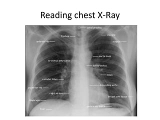
Chest X-rays should be checked for proper positioning and exposure. The lungs should be divided into upper, middle, and lower fields and compared bilaterally. Key areas to examine include the cardiophrenic angles, diaphragm contour, apices, and hilar regions. Vascular markings can indicate cardiac problems if lower lobe vessels are less prominent than upper lobe vessels. Specific findings like Kerley lines or an enlarged carinal angle can suggest conditions such as fluid in the lungs or left atrial enlargement.
4. •check for rt and LT side
•Looks whether is well centralized or not,...check for medial end of Clavicle ...both should be at equi
level from spine..i f not then it difficult to comment about cardiomegaly, medistinal
deviation.. whichever end is more close to spine-----mediastinum is deviated on that
side... 6th ant rib level or 9th post rib level
•look for exposure....if u see four spinal process then its well exposed...if over exposed then it looks
translucent( like emphysema)
•Looks for cardio and costophrenic angle...all should be acute...if nt then its abnormal....also look for
Diaphragmatic contour...if flattened ---then probably some lung disease....Both diaohragm are at
•divide the lungs field into 3 section....Upper: above the anterior end of 2nd rib.....middle: between
2nd anf 4th rib...lower: below 4th rib...compare both lung field...
•check Apical area ...esp behind the Clavicle for TB lesion then check lower lung filed for all 4 angle,,
•check trachea...and trace it upto carina...should be patent
•Cardiac: Let side border is made by Aortic knob, pulm conus, lt atrium and Lt ventricle while rt side
border is made by SVC and rt atrium
•Vascular markings: Lower lobe vessels are more prominent compare to Upper lobe coz of Gravity...If
not then Cardiac problem.. rt side vessels look more prominent than lt side...
Points to be consider
5. • few other points...
• how to say number of thoracic vertebra using chest xray??:--- check
anterior end of 1st rib which is connected to 1st thoracic
vertebera...
• if engorged upper lobe vessels: probably it's puml veins...due to CCF
• Kerly A line: prominent vascular markings in upper lobe
• Kerly B lines: in Lower lobe of lungs on peripheral side ----which
suggest fluid between septa...
• Carinal Ange is around 90...any angle above 90 is abnormal
probably coz of Lt atrial enlargement----( lt atrium lies just below
carina)
• batt wing's appearance: Enlargement of hilar vessels....in Acute
pulm edema..