Vestibular and auditory apparatus-Dr.B.B.Gosai
•
13 gostaram•2,848 visualizações

Functional anatomy of Vestibular and Auditory apparatus presentation by Dr.B.B.Gosai More presentations on https://www.drbbgosai.com/

Recomendados
Mais conteúdo relacionado
Mais procurados
Mais procurados (20)
Destaque
Semelhante a Vestibular and auditory apparatus-Dr.B.B.Gosai
Semelhante a Vestibular and auditory apparatus-Dr.B.B.Gosai (20)
Mais de Dr.B.B. Gosai
Mais de Dr.B.B. Gosai (20)
Último
Último (20)
Vestibular and auditory apparatus-Dr.B.B.Gosai
- 1. 1
- 2. Dr.B.B.Gosai Functional Anatomy of Auditory and Vestibular Apparatus
- 3. What we will learn today? Label the elements of the ossicular chainAND explain the elements can be seen through the tympanic membrane at otoscopy Explain the transformer function of the middle ear and how this can be disturbed? Explain the structure of cochlea Describe the steps involved in the transformation of a fluid movement to nervous activity in the hair cells of the inner ear. Explain the structure of the inner ear Indicate the location of the endolymph and perilymph compartments. Indicate the sensory cells of the vestibular apparatus and mechanism of their stimulation. Explain the detection of linear and rotatory accelerations by vestibular system. Statoconia (Otoconia) Explain the functions of the vestibular system and the sequence of events when vestibular system is stimulated. References: Clinical Oriented Anatomy by Keith Moore : Ch 7 (EAR), pp. 966 to 980
- 4. Parts of Ear: External ear: Pinna, External Acoustic Meatus, tympanic membrane Middle ear: Ear Ossicles (Malleus, Incus, Stapes) Inner ear: Cochlea and vestibular apparatus External and Middle are concerned with the transmission of sound to the inner ear Inner ear converts sound to fluid motion and then to electrical impulses (action potentials)
- 5. External Ear
- 6. External (Outer) Ear •Auricle (pinna): flap of elastic cartilage •External auditory canal •Tympanic membrane (eardrum)
- 7. Auricle (Pinna) Irregular plate of elastic cartilage covered by skin Concha: deepest depression Lobule: important for taking small blood samples and inserting earrings Function: Collection of sound
- 8. External Acoustic Meatus Extends from the pinna to the tympanic membrane: Length: 2-3 cms, Shape: S- shaped Function: Protects the eardrum, Resonator Ceruminous glands secrete cerumen (earwax): trap dust particles
- 9. Tympanic Membrane: Shape: Oval Diameter: 1 cm Attachment:Tympanic sulcus. Parts ofTympanic membrane: The tympanic membrane is divided into 2 parts: Pars flaccida The area of the tympanic membrane superior to the umbo is termed the pars flaccida. It is between anterior and posterior malleolar folds. Pars tensa: the remainder of the tympanic membrane is the pars tensa. The handle of the malleus is firmly attached to the medial tympanic membrane; where the manubrium draws the tympanic membrane medially, a concavity is formed.The apex of this concavity is called the umbo. The tympanic membrane (TM) is thin, semi-transparent membrane that separates the external and middle ear tympanic cavity
- 10. Function:Air vibrations collected by the auricle are transferred to the mobile tympanic membrane, which then transmits the sound to the ossicles. Applied Anatomy: Infection of tympanic membrane is known as Myringitis. During otitis media pus may rupture the membrane and come out through external ear(Perforation).Artificial opening is done in the tympanic membrane to drain the pus from middle ear.This procedure is known as Myrigotomy. Repair of rupture of tympanic membrane is known as Myringoplasty. Myrigotomy.
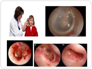
- 11. Otoscopic Examination: Examination of External ear by Otoscope: Handle of Malleus at Umbo Light reflex Parts of membrane
- 12. An annulus fibrosus Lpi long process of incus sometimes visible through a healthy translucent drum Um umbo the end of the malleus handle and the centre of the drum Lr light reflex anteroinferioirly Lp Lateral process of the malleus At Attic also known as pars flaccida Hm handle of the malleus Otoscopic view of External Ear
- 13. Middle Ear
- 14. Middle ear The middle ear located in the petrous portion of the temporal bone and is filled with air secondary to communication with the nasopharynx via the auditory (eustachian) tube. Extent:The tympanic cavity (middle ear) extends from the tympanic membrane to the oval window and contains the bony conduction elements of the malleus, incus, and stapes. Parts: It is divided into two parts: Tympanic cavity proper which is opposite to tympanic membrane and epitympanic recess which is above the level of tympanic membrane. Shape: Biconcave
- 15. Walls of Tympanic Cavity Note: Membranous (lateral) wall formed by Tympanic membrane Medial wall also known as Labyrinthine wall as it separates middle ear from inner ear
- 16. Pharyngotympanic tube (Auditory tube, EustachianTube): connects middle ear cavity with nasopharynx.This tube regularly open by swallowing and yawning Function of auditory tube: equalize pressure in middle ear cavity to atmospheric pressure to allow free movement of tympanic membrane . Contents of Middle Ear cavity: Auditory ossicles (Malleus, Incus, Stapes) Stapedius muscle( prevent excessive movement of stapes) and Tensor tympani muscle (tenses tympanic membrane) ChordaTympani nerve, branch of Facial (VII) cranial nerve Tympanic plexus of nerves
- 17. The ossicular chain: formed by the three smallest bones of the body: the malleus (attached to tympanic membrane), the incus (Middle bone between malleus and stapes), and the stapes (Attached to oval window). The ossicles are held in place by joints, muscles, and ligaments, assist in the transmission of sound. Handle of Malleus is usually visible through tympanic membrane during otoscopy. Ossicular chain amplify the sound 20 X due to leverage.
- 18. Two small fenestrae (oval and round windows), located in the medial wall of the middle ear, separate the middle ear from the inner ear. Fenestra vestibuli: (Oval window): The footplate of the stapes sits in the oval window and the footplate transmits sound to the inner ear (Cochlear organ). Fenestra cochlea: (Round window) covered by a thin membrane, provides an exit for sound vibrations
- 19. Ossicles transmit sound form an air medium to a fluid medium
- 20. Sound transmission through ear
- 21. Quiz Which bone is seen through tympanic membrane in otoscopy attached to Umbo? Oval window is occupied by which bone? Procedure of repair of tympanic perforation is known as:
- 22. Inner Ear
- 24. Inner ear Inner ear contain vestibulocochlear organ for balance and hearing. Bony labyrinth: (cochlea, vestibule and semicircular canals) located in petrous temporal bone and contain perilymph (like CSF) into which membranous labyrinth is suspended. Membranous labyrinth: Vestibular labyrinth: formed by utricle and saccule (for maintenance of balance) Cochlear labyrinth: contain cochlear duct (for hearing) Membranous labyrinth contain endolymph (like ECF).
- 25. Cochlea is shell shaped part of bony labyrinth containing cochlear duct . Spiral canal of cochlea makes 2.5 turns around modiolus. Oval window occupied by Stapes. Round window covered by secondary tympanic membrane Oval window Round window
- 27. Here’s How We Hear Stapes pushes down on perilymph of scala vestibuli. Perilymph pushes vestibular membrane down. Vestibular membrane pushes on endolymph of coclear duct and basilar membrane goes down and up. Secondary tympanic membrane goes out and in. Vestibular membrane separates perilymph of scala vestibuli from endolymph of cochlear duct.
- 28. Here’s How We Hear When hair cells embedded in tectorial membrane vibrate in basilar membrane, this bends stereocilia. Potassium Channels are opened;This depolarizes cells. Action Potential Generated and Neurotransmitters are released. Message is sent to Cochlear Nerve.
- 30. Vestibular Apparatus and Equilibrium Vestibular apparatus maintains the body (mainly the head) at equilibrium (at balance) and stabilizing the eyes relative to the environment Static equilibrium maintenance of the position of the body (mainly the head) relative to the force of gravity Dynamic equilibrium maintenance of the position of the body (mainly the head) in response to sudden movements such as rotation, acceleration, and deceleration. Consists of 2 parts: 1. Utricle and saccule for static equilibrium 2. Semicircular canals for dynamic equilibrium
- 32. Semicircular Canals Anterior, Posterior and Lateral canals Arranged in three different planes Provide information about rotational acceleration. Each canal contains a semicircular duct. At the base is the crista ampullaris enlarged swellings at base of each canal communicating with utricles
- 34. Clinical Aspect Otoscopy: Procedure to examine external ear by straightening external acoustic meatus. In adults by pulling helix up, out and back and in child down and back. Otitis media: infection of middle ear cavity secondary to blockage of auditory tube. Pus collects in the cavity, bulge the tympanic membrane and lead to rupture of tympanic membrane. Pus can be drained by myringotomy
- 35. Clinical Aspect Hearing loss:Tested by tuning fork routinely by Renne’s test andWeber’s test Conductive hearing loss: result from damage to external and middle ear improved by surgery and hearing aid devices Sensorineural hearing loss: result from defects in pathway from cochlea to brain. Can be corrected by cochlear implants Vertigo (Dizziness): due to damage to semicircular ducts. Tinnitus (buzzing or ringing): due to localised damage to cochlear duct. Meniere syndrome: due to blockage of cochlear aqueduct resulting in tinnitus, hearing loss and vertigo.
- 36. Quiz Parts of inner ear? Where is endolymph and perilymph? Vertigo is due to damage to which part of inner ear?
- 37. Summary External ear collects sound and magnified and transformed to inner ear via middle ear ossicular chain. Cochlear organ is responsible for hearing by movements of hair cells. Vestublar apparatus is formed by utricle, saccule and semicircular canals. Function of inner ear is for maintenance of equillibrium. Damage to tympanic membrane (Perforation), Infection of middle ear (Otitis media), Dizziness and hearing defects (Inner ear disorders)