SlideShare uma empresa Scribd logo
1 de 168
Baixar para ler offline
Genetic Dissection of Caulobacter crescentus

                         Surface Colonization


”... It is quite evident that for the most part, water bacteria are not free
          floating organisms, but grow upon submerged surfaces”

                                      Arthur T. Henrics
                                    Journal of Bacteriology
                                       1933, 25: 277-287


           SEM image of Caulobacter crescentus CB15 microcolony grown on borosilicate surface
Table of contents


TABLE OF CONTENTS

Table of contents........................................................................................................... 2

Summary ....................................................................................................................... 6

Overview........................................................................................................................ 8

   What is a biofilm? ............................................................................................................................ 8

   Exopolysaccharides in biofilms...................................................................................................... 10

   Biofilm as a developmental process ............................................................................................... 12

   Structural requirements for biofilm formation ............................................................................... 16

   Regulation of biofilm formation..................................................................................................... 18

   Caulobacter crescentus as a model organism for studying controlled surface attachment and

   biofilm formation............................................................................................................................ 24

   Developmental control of C. crescentus polar appendages............................................................ 25

Aim of thesis................................................................................................................ 29

Chapter 1..................................................................................................................... 30

Abstract ....................................................................................................................... 31

Introduction ................................................................................................................ 33

Materials and Methods .............................................................................................. 36

   Media and Strains ........................................................................................................................... 36

   DNA manipulations........................................................................................................................ 36

   Random Tn5 mutation analysis ...................................................................................................... 37

   Genomic DNA sequencing............................................................................................................. 37

   Construction of deletion mutants.................................................................................................... 37
Table of contents


   Construction of plasmids for chromosomal deletions .................................................................... 38

   Microscopy techniques ................................................................................................................... 40

   Microtiter plate attachment assay ................................................................................................... 40

   Attachment assay with microscopy cover-slides............................................................................ 41

   Holdfast staining and visualization ................................................................................................ 41

   Cellulase and protease assay........................................................................................................... 42

Results.......................................................................................................................... 45

   Isolation and characterization of C. crescentus surface attachment mutants ................................. 45

   Optimal Caulobacter surface attachment correlates with active growth ....................................... 53

   Surface attachment peaks with the coincident exposure of polar organelles ................................. 55

   Optimal surface attachment requires cell differentiation ............................................................... 61

Discussion .................................................................................................................... 64

Acknowledgements..................................................................................................... 70

Chapter 2..................................................................................................................... 71

Abstract ....................................................................................................................... 72

Introduction ................................................................................................................ 73

Materials and methods............................................................................................... 76

   Media and Strains ........................................................................................................................... 76

   Synchronization of Caulobacter crescentus................................................................................... 76

   DNA manipulations........................................................................................................................ 77

   Immunoblots................................................................................................................................... 77

   Construction of chromosomal in-frame deletion mutants and plasmids delivery .......................... 77

                                                                                                                                                   3
Table of contents


   Quantitative reverse transcriptase PCR (QC RT-PCR).................................................................. 79

   Microtiter plate attachment assay ................................................................................................... 80

   Microscopy techniques and image processing ............................................................................... 81

   Overexpression and purification of proteins .................................................................................. 82

   Synthesis and Purification of [33P]cyclic-di-GMP ......................................................................... 83

   DGC (Diguanylate Cyclase) and PDE (Phosphodiesterase) Assays .............................................. 83

Results.......................................................................................................................... 87

   A WecG homolog is required for C. crescentus holdfast formation .............................................. 87

   CC0091 is a c-di-GMP specific phosphodiesterase ....................................................................... 89

   PleD and CC0091 are antagonistic regulators of holdfast biogenesis and surface attachment...... 91

   PleD and CC0091 inversely regulate C. crescentus motility and stalk biogenesis ........................ 95

   CC0095 binds cyclic-di-GMP ........................................................................................................ 99

   Swarmer cell specific expression of CC0091 and C0095 ............................................................ 100

Discussion .................................................................................................................. 102

Acknowledgements................................................................................................... 109

Chapter 3................................................................................................................... 110

Additional results ..................................................................................................... 111

   Biofilm associated C. crescentus cells exhibit increased antibiotic resistance ............................ 111

   Analysis of C. crescentus biofilm maturation in dynamic flow chambers................................... 113

   Identification of Caulobacter crescentus genes specifically expressed during biofilm development

   using recombination-based in vivo expression technology (RIVET) ........................................... 120

Experimental procedures ........................................................................................ 125

                                                                                                                                          4
Table of contents


   Media and Strains ......................................................................................................................... 125

   DNA manipulations...................................................................................................................... 125

   Construction of deletion mutants.................................................................................................. 125

   Construction of plasmids for chromosomal deletions .................................................................. 126

   Construction of plasmids for RIVET analysis.............................................................................. 126

   Biofilm growth for RIVET analysis: ............................................................................................ 127

   Microscopy techniques ................................................................................................................. 128

   Attachment assay with microscopy cover-slides.......................................................................... 128

   Microtiter plate attachment assay ................................................................................................. 128

   Flow chamber experiments........................................................................................................... 129

   Scanning electron microscopy...................................................................................................... 130

Acknowledgement .................................................................................................... 133

Bibliography ............................................................................................................. 134

Addendum................................................................................................................. 147

   Plasmid maps of selected constructs ............................................................................................ 148

   A complete list of strains used in the PhD work .......................................................................... 152

   The complete Tn5 insertion library (surface adherent deficient strains)...................................... 161

   Complete list of plasmids used in the PhD work ......................................................................... 164

Thank –yous.............................................................................................................. 169


                                                                                                                                                 5
Summary


SUMMARY

During its biphasic life cycle Caulobacter crescentus switches from a planktonic to surface attached

life style. This transition requires the continuous remodeling of the cell poles through the temporally

and spatially coordinated assembly and disassembly of polar organelles like the flagellum, pili, and

an adhesive holdfast. A genetic screen for mutants affected in surface binding and colonization led to

the identification of various genes required for motility, pili, and holdfast biogenesis, suggesting a

specific role for all three organelles in C. crescentus surface colonization. Several novel holdfast

genes were identified, which are potentially involved in the synthesis and regulation of the

polysaccharidic component of the holdfast. Quantitative surface binding studies during the C.

crescentus cell cycle revealed that optimal attachment coincides with the presence of flagellum, pili,

and holdfast at the same pole. This indicated that accurate temporal control of polar appendices is

critical for surface colonization of C. crescentus and represents the first example for developmentally

controlled bacterial surface adhesion.

       We have used genetic and biochemical analyzes to demonstrate that di-cyclic guanosine

monophosphate (c-di-GMP) is a central regulatory compound involved in the timing of C. crescentus

pole development. Mutants lacking the diguanylatecyclase PleD show a dramatic delay of holdfast

formation during swarmer cell differentiation. In contrast, cells lacking the GGDEF-EAL composite

protein CC0091 show premature holdfast formation, while overexpression of CC0091 also leads to a

delayed appearance of holdfast. The observation that CC0091 is a c-di-GMP specific

phosphodiesterase indicated that the antagonistic activities of PleD and CC0091 could be responsible

for the correct timing of holdfast formation and flagellum ejection. Finally, our genetic screen

identified a candidate for the c-di-GMP effector protein, which mediates holdfast synthesis in

                                                                                                     6
Summary


response to fluctuating levels of c-di-GMP. The glycosyltransferase CC0095 is strictly required for

holdfast formation and its overexpression leads to premature holdfast synthesis. This and the

observation that CC0095 is able to bind c-di-GMP lead to the hypothesis that holdfast synthesis is

regulated via allosteric control of the CC0095 glycosyltransferase. These data provide the first

example of a developmental process being regulated by the bacterial second messenger, c-di-GMP.


                                                                                                 7
Overview


OVERVIEW

What is a biofilm?

For most of the history of microbiology, microorganisms have primarily been characterized as

planktonic, freely suspended cells and described based on their growth characteristics in nutritionally

rich culture media. However, in the majority of natural environments, bacteria are rarely found in the

planktonic, free-swimming phase. Rather, they are found in association with a biotic or abiotic

surfaces in a structure known as a biofilm (22). It is believed that biofilms are the predominant

microbial lifestyle. Surface association seems to be means for bacteria persisting in biological or

pathogenic microenvironments. For aquatic or soil microorganisms, surface attachment and biofilm

formation may provide an adaptive advantage. For example, high-density communities of attached

bacteria could metabolize insoluble polymeric organic compounds, hemicellulose, or the

exoskeletons of crustaceans and insects. Large negatively charged microbial cell aggregates found in

biofilms may constitute a substratum to concentrate and chelate different limiting nutrients such as

iron. Finally, biofilms are believed to provide protection from toxic compounds, antibiotics, stress

factor and predators (102, 105, 192). It has been speculated that surface attachment and biofilm

formation has evolved as a protective mechanism against grazing protozoan predators (104, 105,

192). The persistence stage of bacterial infections is often associated with biofilm formation, and as a

result of increased resistance to antimicrobial and the scavenging forces of the immune system, is

very difficult to eradicate (39). Persistence of Vibrio cholerae in aquatic environments is thought to

be the main factor for seasonal occurrence of cholera epidemics (105). Biofilm-like colonization of

the lungs of cystic fibrosis (CF) patients by Pseudomonas aeruginosa is considered as the principal


                                                                                                      8
Overview


cause of mortality in CF patients (40). In Yersinia pestis, the biological transmission of plague

depends on blockage of the flea foregut by a biofilm-like cell mass. This blockage is dependent on

the hemin storage (hms) locus. Y. pestis hms mutants, although established long-term infection of the

flea's midgut, failed to colonize the proventriculus. Thus, the hms dependent biofilm formation

affects the course of Y. pestis infection in its insect vector, leading to a change in blood-feeding

behavior and to efficient transmission of plague (28, 63). Another example of biofilm formation role

in pathogenicity comes from Staphylococcus aureus. Recently, Kropec et al. (88) found that in three

mouse models of infection (bacteremia, renal abscess formation, and lethality following high-dose

intraperitoneal infection), using three divergent S. aureus strains, the loss of PNAG by deletion of the

intracellular adhesion (ica) locus had a profound effect on virulence of this microorganism, which

was more susceptible to innate host immune killing (88). Mutant strains showed significantly

reduced abilities to maintain bacterial levels in blood, to spread systemically to the kidneys, or to

induce a moribund/lethal state following intraperitoneal infection (88). Fluckiger et al. (46) have

used a device-related infection model to show that PIA is detectable early in the infection course of

S. epidermidis, and that its production in S. aureus is induced during the course of a device-related

infection. They have shown that PIA production and biofilm formation of both species exist late in

infection, and that the ica genes and biofilm formation are essential for staphylococcal colonization

and endurance on implants (46). Persistence of uropathogenic Escherichia coli as biofilm-like

communities was proposed to be the source for recurrent urinary tract infections (80).

       Biofilm-associated cells can be distinguished from suspended cells by the formation of an

extracellular polymeric substance (EPS) that acts as a matrix for the embedded cells. Biofilm

associated cells often display reduced growth rates and a completely different genetic program


                                                                                                      9
Overview


compared with their planktonic counterpart (reviewed in (38)). Attachment of cells to each other and

to surfaces is a complex process regulated by a diverse range of environmental and possibly host

signals, which are still poorly understood. Attached bacteria may take the form of a dispersed

monolayer of surface-bound cells, they can aggregate on the surface to form microcolonies, or they

may be organized into a well structured three-dimensional biofilm (111).


Exopolysaccharides in biofilms

EPS may account for 50% to 90% of the total organic carbon of biofilms and is considered as the

main matrix material of the biofilm (38). EPS consists of various biopolymers with different

chemical and physical properties; however, it is primarily composed of polysaccharides. Some of

these polysaccharides are polyanionic (174), which allow the association of divalent cations such as

calcium and magnesium that could strengthen the matrix structure by cross linkage. In the case of

some gram-positive bacteria, such as the staphylococci, the chemical composition of EPS may be

quite different and may be primarily cationic (38). EPS is also highly hydrated and thus large

amounts of water can become incorporated into its structure by hydrogen bonding. Sutherland (176)

noted two important properties of EPS for its role in microbial biofilms. First, the chemical

composition and structure of EPS might determine the biofilm conformation (174). For example,

many bacterial EPS possess backbone structures that contain 1,3- or 1,4-β-linked hexose residues and

tend to be relatively rigid and poorly soluble. Second, the EPS of biofilms is not generally

homogeneous but may vary spatially and temporally (174). Leriche et al. exploited the binding

specificity of different lectins to sugars in order to assess the polysaccharide properties during

bacterial biofilm formation of different organisms (98), the results of this study indicated that distinct

organisms produce different amounts of EPS which increases with biofilm development. EPS

                                                                                                       10
Overview


production is affected by the nutrient status of the cells, when an excess in available carbon with

limiting nitrogen, potassium, or phosphate were shown to promote EPS synthesis (174). EPS may

also contribute to the antimicrobial resistance properties of biofilms by impeding the mass transport

of antibiotics through the biofilm, probably by binding directly to these agents (37, 102) .

       In many bacteria, EPS biosynthesis is underlain the regulation of various systems. In V.

cholerae the expression of Vibrio polysaccharide synthesis genes (vps) was shown to be regulated by

VpsR and VpsT, homologous to response regulators of two-component regulatory system (16). The

vps genes expression in this microorganism was shown to be controlled also by absence of the

flagellar structure (190), and also by quorum sensing mediated signals (204). In P. aeruginosa

alginate biosynthesis gene, algC, was shown to be upregulated within 15 minutes following contact

with the surface (29); in addition to alginate synthesis genes, recent studies of the P. aeruginosa

autoaggregative phenotype led to the identification of two genetic loci, psl and pel, that are involved

in the production of two distinct carbohydrate-rich biofilm matrix components. The pel gene cluster

is involved in the production of a glucose-rich matrix material, while the psl gene cluster is involved

in the production of a mannose-rich matrix material (48). Hickman et al. demonstrated that the

expression level of these gene clusters is increased in a wspF mutant, probably due to elevation in the

cellular levels of c-di-GMP which is probably caused by the constitutive activation by

phosphorylation of WspR (62). S. aureus biofilm formation seems to be mediated primarily by the

production of the extracellular polysaccharide PIA/PNAG, which is composed of linear beta-1,6-

linked glucosaminylglycans. The synthesis of PIA/PNAG depends on the expression of the

intercellular adhesion genes icaADBC (55). While most of the S. aureus strains analyzed so far

contain the entire ica gene cluster (23), these genes are only expressed in a few, probably due to the


                                                                                                    11
Overview


regulatory nature and the complex control of these genes. ica genes expression was shown to be

subjected to environmental stimuli such as high osmolarity, anaerobic conditions, high temperature

and certain antibiotics (139). Recent evidence indicates that SarA, a key regulator of S. aureus

virulence factors, is required for the expression of ica genes and the synthesis of PIA/PNAG (183).


Biofilm as a developmental process

The term microbial development was defined as “…changes in form and function that play a

prominent role in the life cycle of the organism…” (119). Recent genetic and molecular approaches

used to study bacterial biofilms, have uncovered various genes and regulatory circuits important for

initial cell-surface interactions and biofilm development. Studies to date suggest that the planktonic-

biofilm transition is, like any other bacterial developmental process, complex and highly regulated.

Biofilm development consists of a series of well-regulated discrete steps: i) reversible attachment, ii)

irreversible attachment, iii) maturation, and iv) dispersion (Figure 1) (155). Reversible attachment

was shown in many organisms to be mediated by flagellar based motility and fimbrial adhesins (64).

Active motility is thought to assist surface binding by helping the cell to overcome the charge barrier

that prevents the negatively charged bacterial cell from reaching certain surfaces. It has been also

postulated that an active flagellar motor could play a part in the regulation switch that upon surface

binding of bacteria, leads to an up-regulation of exopolysaccharide synthesis (96). Irreversible

attachment is mediated mainly by self-made polymeric substances, usually exopolysaccharides,

which not only promote cell-cell and cell-surface contacts, but also construct part of the

encapsulating matrix (33, 108). Flagella-independent motility (gliding or twitching) allows some

bacteria to move on the surface and to form cell-aggregates known as microcolonies (60, 122).

Clonal growth within these microcolonies together with EPS encapsulation results with the

                                                                                                      12
Overview


maturation of a biofilm. Finally, an active dissociation or stream shear forces trigger the dispersal of

sub populations of the biofilm (53, 70, 187). Bacteria within each of these four stages of biofilm

development are physiologically distinct (27). It is obvious that biofilm formation resembles other

adaptive processes of bacterial development like fruiting body formation in Myxococcus xanthus, and

although the molecular and regulatory mechanisms may differ from organism to organism, the stages

of biofilm development seem to be similar in a wide range of microbes (120).


Figure 1) Illustration of the four main stages of biofilm development. Stage i) reversible
attachment of cells to the surface mediated by flagellar motility and adhesive pili. Stage ii)
irreversible cementing of the cells on the surface is a result of EPS production. Stage iii) maturation
of biofilm architecture including water channels and pillars. Stage iv) dispersion of single cells from
the biofilm. This figure was adapted from (173).


      One of the hallmarks of a developmental process is near-complete changes of gene expression

profile of the different stages. In accordance with this, differential stage-specific gene expression has


                                                                                                      13
Overview


been reported during biofilm formation. Genes required for the initial stage of biofilm formation, e.g.

those coding for components of the flagellar motor and adherence pili, are usually repressed in the

mature biofilms of many Gram-negative bacteria simply because although these structures required

during the initial stages of biofilm development, they might destabilize the mature biofilm (154, 156,

195). In contrast, exopolysaccharides synthesis genes, which are critical for the adherence and for the

maintenance of the biofilm structure, exhibit increased expression in biofilm-embedded cells. Thus,

progression from the planktonic to the biofilm state requires a change of the cell’s genetic program.

Several studies have reported on this program change using global analysis of gene expression or

protein synthesis.

      A protein collection of all four stages during P. aeruginosa biofilm formation was established

using 2-D gel electrophoresis (155). On average, consecutive stages differed by 35% of the

detectable proteins. 29% of the protein spots changed upon reversible attachment, and 40% upon

biofilm maturation (155). Escape from biofilm reduced the protein pool by 35% and re-established a

protein profile similar to the one observed for planktonic cells (155). When comparing steady-state

levels of proteins from planktonic and biofilm cells, more than 800 proteins showed a six-fold or

higher change in abundance (155). The identified proteins fall into four main classes: metabolism,

phospholipid and LPS-biosynthesis, protein transport and secretion, as well as adaptation and

protective mechanisms (155). In another study performed with P. aeruginosa, genes responsible for

alginate biosynthesis were shown to be upregulated within 15 minutes after cells adherence to

surfaces, arguing that surface binding might initiate this genetic switch that leads to biofilm

formation (29). A study by Sauer et al. showed that the expression of more than 30 operons was

altered within 6 hours following P. putida surface attachment (154).


                                                                                                    14
Overview


      The comparison of global gene expression profiles of planktonic vs. biofilm cells was

performed with several model organisms. When gene expression in E. coli biofilms grown in a flow

chamber was compared with planktonic cells in stationary or exponential phase, an overall alteration

of more than 600 genes was observed between stationary phase and biofilm cells (156). Only 230

genes were found to be differentially expressed in exponentially growing cells and biofilm cells

(156). Among the genes that showed increased expression in biofilms, several were shown to be

involved in adhesion and autoaggregation. In a parallel study, 38% of a random E. coli lacZ fusion

library showed biofilm-specific expression (137); sessile bacteria showed specific up-regulation of

genes involved in colanic acid biosynthesis (wca locus), while fliC (flagellin) was reduced in

biofilms. Moorthy and Watnick used microarrays to study the transcriptome of V. cholerae during

each stage of biofilm development (113). The transitions from planktonic to monolayer and mature

biofilm identified up to 383 differentially regulated genes. Most of these genes were specific for only

one of the three experimental stages analyzed. These results demonstrated that monolayer and mature

biofilm stages of V. cholerae biofilm development are transcriptionally distinct. A similar analysis

with a clinical isolate of Staphylococcus aureus (UAMS-1) reveled a total of 580 differentially

expressed genes (11). In this study, the largest difference of total numbers of differentially expressed

genes was observed between the biofilms and the exponentially grown planktonic cells (11). Taken

together, these studies make it apparent that biofilms have gene-expression patterns that differ from

those of planktonic bacteria, and telling something about the extensive physiological changes that

occur during biofilm formation. These global gene expression analyzes facilitated the uncovering of

the stage-specific cell physiology and morphology during biofilm development and demonstrated the

complexity of this process.


                                                                                                     15
Overview


Structural requirements for biofilm formation

Initial attachment and microcolony formation

       Pseudomonas aeruginosa- In a pioneering work by O'Toole and Kolter (122), a screen for

the isolation of P. aeruginosa Tn5 insertion mutants defective in the initial steps of biofilm formation

was undertaken, based on the ability of this bacterium to adhere to plastic surface of a microtiter

plate. Two classes of mutants, named sad (surface attachment defective), were described, one class

constitutes flagellar-motility mutants while the other class consists of mutants defective in the

biogenesis of the adhesive type IV pili (122). While a pili mutant was able to form a wild type-like

monolayer of cells on the surface, they were unable to develop into microcolonies. Type-IV pili are

required for twitching motility, a mode of surface locomotion used by P. aeruginosa and other

bacteria in which the polar pili are believed to extend and retract, and thereby propelling bacteria

across a surface. Thus, the findings by O’Toole and Kolter (122) suggested that surface based

motility is required for the second step of biofilm formation.

       Vibrio cholerae- In a similar genetic analysis performed by Watnick and Kolter (189), three

classes of V. cholerae El Tor sad mutants were described (189). The first class of genes is required

for the biosynthesis of the mannose-sensitive haemagglutinin type-IV pilus (MSHA); the second

group of mutants was defective in flagellar motility, including both mutants lacking flagella and

mutants with paralyzed flagella. The third group of sad mutants had transposon insertions in vps

genes. The phenotypes of these mutant classes suggested that pili and flagella accelerate attachment

to and mediate the spread along the abiotic surface, while exopolysaccharide synthesis by the vps

genes is required for the formation of the three-dimensional biofilm architecture. In contrast to


                                                                                                     16
Overview


mutants lacking pili and flagella, EPS mutants were unable to form a detectable biofilm even after

extended incubation time (189).

       E. coli- A study by Pratt and Kolter reveled three classes of attachment-deficient E. coli Tn10

mutants (134). The mutations isolated included flagellar biogenesis and motor function genes, and

genes which were involved in the biogenesis and regulation of type-I pili (134). Interestingly, in a

strain overproducing curli background, flagella were dispensable for initial adhesion and biofilm

development (136), arguing that at least part of the role of flagella in surface colonization might be of

a regulatory nature.


Biofilm maturation

P. aeruginosa- Klausen et al. have shown that flagella and type-IV pili take part in shaping the

architecture of P. aeruginosa biofilms, although they are not essential for biofilm formation (86). The

model which they proposed suggests that the formation of mushroom-shaped structures in P.

aeruginosa biofilms is caused by bacteria which climb on the top of the microcolony stalks using of

type-IV pili mediated twitching motility (85); according to this model, type-IV pili driven bacterial

migration plays a key role in structure formation in the late phase of biofilm development.

       V. cholera- V. cholerae strain which is defective in EPS synthesis fails to form a mature

biofilm architecture (188, 201). Moorthy and Watnick have recently shown that the cell monolayers

formed on surfaces represent a distinct stage of this microorganism biofilm development (111). They

have demonstrated that while MSHA pilus is only required for the monolayer formation, vps is

required for formation and maintenance of the mature biofilm and that the maturation of these


                                                                                                      17
Overview


monolayers to three-dimensional biofilm structure, requires monosaccharides such as mannose,

which induce the expression of vps genes (111).

      E.coli- Molin and co-workers have shown recently that the maturation of E. coli K12 biofilms

requires the presence of an incF plasmids (144). They have demonstrated that while surface

attachment, clonal growth and microcolony formation were not affected in the plasmid plasmid-free

strains, the efficient biofilm maturation could only occurred in strains carrying the conjugation pilus

proficient plasmid (144) and that E. coli strains lacking these plasmids were not able to form the

elaborated three-dimensional biofilm architecture that include pillars and channels (144). They have

shown that the final shape of the mature biofilm seemed to be determined by the pilus configuration,

when various mutants affected in the processing or in the activity of these transfer pili, displayed

differently structured biofilms. In addition to that, flagella, type 1 fimbriae, curli and cell-to-cell

signalling did not seem to be required for biofilm maturation in E. coli K12 carrying the incF

plasmids (144). This work was with a complete agreement with a previous work published by Ghigo

(52), which has demonstrated the involvement of conjugative plasmids in the competence of the

bacterial host to form a biofilm (52) .


Regulation of biofilm formation

Complex regulatory pathways such as the global carbon metabolism regulator (CRC) (121) and

stationary-phase sigma factors (σs) (60, 196) have been shown to play an important role in biofilm

development despite the fact that these systems are not exclusively committed for biofilm

development. High-cell density, high osmolarity, scarce nutrients as well as oxygen limitation are

only some of the situation which a biofilm embedded cell and a stationary phase cell might

encounter; this similarity could explain some of the convergent regulation circuits that control

                                                                                                    18
Overview


biofilm formation in addition to stationary phase and stress respond. Besides being subjected to

global metabolic control, biofilm components underlie specific regulation at the transcriptional and

post-transcriptional level. For example, the Salmonella typhimurium CsgD, a transcriptional regulator

of the LuxR superfamily, has been shown to positively control the expression of cellulose and curli

fimbriae (14). The expression of csgD itself is modulated by a variety of stimuli, including,

osmolarity, oxygen, nutrient availability, pH, temperature, and the subject of control by many

cellular factors, such as RpoS, RpoD, IHF and others (14, 135). In P. aeruginosa GacS/GacA

proteins of the two-component signal transduction system which controls the production of many

secondary metabolites and extracellular enzymes and involved in pathogenicity in plants and animals

(58), were shown to also control biofilm formation when gacA mutant failed to aggregate and form

microcolonies (127). Although the signals that activate the GacS/GacA circuit are not known, it was

demonstrated that the gac genes are activated during the transition from exponential to stationary

phase of the growth (58); and since the expression of rpoS is positively regulated by GacS/GacA,

some of the GacS/GacA-dependent phenotypes may be related to RpoS activity (116, 194). In

addition to the GacA/GacS, a three-component regulatory system specifically required for biofilm

maturation was identified (89). This system is comprised of genes sadARS coding for a putative

sensor histidine kinase and two response regulators; mutations in any of these genes, blocked biofilm

maturation of P. aeruginosa without affecting growth, early biofilm formation, swimming, or

twitching motility (89). The expression of sadR and sadS is very similar in planktonic and biofilm

cells, while sadA expression is slightly decreased ( 2-fold) in biofilm cells. The authors have

postulated that the SadARS system acts as a regulator of both biofilm formation and for genes


                                                                                                  19
Overview


involved in type III secretion (TTSS) and it may function to promote biofilm formation, possibly in

part by repressing the expression of the TTSS (89).

       In addition to the species-specific control mechanisms, biofilm formation is also regulated by

two global signal transduction networks. The first, quorum sensing (QS) allows transmitting

information between cells and has been shown to regulate cellular processes in response to cell

density or crowdedness (128). Since biofilms comprise arrays of dense microbial populations, it was

not surprising to find that QS influences biofilm related processes. Davies et al. (30) showed that P.

aeruginosa PAO1 requires the lasI gene product 3OC12-HSL in order to develop a normal biofilm;

lasI mutant formed flat, undifferentiated biofilms which remain sensitive to SDS (30); interestingly,

mutant biofilms appeared normal when supplemented extracellularly with a synthetic 3OC12 signal

molecule (30). Similarly, Burkholderia cepacia mutants defective in the cep quorum sensing system

were able to form microcolonies on a glass surface, but were unable to develop into a mature biofilm

(68). In E. coli, biofilm formation was shown to be stimulated by the auto-inducer 2 signal (AI-2)

(200). It was suggested that AI-2 stimulates biofilm formation through a regulatory cascade including

novel motility quorum sensing regulator, MqsR, the two component system QseBC which then

promotes cell motility via the master regulon flhDC, stimulating MotA and FliA and leads to biofilm

formation (200). QS-dependent biofilm formation regulation in E. coli was demonstrated also by the

deletion of ydgG (a putative transport protein that either enhances AI-2 secretion or inhibits AI-2

uptake) which increased the intracellular concentration of AI-2 as turn resulted in a 7,000-fold

increase in biofilm thickness and 574-fold increase in biomass in flow cells (59). In contrast, in V.

cholerae, a reciprocal relationship between quorum sensing and biofilm formation was described

(204). V. cholerae strains lacking HapR, a LuxR homolog, forms thicker biofilms; microarray


                                                                                                   20
Overview


analyses of biofilm-associated bacteria showed that the expression of the V. cholerae vps genes is

increased in hapR mutants when CqsA, one of two known autoinducer synthases in V. cholerae, acts

through HapR to repress vps gene expression (204).

       The second global regulator controlling cell adhesiveness and biofilm formation is cyclic

di(3' 5')-guanylic acid (c-di-GMP). C-di-GMP is emerging as a global second messenger in bacteria

controlling “social behavior.” As described above, cell surface appendages mediate bacterial

aggregation and facilitate biofilm formation; flagella and pili which are involved in biofilm formation

were shown to be regulatory targets of c-di-GMP (reviewed in (25, 75, 147)). Genetic studies have

implicated c-di-GMP in the regulation of motility, the production of extracellular polysaccharide,

biofilm establishment and maintenance as well as host persistence in a wide range of bacteria (75,

147). Biochemical studies have reveled that cellular levels of cyclic-di-GMP are inversely controlled

by the activity of diguanylatecyclases (GGDEF domain) and phosphodiesterases (EAL domain)

(Figure 2) (19, 129, 152, 157, 182). GGDEF and EAL domain proteins are abundant and found in

most bacteria, covering all branches of the phylogenetic tree (147). C-di-GMP was first described as

an allosteric activator of the enzyme cellulose synthase of the bacterium Gluconacetobacter xylinum

(150). In Caulobacter crescentus, c-di-GMP was shown to orchestrate the controlled transition of a

flagellated into a “sticky” cell pole which secretes an unknown form of polysaccharide (5, 129). The

production of cellulose or derivatives thereof, is activated by GGDEF domain proteins in several

other bacteria including E. coli, S. enterica, Rhizobium leguminosarum and P. fluorescence (8, 169,

207), in addition to polysaccharides, the biosynthesis of adhesive fimbriae, another component of

extracellular matrix also depends on the activity of GGDEF domain proteins (24, 160). In the current

working model, high levels of c-di-GMP favor the production of adhesive organelles and blocks


                                                                                                    21
Overview


different forms of cell motility (161). Hickman et al. (62) have recently shown that an increase in

cellular levels of c-di-GMP elicited by a specific diguanylatecyclase, WspR, results in higher

expression of the pel and psl EPS gene clusters of P. aeruginosa and led to the formation of mature

biofilms (62). Similarly, vps expression in V. cholerae is controlled by c-di-GMP (182). When the

enzymatic activity of the VieA phosphodiesterase is required to repress EPS production under non-

biofilm conditions (182). The deletion of vieA results in increased cell attachment, probably as a

consequence of up-regulation of VpsR, a positive regulator of vps gene expression (182). Signature-

tagged transposon mutagenesis in Salmonella have led to the identification of CdgR, an EAL domain

protein which its mutagenesis resulted in lower resistance to hydrogen peroxide and accelerated

killing of macrophages in mice model (65). Hoffman et al. have shown that alterations in the

intracellular levels of c-di-GMP caused by the addition of sub inhibitory concentrations of the

antibiotic tobramycin, induced a specific, defensive reaction in both in E. coli and P. aeruginosa

(66). Tobramycin induces the expression of arr phosphodiesterase which results in reduced levels of

c-di-GMP, increased biofilm formation and increased resistance to tobramycin (66). These studies

implicate a complex relationship between c-di-GMP intracellular levels and regulation of biofilm

formation.


                                                                                                22
Overview


Figure 2) The conversion of GTP into c-di-GMP is catalyzed by the diguanylatecyclases, which
reside in the GGDEF domain. Increased intracellular levels of c-di-GMP promote biofilm
formation and the biosynthesis of adhesive organelles and inhibit different types of cell motility
(reviewed in (161) . Degradation of c-di-GMP is catalyzed by the activity of EAL domain of
phosphodiesterases. The illustration was taken from a poster (“Biochemical and genetic identification
of a c-di-GMP binding motif”) presented by Beat and Mathias Christen and Marc Folcher).


                                                                                                  23
Overview


Caulobacter crescentus as a model organism for studying controlled surface

attachment and biofilm formation

The genus Caulobacter consists of a collection of Gram-negative, hetero-oligotrophic aerobe, rod-

like shaped cells that are equipped with a single polar flagellum and polar pili. Caulobacter

crescentus possesses a stalk, a thin cylindrical extension of the cell containing cell wall and

cytoplasm, with an adhesive material, the holdfast, located at its tip. The holdfast mediates strong

irreversible attachment of Caulobacter cells to solid substrates (109). Caulobacter are generally

found in aquatic environments, where they attach to biotic and abiotic surfaces (132, 203) and

participate in biofouling processes (203). The unique life cycle of C. crescentus with its asymmetric

cell division and obligatory cell differentiation has made it one of the preferred model organisms to

study microbial development and the mechanisms underlying bacterial cell cycle control (151). The

dimorphism is established by an asymmetric cell division that gives rise to two genetically identical,

but morphologically and physiologically distinct daughter cells with different developmental

programs: a sessile stalked cell equipped with an adhesive holdfast and a motile swarmer cell bearing

a single flagellum and adhesive pili (15). The stalked cell is competent to start a new replicative

cycle immediately after cell division, whilst the swarmer cell is engaged in chemotaxis while the

replicative program is being blocked. Before the swarmer cell re-enters replication and cell division it

differentiates into a stalked cell, a process during which it loses the flagellum, retracts its pili, and

forms a holdfast and a stalk at the pole previously occupied by the flagellar motor. Dimorphism is

believed to have evolved to allow Caulobacter to cope with life in dilute, nutrient-poor environments

(69). The swarmer cell stage allows rapid dispersal and the scavenging of new nutrients resources,

while the surface adherent form permits growth where nutrients are available.

                                                                                                      24
Overview


The nature of C. crescentus cell poles is constantly changing during its development (Figure 3). Pole

differentiation is regulated by a complex regulatory network which includes several members of two-

component signal transduction proteins (2, 71, 74, 124). Some of these regulators interlink cell-cycle

progression and pole development. E.g., The response regulator CtrA directly controls the initiation

of chromosome replication as well as several aspects of polar morphogenesis and cell division (42).

The intrinsic asymmetry and microscopically visible appendages make it possible to monitor cell

cycle progression and pole differentiation and allow the analysis of temporal and spatial control of

polar organelles like flagellum, pili, holdfast, and stalk.


Developmental control of C. crescentus polar appendages

The synthesis of C. crescentus flagellum requires about 50 different genes. Flagellar gene expression

underlies cell cycle control with the temporal activation of CtrA (36, 143). In addition flagellar gene

transcription is controlled by hierarchical regulatory system in which the expression and productive

assemblage of gene products are required for the expression of gene products which participate

successively in the multistep flagellar assembly (117, 140). This regulatory cascade consists of four

hierarchical classes. The cascade initiates with class I genes, namely CtrA, which promotes the

transcription of the class II genes encoding the MS ring of the basal body, the flagellar switch, and

the flagellum-specific type III secretion system (36, 138). The transcription of the flagellar class III

and IV is dependent on the proper assembly of the class II components (115). In addition, the

expression of class III and IV flagellar genes requires σ54 and the transcriptional activator FlbD,

which in addition to being subjected to cell cycle-regulated phosphorylation (197), FlbD activity is

also subjected to the hierarchical regulation system (114). The ejection of the flagellum during the

swarmer-to-stalked cell transition coincides with the degradation of the FliF flagellar anchor. The

                                                                                                     25
Overview


activity of the diguanylatecyclase response regulator PleD was shown to be required for efficient

removal of FliF, ejection of the flagellum, and stalk biogenesis (3). PleD activity is regulated through

cell-cycle dependent phosphorylation by PleC and DivJ kinases (5, 129).

These elaborate regulatory mechanisms ensure the linking of flagella assembly and disassembly to

the cell cycle and to the development of C. crescentus.

       Pili are extracellular filaments, found in a wide variety of bacteria. Pili were shown to play a

major role in adhesion of bacteria to surfaces, biofilm formation, conjugation, twitching motility, and

host infection (164). Caulobacter crescentus pili are extracellular surface appendages, 1–4 µm in

length and 4 nm in diameter and are located exclusively at the flagellated pole (164). The pili

composed of polymerized pilin subunit (PilA) which is assembled by proteins encoded by a cluster of

pilus assembly genes (cpaA-F) that are closely related to the tight-adherence genes (TAD) from

Actinobacillus actinomycetemcomitans (81, 130). The transcription of cpaB–F is induced in the late

predivisional cell, followed by cpaA and, finally the CtrA-dependent transcription of pilA with peak

of expression in the progeny swarmer cells (95). The timing of pilus assembly can be shifted from the

swarmer cell to the predivisional cell stage by expressing pilA from a constitutive promoter,

suggesting that the temporal transcription is the main type of regulation that prevents premature

assembly of the pili (164). It was demonstrated that the PleC histidine kinase, which is localized to

the piliated pole during the pilus assembly time window, controls the accumulation of PilA (185).

PleC was shown to be responsible for the asymmetric distribution of CpaC (a putative outer

membrane pilus secretion channel) and its assembly factor, CpaE (185).


                                                                                                     26
Overview


The adhesive holdfast is located at the tip of the stalk at the pole previously occupied with the

flagellum. The exact biochemical composition of the holdfast is unknown, however, lectin binding

and glycolytic enzymes sensitivity experiments suggest that the holdfast is composed of

polysaccharides containing N-acetylglucosamine (GlcNAc) oligomers (109). Janakiraman and Brun

used an hfaA-lacZ fusion to show that the transcription of hfaA (part of the hfaA-D gene cluster

which required for holdfast attachment to the cell envelope (21, 92)) is temporally regulated during

the cell cycle. hfaA exhibit maximal transcription levels in predivisional cells (72). The authors

however, have failed to observe the holdfast before differentiation of the swarmer cell had occurred.

How the spatial and temporal regulation of holdfast expression is achieved is still unclear.


                                                                                                    27
Overview


Figure 2) A schematic representation of the Caulobacter crescentus cell cycle. The replication-
incompetent swarmer cell is equipped with a polar flagellum and flp-like pili. After a defined period,
the swarmer cell differentiates into a stalked cell in successive of developmental steps, including the
ejection of the flagellum, the retraction of the pili, the synthesis of the holdfast, and the elongation of
the stalk. Chromosome replication initiation coincides with the formation of the stalked cell. The
timing of several morphogenetic and cell cycle events is shown by the light and dark grey bars
respectively. The flagellated, stalked (ST) and new swarmer (SW) poles are indicated. The relative
duration of each phase is indicated on top as horizontal axis. This figure was adapted from (71)


                                                                                                        28
Aim of thesis


AIM OF THESIS

The aim of this work was to genetically identify components involved in C. crescentus surface

binding and colonization. New structural and regulatory components of C. crescentus pole

development and surface adhesion should be analyzed with respect to their function, their temporal

and spatial coordination, and the specific molecular mechanisms facilitating surface colonization.


                                                                                                     29
Chapter 1


CHAPTER 1

 The coincident exposure of polar organelles optimizes surface attachment during

                         Caulobacter crescentus development


                                 Assaf Levi and Urs Jenal*


            Division of Molecular Microbiology, Biozentrum, University of Basel

                     Klingelbergstrasse 70, CH-4056 Basel, Switzerland


                   In revision of publication in Journal of microbiology

                       Running title: Caulobacter surface attachment

              Keywords: Caulobacter, flagella, pili, holdfast, biofilm, c-di-GMP


 * For Correspondence: Division of Molecular Microbiology, Biozentrum, University of Basel,

                     Klingelbergstrasse 70, CH-4056 Basel, Switzerland


         Tel: +41-61-267-2135; Fax: +41-61-267-2118; e-mail: urs.jenal@unibas.ch


                                                                                              30
Chapter 1


ABSTRACT

During its biphasic life cycle, Caulobacter crescentus oscillates between a planktonic and a surface

attached life style. A hallmark of this transition is the temporally and spatially regulated assembly

and disassembly of polar organelles like flagellum, pili, and an adhesive holdfast. A genetic screen

for mutants affected in surface binding and colonization revealed a large number of known and novel

components of flagellar motility, pili formation, and holdfast biogenesis, arguing that these

organelles are required for optimal surface adhesion of C. crescentus. Several new holdfast genes

were identified, which are potentially involved in the formation and polymerization of

polysaccharide precursors. Together with experiments that implicate a cellulose-like polymer as a

main constituent of holdfast structure and function, this provides the basis for future analyses on the

formation and exact composition of this adhesive organelle. Several lines of evidence suggested that

the coincident exposure of polar organelles optimizes surface attachment during Caulobacter

crescentus development. i) The holdfast is synthesized and exposed on the cell surface very early

during the swarmer-to-stalked cell transition and, during a defined time window, coincides with an

active flagellum and adhesive pili at the same pole. ii) Cell cycle-dependent surface attachment

showed a prominent peak coinciding with the surface exposure of all three polar organelles, and

mutants lacking any one of these subcellular structures exhibited basal levels of attachment. iii)

Active growth, as well as passage through development, greatly enhanced surface colonization. iv) A

delay of holdfast biogenesis observed in a pleD mutant resulted in a strong reduction of surface

binding during development. In cells lacking PleD, a developmentally controlled diguanylate

cyclase, holdfast biogenesis was delayed by almost one third of a cell cycle equivalent, indicating

that PleD and its readout signal, c-di-GMP, are used as timing device for holdfast formation. Based

                                                                                                    31
Chapter 1


on these results we propose a model for C. crescentus surface colonization that involves the

successive and concerted activity of flagella, pili, and holdfast. The model provides a rational

framework for the precise temporal and spatial control of these cellular appendices during

development.


                                                                                             32
Chapter 1


INTRODUCTION

In most natural environments, microbial cells are found attached to surfaces and associated in

communal structures known as biofilms. The formation of biofilms from single planktonic cells,

widely studied in a few model organism (Vibrio cholerae, Pseudomonas aeruginosa, Salmonella

typhimurium, and E. coli), involves several discrete stages, including reversible and irreversible

attachment to surfaces, formation of cell monolayers, microcolony formation, and biofilm maturation

(112, 119, 155, 189). This process is associated with a dramatic change of the cells’ genetic program

and physiology (154-156, 170, 193, 195). Initial stages of surface colonization are facilitated by

cellular appendages like flagella and pili that can mediate initial attachment and accelerate biofilm

development (9, 35, 81, 83, 134, 155). Later stages of biofilm formation are associated with the

formation of an extracellular matrix, which mediates surface anchoring and provides structural

support for the cell community (175). While all major classes of macromolecules can be present in

biofilm matrices, increased synthesis of exopolysaccharide (EPS) is generally associated with biofilm

formation (26, 47, 48).

       The contribution of flagella and pili to various stages of biofilm formation have been

demonstrated independently for several bacteria but it remains to be shown whether flagella, pili and

EPS are part of a coordinated program for surface attachment and colonization rather than

contributing to biofilm formation in a stochastic and independent manner. If these distinct organelles

and mechanisms are indeed interlinked and are part of a program dedicated to surface colonization,

how would these interactions be regulated in time and maybe space? How would cell motility and

adhesive properties be coordinated to optimize surface attachment early during biofilm formation and

to ensure the escape or detachment of cells from biofilms at a later stage? One possibility is that

                                                                                                   33
Chapter 1


different components of this multicellular behavior are co-regulated (148). A number of

environmental signals, including nutrients, temperature, osmolarity, pH, iron, and oxygen influence

biofilm formation (reviewed in: (119)), but little is known about mechanisms that integrate these

inputs and transduce them into an altered bacterial behavior required for surfaces colonization.

       During Caulobacter crescentus development surface adhesion is coupled to cell growth and

division. Each cell division is intrinsically asymmetric and generates a sessile, replicative stalked cell

and a motile, flagellated swarmer cell. A single flagellum is assembled in the predivisional cell at one

pole and is activated prior to cell division (198). Upon separation of the two daughter cells, pili are

formed at the flagellated pole of the swarmer cell (164, 167). The newborn swarmer cell performs

chemotaxis for a defined period (6) before it sheds the flagellum, loses its pili and differentiates into

a stalked cell. During this process, an adhesive holdfast structure and a stalk are assembled at the

pole previously occupied by pili and flagellum. The exact role of the polar pili in C. crescentus and

its temporal and spatial control are unknown but it has been proposed that they might facilitate

surface interaction and cell attachment (12, 167). Irreversible anchoring of C. crescentus cells to

surfaces requires an intact holdfast structure (125). Genetic screens have identified several genes

required for holdfast secretion and anchoring (21, 166). While some of these genes encode homologs

of polysaccharide export components in other gram-negative bacteria, the exact structure and

composition of the holdfast remains unclear (166). Staining and lectin binding experiments had

proposed that it is composed of an acidic polysaccharide, which contains N-acetylglucosamine

(GlcNAc) residues (109, 166, 184).

    The observation that in C. crescentus swarmer cells are able to attach to surfaces (12, 131, 132)

suggested that the model for surface attachment as being mediated by stochastic and independent


                                                                                                       34
Chapter 1


adhesion events might be too simplistic and has indicated that all polar organelles might contribute to

this process in a concerted manner. Here we show that in a static system, flagella, pili, and holdfast

substantially contribute to C. crescentus surface attachment. Using a new method to detect holdfast

we could demonstrate for the first time that holdfast biogenesis occurs much earlier in development

than reported previously. Consequently, all three polar organelles are concomitantly exposed at the

same cell pole during a defined time window of swarmer cell differentiation. This developmental

stage coincides with a sharp peak of surface binding activity during the C. crescentus life cycle. This

attachment peak was reduced or eliminated in mutants lacking pili, flagellum, or holdfast. Moreover,

in a mutant that shows delayed holdfast synthesis during development, attachment is dramatically

reduced. Together with the observation that optimal surface binding is coupled to growth and cell

differentiation this lead us to propose a model for C. crescentus attachment in which rapid surface

binding is optimized by the careful temporal and spatial coordination of all three organelles during

development.


                                                                                                    35
Chapter 1


MATERIALS AND METHODS

Media and Strains

Strains and plasmids used in this study are listed in Table 1. E. coli DH10B and S17-1 were used as

host strain for molecular cloning experiments and as donor strain for conjugational transfer of

plasmids into Caulobacter. E. coli strains were grown at 37°C in Luria-Bertani (LB) broth (153)

supplemented with kanamycin (50 µg/ml) or tetracycline (12.5 µg/ml), when necessary. C.

crescentus strains were grown at 30°C in either PYE complex medium (131) or in M2 minimal

glucose medium (M2G) (78) supplemented with kanamycin (5 µg/ml), tetracycline (2.5 µg/ml),

chloramphenicol (1 µg/ml) or nalidixic acid (20 µg/ml) when necessary. Semisolid agar plates for

motility assays contained 0.3% agar (DIFCO®).

      Synchronization of C. crescentus was done as described earlier (171). Isolated swarmer cells

were released into fresh minimal medium at an OD660 of 0.3. Samples were removed for microscopic

analysis, attachment assays, and holdfast staining at 15 minutes intervals. For surface binding assays,

cells were allowed to attach to polystyrene in microtiter plates for 15 minutes. Cell cycle progression

was monitored by light microscopy.


DNA manipulations

Plasmid and chromosomal DNA preparation, DNA ligation, electroporation, agarose gel

electrophoresis, and PCR amplifications were carried out by using standard techniques (153). All

PCR products used for cloning were amplified with “Expand high-fidelity PCR system®” form

Roche. Restriction enzymes were from New England Biolabs, Inc.


                                                                                                    36
Chapter 1


Random Tn5 mutation analysis

The mini-Tn5 transposon delivery vector pUT_Km2 (32) was inserted into C. crescentus wild type

by conjugation. Approximately 2,000 colonies were grown in 96-well plates in 200 µl of PYE

medium supplemented with kanamycin. Cells were discarded and the microtiter plates were washed

under a gentle stream of distilled water. The plates let to air-dry and surface attached cells were

quantified as described below. Mutants with a reproducible and significant increase or decrease

(>25%) of attachment were selected for further analysis.


Genomic DNA sequencing

Genomic DNA was extracted from C. crescentus wild type or mutant strains using ethanol

precipitation following lysis of cells in 5M Guanidinium thiocyanate, 0.1M EDTA. Tn5 insertion

sites were mapped by direct sequencing of chromosomal DNA using oligonucleotide primers

complementary to the ends of the kanamycin resistance cassette of Tn903: primer #698 (TCT AGA

GTC GAC CTG CAG GC) and #699 (TAC CGA GCT CGA ATT CGG CC). Sequencing reactions

were set up using ~500 ng genomic DNA as template and 10 pmol of sequencing primer in a total

reaction volume of 20 µl following the BigDye Terminator protocol with few modifications:
                                        o
Annealing temperature was raised to 58 C and the number of cycles was increased to 99 (Big Dye;

Perkin-Elmer®). The sequencing reactions were run on an ABI Prism 310 DNA Sequencer or ABI

Prism 3100 Avant Genetic Analyzer (Perkin-Elmer®).


Construction of deletion mutants

In frame deletions of the chromosomal copies of flgH, fliFG, flgDE, flgFG, fliL, CC2277 and

CC0095 were constructed in C. crescentus wild type strain and in UJ590 (∆pilA) using pNPTS138

                                                                                                37
Chapter 1


based constructs carrying in frame deletions in the respective genes. Plasmids (see below) were

introduced into the recipient strains by conjugation and recombinants were selected on PYE plates

supplemented with kanamycin and nalidixic acid. Resulting single colonies were then grown

overnight in liquid PYE medium and plated on PYE containing 3% sucrose. Sucrose-resistant

colonies were screened by PCR for recombinants that had lost the chromosomal copy of the

respective gene.


Construction of plasmids for chromosomal deletions

Plasmid pAL8 was constructed for an in-frame deletion of the complete flgFG coding region. PCR

amplification of a 1.0 kb region upstream of flgFG was done with primers #655 and #656 (5’-GGA

TCC GGC GTT CGA GCT GCT GCT GA-3’ and 5’-GAA TTC TCA CCT GGC GGG TGA GTG

AG-3’). PCR amplification the 1.0 kb region downstream of flgFG was done with primers #657 and

#658 (5’-GAA TTC CGC TCG CCT AAG CGA ACG TC-3’ and 5’-ACT AGT GGC CGA GAT

CTT GCC GTC GA-3’). Ligation of both fragments into pNPTS138 (SphI/SpeI) resulted in plasmid

pAL8.

        Plasmid pAL2 was constructed for an in-frame deletion of the complete flgH coding region.

PCR amplification of a 1.0 kb region upstream of flgH was done with primers #637 and #638 (5’-

GGA CTA GTC CCG GCG ACA ACC TGT TCC TGG-3’ and 5’-CGG GAT CCC GGA CGA

CGC ATG ATC TGG TCC-3’). PCR amplification a 1.0 kb region downstream of flgH was done

with primers #639 and #640 (5’- CGG GAT CCC GGG TCG AGA AGT TCT CGC CCT-3’ and 5’-

CGG AAT TCC GGG AGC GCA TTC GAC GTC TGG-3’). Ligation of both fragments into

pNPTS138 (SpeI/EcoRI) resulted in plasmid pAL2.


                                                                                              38
Chapter 1


Plasmid pAL6 was constructed in order to create an in-frame deletion of the complete fliL coding

region. PCR amplification of a 1.0 kb region upstream the fliL was done with primers #710 and #711

(5’-ACT AGT CCA TCA TCT TGG CGA CGC-3’ and 5’- GAA TTC CGT ACT CAT GCG CGA

AGC -3’). PCR amplification a 1.0 kb region downstream of fliL was done with primers #712 and

#713 (5’- GAA TTC GCG AAC GAT CAT GGC GGA-3’ and 5’-GCA TGC ACC TGC ATG TTC

AGC ACG-3’). Ligation of both fragments into pNPTS138 (SpeI/SphI) resulted in plasmid pAL6.

       Plasmid pAL7 was constructed in order to create an in-frame deletion of the complete flgDE

coding region. PCR amplification of a 1.0 kb region upstream the flgDE coding sequences was done

with primers #563 and #564 (5’- GCA TGC GAC CGC CGC GAC CGC CCC GC-3’ and 5’- GAA

TTC CCG GTC AAG GAC CGA GGC GG -3’). PCR amplification a 1.0 kb region downstream of

fliL was done with primers #565 and #566 (5’- GAA TTC TCG AAG ATC ATC ACG ACC GC-3’

and 5’- CTA GTT GGC GAC CTT GTC GCG CGG C-3’). Ligation of both fragments into

pNPTS138 (SpeI/SphI) resulted in plasmid pAL7.

An in-frame deletion of the chromosomal copies of fliF and fliG genes was generated in wild-type

strain CB15 using plasmid pBG22 (56).

       Plasmid pAL21 was constructed in order to create an in-frame deletion of the complete

CC2277 coding region. PCR amplification of a 1.0 kb region upstream the CC2277 was done with

primers #625 and #626 (5’-cgg aat tcC GGG CTT CCT GCC GTT CCA CCG-3’ and 5’- gct cta gaG

CGC GCC ATC AGG CCT CCG TGT-3’). PCR amplification a 1.0 kb region downstream of

CC2277 was done with primers #627 and #628 (5’- gct cta gaG CCG CGT GAG CGT GAT CAC

CGA-3’ and 5’-ggg gta ccC CCC AGG CCA AAG GTG ACC GGC-3’). Ligation of both fragments

into pNPTS138 (EcoRI/KpnI) resulted in plasmid pAL21.


                                                                                               39
Chapter 1


Plasmid pDM25 was constructed in order to create an in-frame deletion of the complete CC0095

coding region. PCR amplification of a 550 bp region upstream of CC0095 was done with primers

#1380 and #1381 (5’- GAA TTC TTC GAC CGT TCC CAG CCC-3’ and 5’- GGA TCC CGC TGT

CCA GAC GCT CTA-3’). PCR amplification a 550 bp region downstream of CC0095 was done

with primers #1382 and #1383 (5’- GGA TCC TGA GGA ACG AAC ATC TCC GCA G-3’ and 5’-

AAG CTT CGA CAA GGA CGG CCA GAA GGA-3’). Ligation of both fragments into pNPTS138

(EcoRI/HindIII) resulted in plasmid pDM25.


Microscopy techniques

Cell morphology, motility, and rosette formation were analyzed by light microscopy using a Nikon

Eclipse 6000 with a planApo 100x phase contrast objective or an Olympus AX70 with an UplanApo

100x phase contrast objective. Pictures were taken with a charge-coupled device camera

(Hamamatsu®) connected to the Olympus microscope and analyzed with Open-lab (Improvision®)

software.


Microtiter plate attachment assay

For C. crescentus surface attachment assays the protocol described by O’Toole was slightly

modified (123). Stationary-phase cultures were diluted with fresh PYE (plus supplements when

mentioned) into 96, 24 or 12 well polystyrene microtiter plates (at final volumes of 0.2, 1.0, or 2.0

ml, respectively) to an OD660 of 0.05 and incubated at 30ºC on a shaker (200 rpm) until cultures

reached an OD660 between 0.9-1.2. Cells were discarded and the wells were washed gently under a

stream of distilled H2O to remove unattached cells. Plates were air dried and a culture-volume of

0.1% crystal violet (CV) was added and incubated with shaking for 15 min. Wells were washed again


                                                                                                  40
Chapter 1


several times with distilled H2O and CV was dissolved in 20% acetic acid. The color intensity was

measured with a microplate reader spectrophotometer at 600 nm. Rapid attachment assays were

performed as described above with the following modifications: Aliquots of 150 µl of mid-

logarithmic phase cultures (OD660 of 0.4-0.6) were transferred to microtiter plates and incubated for

short period (15-120 minutes, as indicated) at room temperature. CV stain (0.1%, final concentration)

was added to the cells in order to fix and stain the cells. The wells were washed and analyzed as

described above.


Attachment assay with microscopy cover-slides

Sterile cover-slides (Ø 18 mm) were placed in 12 well polystyrene microtiter plates (Falcon®) and 1

ml of culture (OD600 ∼ 0.05) was added. The plates were incubated at 30º C with shaking (100 RPM)

for different periods. Calcofluor white stain (Sigma-Aldrich®) was added when needed to a final

concentration of 0.002%. Cover-slides were washed several times in distilled H2O to remove

unattached cells. For calcofluor staining experiments, cells were incubated for 15 minutes in the dark,

washed again with distilled H2O, and analyzed microscopically.


Holdfast staining and visualization

A mixture of Oregon green 488 conjugated wheat-germ agglutinin (Molecular Probes®) and

calcofluor white (Sigma) at final concentrations of 0.2 mg/ml and 0.1 mg/ml, respectively, were

added to a C. crescentus liquid cultures and incubated at room temperature for 15 minutes in the dark

with occasional manual stirring. The cells were washed with distilled water and were resuspended

with SlowFade® antifade (Molecular Probes). Stained holdfasts were visualized and recorded

microscopically as indicated above, using DAPI filter setting.


                                                                                                    41
Chapter 1


Cellulase and protease assay

Cellulase (1,4-(1,3:1,4)-β-D-Glucan 4-glucano-hydrolase) from Trichoderma reesei ATCC 26921

(Sigma-Aldrich) was added to cell cultures or to a glass cover-slide assay at dilutions of 1:100 (≥

1.5U; 1U=1 µmol glucose from methylcellulose per minute at 40ºC). Removal of attached cells with
                                                                         o
cellulase was done in 50 mM sodium acetate buffer pH 5.5 @ 37 C for 5 hrs. Proteinase K

(Endopeptidase K, recombinant from Pichia pastoris, PCR grade; Roche Diagnostics, Mannheim,

Germany) was added to cell cultures or to a glass cover-slide assay at a final concentration of 0.01

mg /ml. Removal of attached cells with Proteinase K was done in 50 mM sodium acetate buffer pH
         o
5.5 @ 37 C for 5h. All other hydrolytic enzyme were tested as described above for the cellulase, i.e.,

enzymes were added to the growth media or assayed with 50 mM sodium acetate buffer. All enzymes

were used at concentrations that did not affect growth or viability of C. crescentus.


                                                                                                   42
Chapter 1


Table 1: Strains and plasmids used in this study

 Strain/ plasmid Description                                        Source/reference

 Strains:

 E. coli

 S17-1             M294::RP4-2 (tet::Mu) (Kan::Tn7)                 (162)
 DH10B             F-mcrA ∆(mrr- hsd RMS-mcrBC) φ80dlacZ∆M15        GIBCO BRL®
                   ∆lacX74 endA1 recA1 deoR ∆(ara, leu)7697
                   araD139 galU galK nupG rpsL
 UJ1288            E. coli S17-1 with pUT_Km2                       (32)
 UJ606             MT607 containing pRK600                          (159)
 C. crescentus
 CB15              Caulobacter wild type                            (131)
 NA1000            Synchronizable mutant of CB15 lacking holdfast   (45)
 YB2862            CB15 ∆hfsB                                       (166)
 UJ590             CB15 ∆pilA                                       M. Ackermann
 UJ730             CB15 ∆pleD                                       (5)
 UJ1847            CB15 ∆fliFG                                      This work
 UJ2430            CB15 ∆flgH                                       This work
 UJ2438            CB15 ∆flgDE                                      This work
 UJ2440            CB15 ∆flgFG                                      This work
 UJ2441            CB15 ∆pilA ∆flgFG                                This work
 UJ2442            CB15 ∆fliL                                       This work
 UJ2591            CB15 motA::Tn5                                   This work
 UJ2982            CB15 ∆CC0095                                     This work
 UJ2984            CB15 ∆CC2277                                     This work
 Plasmids:
 pAL2              pNPTS138 with SpeI/EcoRI 2kb fragment            This work
                   designed for in-frame deletion of flgH
 pAL6              pNPTS138 with SpeI/SphI 1.8kb fragment           This work
                   designed for in-frame deletion of fliL
 pAL7              pNPTS138 with SphI/SpeI 2.1kb fragment           This work
                   designed for in-frame deletion of flgDE


                                                                                             43
Chapter 1


pAL8       pNPTS138 with SphI/SpeI 2.2kb fragment         This work
           designed for in-frame deletion of flgFG
pAL21      pNPTS138 with EcoRI/KpnI 1.9 kb fragment       This work
           designed for in-frame deletion of CC2277
pDM25      pNPTS138 with EcoRI/HindIII 1.05 kb fragment   This work
           designed for in-frame deletion of CC0095
pNPTS138   KanR pLitmus38 derived vector with oriT and    Dickon Alley
           sacB
pUT_Km2    Mini-Tn5 transposon delivery vector            (32)


                                                                               44
Chapter 1


RESULTS

Isolation and characterization of C. crescentus surface attachment mutants

In order to identify components involved in surface colonization, a Tn5 insertion library of C.

crescentus wild type strain was screened for mutants with an altered ability to attach to a plastic

surface using the assay first described by O’Toole and Kolter (123). A total of 96 mutants were

isolated that were not affected in growth but showed significantly altered surface binding efficiency.

For 65 of these mutants the transposon insertion site was mapped to a total of 49 independent genes

by direct sequencing of chromosomal DNA (see Materials and Methods). The identity of the mutated

genes and the relative attachment of the corresponding mutant strains are shown in Fig. 1A. In many

cases, polar effects on clustered downstream genes cannot be excluded. Strikingly, a large fraction of

the insertions affected polar organelle function or biogenesis with the largest subgroup mapping to

flagellar genes. All motility mutants showed a similar surface attachment phenotype with a reduction

of 50-70% compared to wild type (Fig. 1A). Most of these mutants affected the assembly of the

hook-basal-body structure or, in the case of motA, resulted in fully assembled but paralyzed

flagellum.

       Several novel genes required for motility were identified: CC0934, CC1064, CC2058, and

CC2059 (Fig. 1A). While CC1064 codes for a hypothetical protein with no known homologs,

CC2058 and CC2059 have orthologs in Rhodopseudomonas palustris, Bradyrhizobium japonicum,

and other α-purple bacteria (COG0457 and COG3334). In all three species these two orthologous

genes are part of a chromosomal gene cluster that also contains the known flagellar genes fliL and

fliM (202). This suggested that CC2058 and CC2059 represent novel flagellar genes specific for the


                                                                                                   45
Chapter 1


α-purple group of gram-negative bacteria. The observation that insertions in CC2059 result in a fla-

phenotype while CC2058 mutants are mot- indicated that both genes have distinct roles in flagellar

assembly or function (data not shown). A transposon insertion in CC0934, which encodes one of the

61 C. crescentus histidine protein kinase paralogs (118), resulted in cells lacking a polar flagellum

(data not shown). The observation that the downstream genes of both CC0934 and CC1064 have

opposite orientations, excludes polar effects of the Tn insertions mapped in these two genes.

       To confirm the effect of motility mutants on surface binding, defined in-frame deletions of

selected flagellar genes were generated in wild type strain. Mutants lacking inner ring (fliFG), rod

(flgFG), hook (flgDE), or outer ring (flgH) components of the flagellum were all non-motile (data not

shown) and showed a 50-70% reduction in surface attachment (Fig. 1B). A fliL deletion mutant, that

assembles a complete but paralyzed flagellum (77), showed a similar reduction in surface binding.

Together, this indicated that motility provides a strong benefit for surface colonization and confirms

the earlier finding that it is not the flagellum per se that is necessary for optimal attachment but the

motility imparted by the flagellum (12).

       Skerker and Shapiro (164) had genetically identified a seven clustered genes (CC2948-

CC2942) required for the biogenesis of swarmer pole-specific pili. Our screen for altered surface

binding identified mutational insertions in CC2948, the gene coding for the major pilin subunit PilA

(164), and in two flanking genes of this cluster, which are also potentially involved in pili biogenesis

(Fig. 1A). CC2941 and CC2950 code for homologs of the tadB and tadG genes, both of which are

required for the synthesis of Flp pili (81). Mutations in all three genes reduced surface attachment by

about 60% (Fig. 1A) and resulted in resistance to bacteriophage CbK (data not shown), indicative of

a failure to assemble functional polar pili (93). Similarly, an in-frame deletion of pilA (CC2948)


                                                                                                     46
Chapter 1


encoding the major pilin subunit (164) reduced surface binding efficiency by about 90% (Fig. 1B).

These findings suggest that polar pili are required for optimal surface attachment and are in

agreement with the observation that a C. crescentus mutant lacking the pre-pilin peptidase CpaA also

showed significantly lower binding affinity to polystyrene (12).

       A third class of polar organelle mutants mapped to genes involved in holdfast synthesis and

showed the strongest change in surface binding (Fig. 1A). While some of these insertions were

mapped to regulatory genes known to be required for polar development (pleC (186), podJ (41)), two

insertions were found in genes identified recently as being involved in holdfast synthesis (hfsD,

CC2432) (166) and anchoring (hfaB, CC2630) (21). Two additional Tn insertions mapped to novel

genes required for holdfast synthesis. The first, CC2277, codes for an ortholog of ExoM, a glycosyl

transferase 2 family protein involved in succinoglycan biosynthesis in Sinorhizobium meliloti (145).

The second, CC0095, is part of a chromosomal region, which contains several candidate genes for

oligosaccharide biosynthesis, including a putative UDP-N-acetylmannosaminuronic acid transferase

(CC0095, COG1922), UDP-glucose epimerase (CC0092, COG1087), and a GGDEF/EAL family

protein (CC0091, COG2200), orthologs of which have been implicated in the control of cellulose

polymerization in Acetobacter xylinum (177). Both mutant strains failed to synthesize a visible

holdfast structure (Fig. 1C) and were unable to attach to a plastic (Fig. 1A) and glass surface (Fig. 2,

data not shown).

       We also identified mutants with an increased propensity to attach (Fig. 1A) and most of these

displayed a more intense holdfast staining. Two of the mutated genes, orthologs of manB (CC2264,

COG1109) and manC (CC3618, COG836), are of particular interest, because their insertional

inactivation had a dramatic effect on holdfast formation (Fig. 1C). The manB gene codes for


                                                                                                     47
Chapter 1


phosphomannomutase (converts mannose-6-P into mannose-1-P) and manC codes for mannose-1-P

guanylyltransferase (converts mannose-1-P into GDP-mannose). Since we observed a comparable

increase in attachment and holdfast formation when mannose was added to the growth medium (data

not shown), we conclude that mannose or mannose-6-P is a precursor for holdfast biogenesis and that

manC and manB mutants show an increased holdfast formation because they accumulate mannose or

mannose-6-P.

       The identification and analysis of a large number of surface colonization mutants confirm

other findings on the importance of polar organelles for surface attachment of C. crescentus and

emphasize that the holdfast structure is most critical but not sufficient for optimal surface binding

(12). The requirement for these polar organelles is not specific for plastic surface. As shown in Fig.

2, cells that lacked a functional flagellum, polar pili, or holdfast showed decreased colonization of

glass surface at the broth/air interphase in non-shaking cultures. This result is similar to the

attachment phenotype observed for plastic surfaces and is consistent with a general role for the three

polar appendices in surface colonization.


                                                                                                   48
Chapter 1


     A


     B                             C


Figure 1) Surface attachment and holdfast formation of C. crescentus wild type and
transposon-derived mutants. A) Relative attachment of C. crescentus wild type and selected
transposon mutants. Cultures were grown overnight in polystyrene 96–wells microtiter plates and
surface attachment was quantified as described in the Methods section. Attachment is shown as
relative value of wild type. Mutated genes are indicated with the standard C. crescentus gene
designation (CC numbers). For Tn5 insertions in known genes or in paralogs with significant


                                                                                            49
Chapter 1


sequence conservation, gene names are indicated in brackets. The attachment of wild type (blue bar)
was arbitrarily set to 1.0. Gradient bars indicate mutants with a motility defect. B) Relative
attachment of C. crescentus wild type and selected in-frame deletion mutants. C) Holdfast staining of
C. crescentus wild type and selected mutants. Holdfasts were stained and visualized as described in
the Methods section. Arrows indicate holdfast staining in wild type. Mutants with a deletion in
CC0095 and CC2277 have no detectable holdfast, while manB and manC mutants show a dramatic
overproduction of holdfast polysaccharide.


                                                                 Figure     2)     Polar     organelles
                                                                 contribute to the attachment of C.
                                                                 crescentus to glass. Cultures of
                                                                 CB15 wild type and mutants lacking
                                                                 flagellum, pili or holdfast were
                                                                 grown for 72 hrs in glass tubes with
                                                                 shaking   (left   panel)   or   without
                                                                 shaking (right panels). Cells were
                                                                 discarded, tubes were washed with
                                                                 water, and surface attachment was
                                                                 visualized by crystal violet staining.
Attachment is uniformly distributed over the entire glass surface in shaking cultures. In contrast, non-
shaking conditions cause C. crescentus to attach at the air-broth interphase. A cellulose-like polymer
contributes to irreversible surface anchoring of C. crescentus cells.


                                                                                                     50
Chapter 1


Holdfast tightly binds the lectin wheat germ agglutinin (WGA) (109) and specifically binds to

calcofluor white (this work). To demonstrate the importance of holdfast for C. crescentus surface

adhesion and to probe holdfast composition we measured attachment in the presence of compounds

that might bind to or interfere with the integrity of the holdfast. When cells were allowed to adsorb to

polystyrene for one hour in the presence of calcofluor (Fig. 3A) or WGA (data not shown) surface

attachment was significantly decreased. In contrast, Congo red (data not shown) and concanavalin A

(Fig. 3A), both that compounds bind different polysaccharides but do not bind holdfast (109), did not

interfere with attachment. In addition, attachment of cells in the presence of chitinase or α-amylase

did not reduce surface binding. In contrast, when cellulase or Proteinase K was present at

concentrations that did not affect growth, attachment to plastic was strongly reduced (Fig. 3A).

Similarly, when polystyrene wells were first colonized with C. crescentus cells for 12 hours and then

treated with cellulase, the total number of attached cells was also significantly reduced (Fig. 3A),

indicating that the hydrolytic enzyme is able to release already surface anchored cells. A similar

reduction was observed when cells attached to glass surface were treated with cellulase (Fig. 3B).

Finally, treatment with either cellulase or proteinase almost completely abolished rosette formation

(Fig. 3C), arguing that the reduction of attachment observed in Fig. 3A and 3B is indeed the result of

a damaged component of the holdfast structure. While treatment with cellulase clearly reduced the

intensity of holdfast staining with calcofluor and fluorescent WGA (Fig. 3C), treatment with

proteinase did not diminish fluorescent labeling of the holdfast, but holdfast structures were often

found detached from cells (Fig. 3C). This is consistent with the idea that holdfast is composed of at

least two different components, a cellulase-sensitive polysaccharide and a proteinaceous component

that might help to anchor the sugar moiety in the cell envelope.


                                                                                                     51
Chapter 1


Figure 3) Holdfast mediated surface attachment of C. crescentus is sensitive to cellulase and
proteinase K. A) A culture of C. crescentus wild-type strain was harvested in the logarithmic growth
phase, transferred to microtiter plates and incubated for one hour in the presence of the following
compounds: ConA: 0.02% concanavalin A; Calc: 0.1% calcofluor; Chit: 0.03% chitinase; Amyl: 2%
                                                                        amylase;      Cell:     5%
                                                                        cellulase;   Prot:    0.02%
                                                                        proteinase K. Cells were
                                                                        then washed and surface
                                                                        attachment was quantified
                                                                        by crystal violet staining.
                                                                        The attachment level of
                                                                        CB15 wild type (blue bar)
                                                                        was arbitrarily set to one.
                                                                        B) Phase-contrast images
                                                                        of C. crescentus wild type
strain CB15 attached to a glass surface. Cells were allowed to attach to microscopy cover-slides in


                                                                                                 52
Caulobacter crescentus Surface Adherence As A Developmental Process  A Ph D Thesis.

Mais conteúdo relacionado

Mais procurados

Patterns of Reading Impairments in Cases of Anomia - Dr Christopher Williams
Patterns of Reading Impairments in Cases of Anomia - Dr Christopher WilliamsPatterns of Reading Impairments in Cases of Anomia - Dr Christopher Williams
Patterns of Reading Impairments in Cases of Anomia - Dr Christopher WilliamsDr Christopher Williams
 
Auditoría de TrueCrypt: Informe final fase II
Auditoría de TrueCrypt: Informe final fase IIAuditoría de TrueCrypt: Informe final fase II
Auditoría de TrueCrypt: Informe final fase IIChema Alonso
 
Quick testprofessional book_preview
Quick testprofessional book_previewQuick testprofessional book_preview
Quick testprofessional book_previewSaurabh Singh
 
Complete book of phone scripts
Complete book of phone scriptsComplete book of phone scripts
Complete book of phone scriptsDinh Giang Le
 
Hibernate Reference
Hibernate ReferenceHibernate Reference
Hibernate ReferenceSyed Shahul
 
538352main sts134 presskit_508
538352main sts134 presskit_508538352main sts134 presskit_508
538352main sts134 presskit_508Sérgio Sacani
 
Clojure forbeginners
Clojure forbeginnersClojure forbeginners
Clojure forbeginnersKwanzoo Dev
 
Native Plant Revegetation Guide For Colorado
Native Plant Revegetation Guide For ColoradoNative Plant Revegetation Guide For Colorado
Native Plant Revegetation Guide For ColoradoFiona9864
 
Tu dien tranh tau thuy
Tu dien tranh tau thuyTu dien tranh tau thuy
Tu dien tranh tau thuyAnna Ho
 
CALM DURING THE STORM:Best Practices in Multicast Security
CALM DURING THE STORM:Best Practices in Multicast SecurityCALM DURING THE STORM:Best Practices in Multicast Security
CALM DURING THE STORM:Best Practices in Multicast SecurityJohnson Liu
 
Creating A Business Advantage With Offshore Resources
Creating A Business Advantage With Offshore ResourcesCreating A Business Advantage With Offshore Resources
Creating A Business Advantage With Offshore ResourcesKPI Partners
 
Introduction to Methods of Applied Mathematics
Introduction to Methods of Applied MathematicsIntroduction to Methods of Applied Mathematics
Introduction to Methods of Applied Mathematics鍾誠 陳鍾誠
 
Cấu trúc dữ liệu và giải thuật[bookbooming.com]
Cấu trúc dữ liệu và giải thuật[bookbooming.com]Cấu trúc dữ liệu và giải thuật[bookbooming.com]
Cấu trúc dữ liệu và giải thuật[bookbooming.com]bookbooming1
 
iOS App Reverse Engineering
iOS App Reverse EngineeringiOS App Reverse Engineering
iOS App Reverse EngineeringZishe Sha
 

Mais procurados (16)

Patterns of Reading Impairments in Cases of Anomia - Dr Christopher Williams
Patterns of Reading Impairments in Cases of Anomia - Dr Christopher WilliamsPatterns of Reading Impairments in Cases of Anomia - Dr Christopher Williams
Patterns of Reading Impairments in Cases of Anomia - Dr Christopher Williams
 
Auditoría de TrueCrypt: Informe final fase II
Auditoría de TrueCrypt: Informe final fase IIAuditoría de TrueCrypt: Informe final fase II
Auditoría de TrueCrypt: Informe final fase II
 
TeamD_final_report
TeamD_final_reportTeamD_final_report
TeamD_final_report
 
Quick testprofessional book_preview
Quick testprofessional book_previewQuick testprofessional book_preview
Quick testprofessional book_preview
 
Complete book of phone scripts
Complete book of phone scriptsComplete book of phone scripts
Complete book of phone scripts
 
Hacking.pdf
Hacking.pdfHacking.pdf
Hacking.pdf
 
Hibernate Reference
Hibernate ReferenceHibernate Reference
Hibernate Reference
 
538352main sts134 presskit_508
538352main sts134 presskit_508538352main sts134 presskit_508
538352main sts134 presskit_508
 
Clojure forbeginners
Clojure forbeginnersClojure forbeginners
Clojure forbeginners
 
Native Plant Revegetation Guide For Colorado
Native Plant Revegetation Guide For ColoradoNative Plant Revegetation Guide For Colorado
Native Plant Revegetation Guide For Colorado
 
Tu dien tranh tau thuy
Tu dien tranh tau thuyTu dien tranh tau thuy
Tu dien tranh tau thuy
 
CALM DURING THE STORM:Best Practices in Multicast Security
CALM DURING THE STORM:Best Practices in Multicast SecurityCALM DURING THE STORM:Best Practices in Multicast Security
CALM DURING THE STORM:Best Practices in Multicast Security
 
Creating A Business Advantage With Offshore Resources
Creating A Business Advantage With Offshore ResourcesCreating A Business Advantage With Offshore Resources
Creating A Business Advantage With Offshore Resources
 
Introduction to Methods of Applied Mathematics
Introduction to Methods of Applied MathematicsIntroduction to Methods of Applied Mathematics
Introduction to Methods of Applied Mathematics
 
Cấu trúc dữ liệu và giải thuật[bookbooming.com]
Cấu trúc dữ liệu và giải thuật[bookbooming.com]Cấu trúc dữ liệu và giải thuật[bookbooming.com]
Cấu trúc dữ liệu và giải thuật[bookbooming.com]
 
iOS App Reverse Engineering
iOS App Reverse EngineeringiOS App Reverse Engineering
iOS App Reverse Engineering
 

Semelhante a Caulobacter crescentus Surface Adherence As A Developmental Process A Ph D Thesis.

Advanced Analytical Techniques in Dairy Chemistry.pdf
Advanced Analytical Techniques in Dairy Chemistry.pdfAdvanced Analytical Techniques in Dairy Chemistry.pdf
Advanced Analytical Techniques in Dairy Chemistry.pdfANAYNALLELYLAPON
 
BizTalk Practical Course Preview
BizTalk Practical Course PreviewBizTalk Practical Course Preview
BizTalk Practical Course PreviewMoustafaRefaat
 
ICSA17 Imunologia - Manual eletroforese
ICSA17 Imunologia - Manual eletroforeseICSA17 Imunologia - Manual eletroforese
ICSA17 Imunologia - Manual eletroforeseRicardo Portela
 
4473439C_Smartexample
4473439C_Smartexample4473439C_Smartexample
4473439C_SmartexampleNora Smart
 
Guide to Hydrological Practices: Data Acquisition and Processing, Analysis, F...
Guide to Hydrological Practices: Data Acquisition and Processing, Analysis, F...Guide to Hydrological Practices: Data Acquisition and Processing, Analysis, F...
Guide to Hydrological Practices: Data Acquisition and Processing, Analysis, F...indiawrm
 
Specification of the Linked Media Layer
Specification of the Linked Media LayerSpecification of the Linked Media Layer
Specification of the Linked Media LayerLinkedTV
 
Herbarium Technique.pdf
Herbarium Technique.pdfHerbarium Technique.pdf
Herbarium Technique.pdfgramushi24
 
xMAP Cookbook 2nd Edition
xMAP Cookbook 2nd EditionxMAP Cookbook 2nd Edition
xMAP Cookbook 2nd EditionCharles Martin
 
Yahoo Web Analytics API Reference Guide
Yahoo Web Analytics API Reference GuideYahoo Web Analytics API Reference Guide
Yahoo Web Analytics API Reference GuideAndrew Talcott
 
Skripsi - Daftar Isi
Skripsi - Daftar IsiSkripsi - Daftar Isi
Skripsi - Daftar IsiRian Maulana
 
nasa-safer-using-b-method
nasa-safer-using-b-methodnasa-safer-using-b-method
nasa-safer-using-b-methodSylvain Verly
 
X map.cookbook.3rdedition.wr
X map.cookbook.3rdedition.wrX map.cookbook.3rdedition.wr
X map.cookbook.3rdedition.wrcadich
 
Protective Device Coordination
Protective Device CoordinationProtective Device Coordination
Protective Device Coordinationjoeengi
 
E-waste overview
E-waste overviewE-waste overview
E-waste overviewAbhilashgpn
 
QbD Model Case Study of MONOCLONAL ANTIBODY : A-Mab.
QbD Model Case Study of MONOCLONAL ANTIBODY : A-Mab.QbD Model Case Study of MONOCLONAL ANTIBODY : A-Mab.
QbD Model Case Study of MONOCLONAL ANTIBODY : A-Mab.Shivang Chaudhary
 
1640 99 004 6 18.04.2011 tattoo-star usermanual
1640 99 004 6 18.04.2011 tattoo-star usermanual1640 99 004 6 18.04.2011 tattoo-star usermanual
1640 99 004 6 18.04.2011 tattoo-star usermanualgalex85
 
Rafal_Malanij_MSc_Dissertation
Rafal_Malanij_MSc_DissertationRafal_Malanij_MSc_Dissertation
Rafal_Malanij_MSc_DissertationRafał Małanij
 
Outhwaite-Aaron-MASc-PEAS-August-2015
Outhwaite-Aaron-MASc-PEAS-August-2015Outhwaite-Aaron-MASc-PEAS-August-2015
Outhwaite-Aaron-MASc-PEAS-August-2015Aaron Outhwaite
 

Semelhante a Caulobacter crescentus Surface Adherence As A Developmental Process A Ph D Thesis. (20)

Advanced Analytical Techniques in Dairy Chemistry.pdf
Advanced Analytical Techniques in Dairy Chemistry.pdfAdvanced Analytical Techniques in Dairy Chemistry.pdf
Advanced Analytical Techniques in Dairy Chemistry.pdf
 
BizTalk Practical Course Preview
BizTalk Practical Course PreviewBizTalk Practical Course Preview
BizTalk Practical Course Preview
 
ICSA17 Imunologia - Manual eletroforese
ICSA17 Imunologia - Manual eletroforeseICSA17 Imunologia - Manual eletroforese
ICSA17 Imunologia - Manual eletroforese
 
4473439C_Smartexample
4473439C_Smartexample4473439C_Smartexample
4473439C_Smartexample
 
Guide to Hydrological Practices: Data Acquisition and Processing, Analysis, F...
Guide to Hydrological Practices: Data Acquisition and Processing, Analysis, F...Guide to Hydrological Practices: Data Acquisition and Processing, Analysis, F...
Guide to Hydrological Practices: Data Acquisition and Processing, Analysis, F...
 
Specification of the Linked Media Layer
Specification of the Linked Media LayerSpecification of the Linked Media Layer
Specification of the Linked Media Layer
 
Herbarium Technique.pdf
Herbarium Technique.pdfHerbarium Technique.pdf
Herbarium Technique.pdf
 
xMAP Cookbook 2nd Edition
xMAP Cookbook 2nd EditionxMAP Cookbook 2nd Edition
xMAP Cookbook 2nd Edition
 
Yahoo Web Analytics API Reference Guide
Yahoo Web Analytics API Reference GuideYahoo Web Analytics API Reference Guide
Yahoo Web Analytics API Reference Guide
 
Skripsi - Daftar Isi
Skripsi - Daftar IsiSkripsi - Daftar Isi
Skripsi - Daftar Isi
 
Gemini Manual
Gemini ManualGemini Manual
Gemini Manual
 
nasa-safer-using-b-method
nasa-safer-using-b-methodnasa-safer-using-b-method
nasa-safer-using-b-method
 
X map.cookbook.3rdedition.wr
X map.cookbook.3rdedition.wrX map.cookbook.3rdedition.wr
X map.cookbook.3rdedition.wr
 
Protective Device Coordination
Protective Device CoordinationProtective Device Coordination
Protective Device Coordination
 
E-waste overview
E-waste overviewE-waste overview
E-waste overview
 
QbD Model Case Study of MONOCLONAL ANTIBODY : A-Mab.
QbD Model Case Study of MONOCLONAL ANTIBODY : A-Mab.QbD Model Case Study of MONOCLONAL ANTIBODY : A-Mab.
QbD Model Case Study of MONOCLONAL ANTIBODY : A-Mab.
 
1640 99 004 6 18.04.2011 tattoo-star usermanual
1640 99 004 6 18.04.2011 tattoo-star usermanual1640 99 004 6 18.04.2011 tattoo-star usermanual
1640 99 004 6 18.04.2011 tattoo-star usermanual
 
It project development fundamentals
It project development fundamentalsIt project development fundamentals
It project development fundamentals
 
Rafal_Malanij_MSc_Dissertation
Rafal_Malanij_MSc_DissertationRafal_Malanij_MSc_Dissertation
Rafal_Malanij_MSc_Dissertation
 
Outhwaite-Aaron-MASc-PEAS-August-2015
Outhwaite-Aaron-MASc-PEAS-August-2015Outhwaite-Aaron-MASc-PEAS-August-2015
Outhwaite-Aaron-MASc-PEAS-August-2015
 

Caulobacter crescentus Surface Adherence As A Developmental Process A Ph D Thesis.

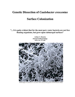
  • 1. Genetic Dissection of Caulobacter crescentus Surface Colonization ”... It is quite evident that for the most part, water bacteria are not free floating organisms, but grow upon submerged surfaces” Arthur T. Henrics Journal of Bacteriology 1933, 25: 277-287 SEM image of Caulobacter crescentus CB15 microcolony grown on borosilicate surface
  • 2. Table of contents TABLE OF CONTENTS Table of contents........................................................................................................... 2 Summary ....................................................................................................................... 6 Overview........................................................................................................................ 8 What is a biofilm? ............................................................................................................................ 8 Exopolysaccharides in biofilms...................................................................................................... 10 Biofilm as a developmental process ............................................................................................... 12 Structural requirements for biofilm formation ............................................................................... 16 Regulation of biofilm formation..................................................................................................... 18 Caulobacter crescentus as a model organism for studying controlled surface attachment and biofilm formation............................................................................................................................ 24 Developmental control of C. crescentus polar appendages............................................................ 25 Aim of thesis................................................................................................................ 29 Chapter 1..................................................................................................................... 30 Abstract ....................................................................................................................... 31 Introduction ................................................................................................................ 33 Materials and Methods .............................................................................................. 36 Media and Strains ........................................................................................................................... 36 DNA manipulations........................................................................................................................ 36 Random Tn5 mutation analysis ...................................................................................................... 37 Genomic DNA sequencing............................................................................................................. 37 Construction of deletion mutants.................................................................................................... 37
  • 3. Table of contents Construction of plasmids for chromosomal deletions .................................................................... 38 Microscopy techniques ................................................................................................................... 40 Microtiter plate attachment assay ................................................................................................... 40 Attachment assay with microscopy cover-slides............................................................................ 41 Holdfast staining and visualization ................................................................................................ 41 Cellulase and protease assay........................................................................................................... 42 Results.......................................................................................................................... 45 Isolation and characterization of C. crescentus surface attachment mutants ................................. 45 Optimal Caulobacter surface attachment correlates with active growth ....................................... 53 Surface attachment peaks with the coincident exposure of polar organelles ................................. 55 Optimal surface attachment requires cell differentiation ............................................................... 61 Discussion .................................................................................................................... 64 Acknowledgements..................................................................................................... 70 Chapter 2..................................................................................................................... 71 Abstract ....................................................................................................................... 72 Introduction ................................................................................................................ 73 Materials and methods............................................................................................... 76 Media and Strains ........................................................................................................................... 76 Synchronization of Caulobacter crescentus................................................................................... 76 DNA manipulations........................................................................................................................ 77 Immunoblots................................................................................................................................... 77 Construction of chromosomal in-frame deletion mutants and plasmids delivery .......................... 77 3
  • 4. Table of contents Quantitative reverse transcriptase PCR (QC RT-PCR).................................................................. 79 Microtiter plate attachment assay ................................................................................................... 80 Microscopy techniques and image processing ............................................................................... 81 Overexpression and purification of proteins .................................................................................. 82 Synthesis and Purification of [33P]cyclic-di-GMP ......................................................................... 83 DGC (Diguanylate Cyclase) and PDE (Phosphodiesterase) Assays .............................................. 83 Results.......................................................................................................................... 87 A WecG homolog is required for C. crescentus holdfast formation .............................................. 87 CC0091 is a c-di-GMP specific phosphodiesterase ....................................................................... 89 PleD and CC0091 are antagonistic regulators of holdfast biogenesis and surface attachment...... 91 PleD and CC0091 inversely regulate C. crescentus motility and stalk biogenesis ........................ 95 CC0095 binds cyclic-di-GMP ........................................................................................................ 99 Swarmer cell specific expression of CC0091 and C0095 ............................................................ 100 Discussion .................................................................................................................. 102 Acknowledgements................................................................................................... 109 Chapter 3................................................................................................................... 110 Additional results ..................................................................................................... 111 Biofilm associated C. crescentus cells exhibit increased antibiotic resistance ............................ 111 Analysis of C. crescentus biofilm maturation in dynamic flow chambers................................... 113 Identification of Caulobacter crescentus genes specifically expressed during biofilm development using recombination-based in vivo expression technology (RIVET) ........................................... 120 Experimental procedures ........................................................................................ 125 4
  • 5. Table of contents Media and Strains ......................................................................................................................... 125 DNA manipulations...................................................................................................................... 125 Construction of deletion mutants.................................................................................................. 125 Construction of plasmids for chromosomal deletions .................................................................. 126 Construction of plasmids for RIVET analysis.............................................................................. 126 Biofilm growth for RIVET analysis: ............................................................................................ 127 Microscopy techniques ................................................................................................................. 128 Attachment assay with microscopy cover-slides.......................................................................... 128 Microtiter plate attachment assay ................................................................................................. 128 Flow chamber experiments........................................................................................................... 129 Scanning electron microscopy...................................................................................................... 130 Acknowledgement .................................................................................................... 133 Bibliography ............................................................................................................. 134 Addendum................................................................................................................. 147 Plasmid maps of selected constructs ............................................................................................ 148 A complete list of strains used in the PhD work .......................................................................... 152 The complete Tn5 insertion library (surface adherent deficient strains)...................................... 161 Complete list of plasmids used in the PhD work ......................................................................... 164 Thank –yous.............................................................................................................. 169 5
  • 6. Summary SUMMARY During its biphasic life cycle Caulobacter crescentus switches from a planktonic to surface attached life style. This transition requires the continuous remodeling of the cell poles through the temporally and spatially coordinated assembly and disassembly of polar organelles like the flagellum, pili, and an adhesive holdfast. A genetic screen for mutants affected in surface binding and colonization led to the identification of various genes required for motility, pili, and holdfast biogenesis, suggesting a specific role for all three organelles in C. crescentus surface colonization. Several novel holdfast genes were identified, which are potentially involved in the synthesis and regulation of the polysaccharidic component of the holdfast. Quantitative surface binding studies during the C. crescentus cell cycle revealed that optimal attachment coincides with the presence of flagellum, pili, and holdfast at the same pole. This indicated that accurate temporal control of polar appendices is critical for surface colonization of C. crescentus and represents the first example for developmentally controlled bacterial surface adhesion. We have used genetic and biochemical analyzes to demonstrate that di-cyclic guanosine monophosphate (c-di-GMP) is a central regulatory compound involved in the timing of C. crescentus pole development. Mutants lacking the diguanylatecyclase PleD show a dramatic delay of holdfast formation during swarmer cell differentiation. In contrast, cells lacking the GGDEF-EAL composite protein CC0091 show premature holdfast formation, while overexpression of CC0091 also leads to a delayed appearance of holdfast. The observation that CC0091 is a c-di-GMP specific phosphodiesterase indicated that the antagonistic activities of PleD and CC0091 could be responsible for the correct timing of holdfast formation and flagellum ejection. Finally, our genetic screen identified a candidate for the c-di-GMP effector protein, which mediates holdfast synthesis in 6
  • 7. Summary response to fluctuating levels of c-di-GMP. The glycosyltransferase CC0095 is strictly required for holdfast formation and its overexpression leads to premature holdfast synthesis. This and the observation that CC0095 is able to bind c-di-GMP lead to the hypothesis that holdfast synthesis is regulated via allosteric control of the CC0095 glycosyltransferase. These data provide the first example of a developmental process being regulated by the bacterial second messenger, c-di-GMP. 7
  • 8. Overview OVERVIEW What is a biofilm? For most of the history of microbiology, microorganisms have primarily been characterized as planktonic, freely suspended cells and described based on their growth characteristics in nutritionally rich culture media. However, in the majority of natural environments, bacteria are rarely found in the planktonic, free-swimming phase. Rather, they are found in association with a biotic or abiotic surfaces in a structure known as a biofilm (22). It is believed that biofilms are the predominant microbial lifestyle. Surface association seems to be means for bacteria persisting in biological or pathogenic microenvironments. For aquatic or soil microorganisms, surface attachment and biofilm formation may provide an adaptive advantage. For example, high-density communities of attached bacteria could metabolize insoluble polymeric organic compounds, hemicellulose, or the exoskeletons of crustaceans and insects. Large negatively charged microbial cell aggregates found in biofilms may constitute a substratum to concentrate and chelate different limiting nutrients such as iron. Finally, biofilms are believed to provide protection from toxic compounds, antibiotics, stress factor and predators (102, 105, 192). It has been speculated that surface attachment and biofilm formation has evolved as a protective mechanism against grazing protozoan predators (104, 105, 192). The persistence stage of bacterial infections is often associated with biofilm formation, and as a result of increased resistance to antimicrobial and the scavenging forces of the immune system, is very difficult to eradicate (39). Persistence of Vibrio cholerae in aquatic environments is thought to be the main factor for seasonal occurrence of cholera epidemics (105). Biofilm-like colonization of the lungs of cystic fibrosis (CF) patients by Pseudomonas aeruginosa is considered as the principal 8
  • 9. Overview cause of mortality in CF patients (40). In Yersinia pestis, the biological transmission of plague depends on blockage of the flea foregut by a biofilm-like cell mass. This blockage is dependent on the hemin storage (hms) locus. Y. pestis hms mutants, although established long-term infection of the flea's midgut, failed to colonize the proventriculus. Thus, the hms dependent biofilm formation affects the course of Y. pestis infection in its insect vector, leading to a change in blood-feeding behavior and to efficient transmission of plague (28, 63). Another example of biofilm formation role in pathogenicity comes from Staphylococcus aureus. Recently, Kropec et al. (88) found that in three mouse models of infection (bacteremia, renal abscess formation, and lethality following high-dose intraperitoneal infection), using three divergent S. aureus strains, the loss of PNAG by deletion of the intracellular adhesion (ica) locus had a profound effect on virulence of this microorganism, which was more susceptible to innate host immune killing (88). Mutant strains showed significantly reduced abilities to maintain bacterial levels in blood, to spread systemically to the kidneys, or to induce a moribund/lethal state following intraperitoneal infection (88). Fluckiger et al. (46) have used a device-related infection model to show that PIA is detectable early in the infection course of S. epidermidis, and that its production in S. aureus is induced during the course of a device-related infection. They have shown that PIA production and biofilm formation of both species exist late in infection, and that the ica genes and biofilm formation are essential for staphylococcal colonization and endurance on implants (46). Persistence of uropathogenic Escherichia coli as biofilm-like communities was proposed to be the source for recurrent urinary tract infections (80). Biofilm-associated cells can be distinguished from suspended cells by the formation of an extracellular polymeric substance (EPS) that acts as a matrix for the embedded cells. Biofilm associated cells often display reduced growth rates and a completely different genetic program 9
  • 10. Overview compared with their planktonic counterpart (reviewed in (38)). Attachment of cells to each other and to surfaces is a complex process regulated by a diverse range of environmental and possibly host signals, which are still poorly understood. Attached bacteria may take the form of a dispersed monolayer of surface-bound cells, they can aggregate on the surface to form microcolonies, or they may be organized into a well structured three-dimensional biofilm (111). Exopolysaccharides in biofilms EPS may account for 50% to 90% of the total organic carbon of biofilms and is considered as the main matrix material of the biofilm (38). EPS consists of various biopolymers with different chemical and physical properties; however, it is primarily composed of polysaccharides. Some of these polysaccharides are polyanionic (174), which allow the association of divalent cations such as calcium and magnesium that could strengthen the matrix structure by cross linkage. In the case of some gram-positive bacteria, such as the staphylococci, the chemical composition of EPS may be quite different and may be primarily cationic (38). EPS is also highly hydrated and thus large amounts of water can become incorporated into its structure by hydrogen bonding. Sutherland (176) noted two important properties of EPS for its role in microbial biofilms. First, the chemical composition and structure of EPS might determine the biofilm conformation (174). For example, many bacterial EPS possess backbone structures that contain 1,3- or 1,4-β-linked hexose residues and tend to be relatively rigid and poorly soluble. Second, the EPS of biofilms is not generally homogeneous but may vary spatially and temporally (174). Leriche et al. exploited the binding specificity of different lectins to sugars in order to assess the polysaccharide properties during bacterial biofilm formation of different organisms (98), the results of this study indicated that distinct organisms produce different amounts of EPS which increases with biofilm development. EPS 10
  • 11. Overview production is affected by the nutrient status of the cells, when an excess in available carbon with limiting nitrogen, potassium, or phosphate were shown to promote EPS synthesis (174). EPS may also contribute to the antimicrobial resistance properties of biofilms by impeding the mass transport of antibiotics through the biofilm, probably by binding directly to these agents (37, 102) . In many bacteria, EPS biosynthesis is underlain the regulation of various systems. In V. cholerae the expression of Vibrio polysaccharide synthesis genes (vps) was shown to be regulated by VpsR and VpsT, homologous to response regulators of two-component regulatory system (16). The vps genes expression in this microorganism was shown to be controlled also by absence of the flagellar structure (190), and also by quorum sensing mediated signals (204). In P. aeruginosa alginate biosynthesis gene, algC, was shown to be upregulated within 15 minutes following contact with the surface (29); in addition to alginate synthesis genes, recent studies of the P. aeruginosa autoaggregative phenotype led to the identification of two genetic loci, psl and pel, that are involved in the production of two distinct carbohydrate-rich biofilm matrix components. The pel gene cluster is involved in the production of a glucose-rich matrix material, while the psl gene cluster is involved in the production of a mannose-rich matrix material (48). Hickman et al. demonstrated that the expression level of these gene clusters is increased in a wspF mutant, probably due to elevation in the cellular levels of c-di-GMP which is probably caused by the constitutive activation by phosphorylation of WspR (62). S. aureus biofilm formation seems to be mediated primarily by the production of the extracellular polysaccharide PIA/PNAG, which is composed of linear beta-1,6- linked glucosaminylglycans. The synthesis of PIA/PNAG depends on the expression of the intercellular adhesion genes icaADBC (55). While most of the S. aureus strains analyzed so far contain the entire ica gene cluster (23), these genes are only expressed in a few, probably due to the 11
  • 12. Overview regulatory nature and the complex control of these genes. ica genes expression was shown to be subjected to environmental stimuli such as high osmolarity, anaerobic conditions, high temperature and certain antibiotics (139). Recent evidence indicates that SarA, a key regulator of S. aureus virulence factors, is required for the expression of ica genes and the synthesis of PIA/PNAG (183). Biofilm as a developmental process The term microbial development was defined as “…changes in form and function that play a prominent role in the life cycle of the organism…” (119). Recent genetic and molecular approaches used to study bacterial biofilms, have uncovered various genes and regulatory circuits important for initial cell-surface interactions and biofilm development. Studies to date suggest that the planktonic- biofilm transition is, like any other bacterial developmental process, complex and highly regulated. Biofilm development consists of a series of well-regulated discrete steps: i) reversible attachment, ii) irreversible attachment, iii) maturation, and iv) dispersion (Figure 1) (155). Reversible attachment was shown in many organisms to be mediated by flagellar based motility and fimbrial adhesins (64). Active motility is thought to assist surface binding by helping the cell to overcome the charge barrier that prevents the negatively charged bacterial cell from reaching certain surfaces. It has been also postulated that an active flagellar motor could play a part in the regulation switch that upon surface binding of bacteria, leads to an up-regulation of exopolysaccharide synthesis (96). Irreversible attachment is mediated mainly by self-made polymeric substances, usually exopolysaccharides, which not only promote cell-cell and cell-surface contacts, but also construct part of the encapsulating matrix (33, 108). Flagella-independent motility (gliding or twitching) allows some bacteria to move on the surface and to form cell-aggregates known as microcolonies (60, 122). Clonal growth within these microcolonies together with EPS encapsulation results with the 12
  • 13. Overview maturation of a biofilm. Finally, an active dissociation or stream shear forces trigger the dispersal of sub populations of the biofilm (53, 70, 187). Bacteria within each of these four stages of biofilm development are physiologically distinct (27). It is obvious that biofilm formation resembles other adaptive processes of bacterial development like fruiting body formation in Myxococcus xanthus, and although the molecular and regulatory mechanisms may differ from organism to organism, the stages of biofilm development seem to be similar in a wide range of microbes (120). Figure 1) Illustration of the four main stages of biofilm development. Stage i) reversible attachment of cells to the surface mediated by flagellar motility and adhesive pili. Stage ii) irreversible cementing of the cells on the surface is a result of EPS production. Stage iii) maturation of biofilm architecture including water channels and pillars. Stage iv) dispersion of single cells from the biofilm. This figure was adapted from (173). One of the hallmarks of a developmental process is near-complete changes of gene expression profile of the different stages. In accordance with this, differential stage-specific gene expression has 13
  • 14. Overview been reported during biofilm formation. Genes required for the initial stage of biofilm formation, e.g. those coding for components of the flagellar motor and adherence pili, are usually repressed in the mature biofilms of many Gram-negative bacteria simply because although these structures required during the initial stages of biofilm development, they might destabilize the mature biofilm (154, 156, 195). In contrast, exopolysaccharides synthesis genes, which are critical for the adherence and for the maintenance of the biofilm structure, exhibit increased expression in biofilm-embedded cells. Thus, progression from the planktonic to the biofilm state requires a change of the cell’s genetic program. Several studies have reported on this program change using global analysis of gene expression or protein synthesis. A protein collection of all four stages during P. aeruginosa biofilm formation was established using 2-D gel electrophoresis (155). On average, consecutive stages differed by 35% of the detectable proteins. 29% of the protein spots changed upon reversible attachment, and 40% upon biofilm maturation (155). Escape from biofilm reduced the protein pool by 35% and re-established a protein profile similar to the one observed for planktonic cells (155). When comparing steady-state levels of proteins from planktonic and biofilm cells, more than 800 proteins showed a six-fold or higher change in abundance (155). The identified proteins fall into four main classes: metabolism, phospholipid and LPS-biosynthesis, protein transport and secretion, as well as adaptation and protective mechanisms (155). In another study performed with P. aeruginosa, genes responsible for alginate biosynthesis were shown to be upregulated within 15 minutes after cells adherence to surfaces, arguing that surface binding might initiate this genetic switch that leads to biofilm formation (29). A study by Sauer et al. showed that the expression of more than 30 operons was altered within 6 hours following P. putida surface attachment (154). 14
  • 15. Overview The comparison of global gene expression profiles of planktonic vs. biofilm cells was performed with several model organisms. When gene expression in E. coli biofilms grown in a flow chamber was compared with planktonic cells in stationary or exponential phase, an overall alteration of more than 600 genes was observed between stationary phase and biofilm cells (156). Only 230 genes were found to be differentially expressed in exponentially growing cells and biofilm cells (156). Among the genes that showed increased expression in biofilms, several were shown to be involved in adhesion and autoaggregation. In a parallel study, 38% of a random E. coli lacZ fusion library showed biofilm-specific expression (137); sessile bacteria showed specific up-regulation of genes involved in colanic acid biosynthesis (wca locus), while fliC (flagellin) was reduced in biofilms. Moorthy and Watnick used microarrays to study the transcriptome of V. cholerae during each stage of biofilm development (113). The transitions from planktonic to monolayer and mature biofilm identified up to 383 differentially regulated genes. Most of these genes were specific for only one of the three experimental stages analyzed. These results demonstrated that monolayer and mature biofilm stages of V. cholerae biofilm development are transcriptionally distinct. A similar analysis with a clinical isolate of Staphylococcus aureus (UAMS-1) reveled a total of 580 differentially expressed genes (11). In this study, the largest difference of total numbers of differentially expressed genes was observed between the biofilms and the exponentially grown planktonic cells (11). Taken together, these studies make it apparent that biofilms have gene-expression patterns that differ from those of planktonic bacteria, and telling something about the extensive physiological changes that occur during biofilm formation. These global gene expression analyzes facilitated the uncovering of the stage-specific cell physiology and morphology during biofilm development and demonstrated the complexity of this process. 15
  • 16. Overview Structural requirements for biofilm formation Initial attachment and microcolony formation Pseudomonas aeruginosa- In a pioneering work by O'Toole and Kolter (122), a screen for the isolation of P. aeruginosa Tn5 insertion mutants defective in the initial steps of biofilm formation was undertaken, based on the ability of this bacterium to adhere to plastic surface of a microtiter plate. Two classes of mutants, named sad (surface attachment defective), were described, one class constitutes flagellar-motility mutants while the other class consists of mutants defective in the biogenesis of the adhesive type IV pili (122). While a pili mutant was able to form a wild type-like monolayer of cells on the surface, they were unable to develop into microcolonies. Type-IV pili are required for twitching motility, a mode of surface locomotion used by P. aeruginosa and other bacteria in which the polar pili are believed to extend and retract, and thereby propelling bacteria across a surface. Thus, the findings by O’Toole and Kolter (122) suggested that surface based motility is required for the second step of biofilm formation. Vibrio cholerae- In a similar genetic analysis performed by Watnick and Kolter (189), three classes of V. cholerae El Tor sad mutants were described (189). The first class of genes is required for the biosynthesis of the mannose-sensitive haemagglutinin type-IV pilus (MSHA); the second group of mutants was defective in flagellar motility, including both mutants lacking flagella and mutants with paralyzed flagella. The third group of sad mutants had transposon insertions in vps genes. The phenotypes of these mutant classes suggested that pili and flagella accelerate attachment to and mediate the spread along the abiotic surface, while exopolysaccharide synthesis by the vps genes is required for the formation of the three-dimensional biofilm architecture. In contrast to 16
  • 17. Overview mutants lacking pili and flagella, EPS mutants were unable to form a detectable biofilm even after extended incubation time (189). E. coli- A study by Pratt and Kolter reveled three classes of attachment-deficient E. coli Tn10 mutants (134). The mutations isolated included flagellar biogenesis and motor function genes, and genes which were involved in the biogenesis and regulation of type-I pili (134). Interestingly, in a strain overproducing curli background, flagella were dispensable for initial adhesion and biofilm development (136), arguing that at least part of the role of flagella in surface colonization might be of a regulatory nature. Biofilm maturation P. aeruginosa- Klausen et al. have shown that flagella and type-IV pili take part in shaping the architecture of P. aeruginosa biofilms, although they are not essential for biofilm formation (86). The model which they proposed suggests that the formation of mushroom-shaped structures in P. aeruginosa biofilms is caused by bacteria which climb on the top of the microcolony stalks using of type-IV pili mediated twitching motility (85); according to this model, type-IV pili driven bacterial migration plays a key role in structure formation in the late phase of biofilm development. V. cholera- V. cholerae strain which is defective in EPS synthesis fails to form a mature biofilm architecture (188, 201). Moorthy and Watnick have recently shown that the cell monolayers formed on surfaces represent a distinct stage of this microorganism biofilm development (111). They have demonstrated that while MSHA pilus is only required for the monolayer formation, vps is required for formation and maintenance of the mature biofilm and that the maturation of these 17
  • 18. Overview monolayers to three-dimensional biofilm structure, requires monosaccharides such as mannose, which induce the expression of vps genes (111). E.coli- Molin and co-workers have shown recently that the maturation of E. coli K12 biofilms requires the presence of an incF plasmids (144). They have demonstrated that while surface attachment, clonal growth and microcolony formation were not affected in the plasmid plasmid-free strains, the efficient biofilm maturation could only occurred in strains carrying the conjugation pilus proficient plasmid (144) and that E. coli strains lacking these plasmids were not able to form the elaborated three-dimensional biofilm architecture that include pillars and channels (144). They have shown that the final shape of the mature biofilm seemed to be determined by the pilus configuration, when various mutants affected in the processing or in the activity of these transfer pili, displayed differently structured biofilms. In addition to that, flagella, type 1 fimbriae, curli and cell-to-cell signalling did not seem to be required for biofilm maturation in E. coli K12 carrying the incF plasmids (144). This work was with a complete agreement with a previous work published by Ghigo (52), which has demonstrated the involvement of conjugative plasmids in the competence of the bacterial host to form a biofilm (52) . Regulation of biofilm formation Complex regulatory pathways such as the global carbon metabolism regulator (CRC) (121) and stationary-phase sigma factors (σs) (60, 196) have been shown to play an important role in biofilm development despite the fact that these systems are not exclusively committed for biofilm development. High-cell density, high osmolarity, scarce nutrients as well as oxygen limitation are only some of the situation which a biofilm embedded cell and a stationary phase cell might encounter; this similarity could explain some of the convergent regulation circuits that control 18
  • 19. Overview biofilm formation in addition to stationary phase and stress respond. Besides being subjected to global metabolic control, biofilm components underlie specific regulation at the transcriptional and post-transcriptional level. For example, the Salmonella typhimurium CsgD, a transcriptional regulator of the LuxR superfamily, has been shown to positively control the expression of cellulose and curli fimbriae (14). The expression of csgD itself is modulated by a variety of stimuli, including, osmolarity, oxygen, nutrient availability, pH, temperature, and the subject of control by many cellular factors, such as RpoS, RpoD, IHF and others (14, 135). In P. aeruginosa GacS/GacA proteins of the two-component signal transduction system which controls the production of many secondary metabolites and extracellular enzymes and involved in pathogenicity in plants and animals (58), were shown to also control biofilm formation when gacA mutant failed to aggregate and form microcolonies (127). Although the signals that activate the GacS/GacA circuit are not known, it was demonstrated that the gac genes are activated during the transition from exponential to stationary phase of the growth (58); and since the expression of rpoS is positively regulated by GacS/GacA, some of the GacS/GacA-dependent phenotypes may be related to RpoS activity (116, 194). In addition to the GacA/GacS, a three-component regulatory system specifically required for biofilm maturation was identified (89). This system is comprised of genes sadARS coding for a putative sensor histidine kinase and two response regulators; mutations in any of these genes, blocked biofilm maturation of P. aeruginosa without affecting growth, early biofilm formation, swimming, or twitching motility (89). The expression of sadR and sadS is very similar in planktonic and biofilm cells, while sadA expression is slightly decreased ( 2-fold) in biofilm cells. The authors have postulated that the SadARS system acts as a regulator of both biofilm formation and for genes 19
  • 20. Overview involved in type III secretion (TTSS) and it may function to promote biofilm formation, possibly in part by repressing the expression of the TTSS (89). In addition to the species-specific control mechanisms, biofilm formation is also regulated by two global signal transduction networks. The first, quorum sensing (QS) allows transmitting information between cells and has been shown to regulate cellular processes in response to cell density or crowdedness (128). Since biofilms comprise arrays of dense microbial populations, it was not surprising to find that QS influences biofilm related processes. Davies et al. (30) showed that P. aeruginosa PAO1 requires the lasI gene product 3OC12-HSL in order to develop a normal biofilm; lasI mutant formed flat, undifferentiated biofilms which remain sensitive to SDS (30); interestingly, mutant biofilms appeared normal when supplemented extracellularly with a synthetic 3OC12 signal molecule (30). Similarly, Burkholderia cepacia mutants defective in the cep quorum sensing system were able to form microcolonies on a glass surface, but were unable to develop into a mature biofilm (68). In E. coli, biofilm formation was shown to be stimulated by the auto-inducer 2 signal (AI-2) (200). It was suggested that AI-2 stimulates biofilm formation through a regulatory cascade including novel motility quorum sensing regulator, MqsR, the two component system QseBC which then promotes cell motility via the master regulon flhDC, stimulating MotA and FliA and leads to biofilm formation (200). QS-dependent biofilm formation regulation in E. coli was demonstrated also by the deletion of ydgG (a putative transport protein that either enhances AI-2 secretion or inhibits AI-2 uptake) which increased the intracellular concentration of AI-2 as turn resulted in a 7,000-fold increase in biofilm thickness and 574-fold increase in biomass in flow cells (59). In contrast, in V. cholerae, a reciprocal relationship between quorum sensing and biofilm formation was described (204). V. cholerae strains lacking HapR, a LuxR homolog, forms thicker biofilms; microarray 20
  • 21. Overview analyses of biofilm-associated bacteria showed that the expression of the V. cholerae vps genes is increased in hapR mutants when CqsA, one of two known autoinducer synthases in V. cholerae, acts through HapR to repress vps gene expression (204). The second global regulator controlling cell adhesiveness and biofilm formation is cyclic di(3' 5')-guanylic acid (c-di-GMP). C-di-GMP is emerging as a global second messenger in bacteria controlling “social behavior.” As described above, cell surface appendages mediate bacterial aggregation and facilitate biofilm formation; flagella and pili which are involved in biofilm formation were shown to be regulatory targets of c-di-GMP (reviewed in (25, 75, 147)). Genetic studies have implicated c-di-GMP in the regulation of motility, the production of extracellular polysaccharide, biofilm establishment and maintenance as well as host persistence in a wide range of bacteria (75, 147). Biochemical studies have reveled that cellular levels of cyclic-di-GMP are inversely controlled by the activity of diguanylatecyclases (GGDEF domain) and phosphodiesterases (EAL domain) (Figure 2) (19, 129, 152, 157, 182). GGDEF and EAL domain proteins are abundant and found in most bacteria, covering all branches of the phylogenetic tree (147). C-di-GMP was first described as an allosteric activator of the enzyme cellulose synthase of the bacterium Gluconacetobacter xylinum (150). In Caulobacter crescentus, c-di-GMP was shown to orchestrate the controlled transition of a flagellated into a “sticky” cell pole which secretes an unknown form of polysaccharide (5, 129). The production of cellulose or derivatives thereof, is activated by GGDEF domain proteins in several other bacteria including E. coli, S. enterica, Rhizobium leguminosarum and P. fluorescence (8, 169, 207), in addition to polysaccharides, the biosynthesis of adhesive fimbriae, another component of extracellular matrix also depends on the activity of GGDEF domain proteins (24, 160). In the current working model, high levels of c-di-GMP favor the production of adhesive organelles and blocks 21
  • 22. Overview different forms of cell motility (161). Hickman et al. (62) have recently shown that an increase in cellular levels of c-di-GMP elicited by a specific diguanylatecyclase, WspR, results in higher expression of the pel and psl EPS gene clusters of P. aeruginosa and led to the formation of mature biofilms (62). Similarly, vps expression in V. cholerae is controlled by c-di-GMP (182). When the enzymatic activity of the VieA phosphodiesterase is required to repress EPS production under non- biofilm conditions (182). The deletion of vieA results in increased cell attachment, probably as a consequence of up-regulation of VpsR, a positive regulator of vps gene expression (182). Signature- tagged transposon mutagenesis in Salmonella have led to the identification of CdgR, an EAL domain protein which its mutagenesis resulted in lower resistance to hydrogen peroxide and accelerated killing of macrophages in mice model (65). Hoffman et al. have shown that alterations in the intracellular levels of c-di-GMP caused by the addition of sub inhibitory concentrations of the antibiotic tobramycin, induced a specific, defensive reaction in both in E. coli and P. aeruginosa (66). Tobramycin induces the expression of arr phosphodiesterase which results in reduced levels of c-di-GMP, increased biofilm formation and increased resistance to tobramycin (66). These studies implicate a complex relationship between c-di-GMP intracellular levels and regulation of biofilm formation. 22
  • 23. Overview Figure 2) The conversion of GTP into c-di-GMP is catalyzed by the diguanylatecyclases, which reside in the GGDEF domain. Increased intracellular levels of c-di-GMP promote biofilm formation and the biosynthesis of adhesive organelles and inhibit different types of cell motility (reviewed in (161) . Degradation of c-di-GMP is catalyzed by the activity of EAL domain of phosphodiesterases. The illustration was taken from a poster (“Biochemical and genetic identification of a c-di-GMP binding motif”) presented by Beat and Mathias Christen and Marc Folcher). 23
  • 24. Overview Caulobacter crescentus as a model organism for studying controlled surface attachment and biofilm formation The genus Caulobacter consists of a collection of Gram-negative, hetero-oligotrophic aerobe, rod- like shaped cells that are equipped with a single polar flagellum and polar pili. Caulobacter crescentus possesses a stalk, a thin cylindrical extension of the cell containing cell wall and cytoplasm, with an adhesive material, the holdfast, located at its tip. The holdfast mediates strong irreversible attachment of Caulobacter cells to solid substrates (109). Caulobacter are generally found in aquatic environments, where they attach to biotic and abiotic surfaces (132, 203) and participate in biofouling processes (203). The unique life cycle of C. crescentus with its asymmetric cell division and obligatory cell differentiation has made it one of the preferred model organisms to study microbial development and the mechanisms underlying bacterial cell cycle control (151). The dimorphism is established by an asymmetric cell division that gives rise to two genetically identical, but morphologically and physiologically distinct daughter cells with different developmental programs: a sessile stalked cell equipped with an adhesive holdfast and a motile swarmer cell bearing a single flagellum and adhesive pili (15). The stalked cell is competent to start a new replicative cycle immediately after cell division, whilst the swarmer cell is engaged in chemotaxis while the replicative program is being blocked. Before the swarmer cell re-enters replication and cell division it differentiates into a stalked cell, a process during which it loses the flagellum, retracts its pili, and forms a holdfast and a stalk at the pole previously occupied by the flagellar motor. Dimorphism is believed to have evolved to allow Caulobacter to cope with life in dilute, nutrient-poor environments (69). The swarmer cell stage allows rapid dispersal and the scavenging of new nutrients resources, while the surface adherent form permits growth where nutrients are available. 24
  • 25. Overview The nature of C. crescentus cell poles is constantly changing during its development (Figure 3). Pole differentiation is regulated by a complex regulatory network which includes several members of two- component signal transduction proteins (2, 71, 74, 124). Some of these regulators interlink cell-cycle progression and pole development. E.g., The response regulator CtrA directly controls the initiation of chromosome replication as well as several aspects of polar morphogenesis and cell division (42). The intrinsic asymmetry and microscopically visible appendages make it possible to monitor cell cycle progression and pole differentiation and allow the analysis of temporal and spatial control of polar organelles like flagellum, pili, holdfast, and stalk. Developmental control of C. crescentus polar appendages The synthesis of C. crescentus flagellum requires about 50 different genes. Flagellar gene expression underlies cell cycle control with the temporal activation of CtrA (36, 143). In addition flagellar gene transcription is controlled by hierarchical regulatory system in which the expression and productive assemblage of gene products are required for the expression of gene products which participate successively in the multistep flagellar assembly (117, 140). This regulatory cascade consists of four hierarchical classes. The cascade initiates with class I genes, namely CtrA, which promotes the transcription of the class II genes encoding the MS ring of the basal body, the flagellar switch, and the flagellum-specific type III secretion system (36, 138). The transcription of the flagellar class III and IV is dependent on the proper assembly of the class II components (115). In addition, the expression of class III and IV flagellar genes requires σ54 and the transcriptional activator FlbD, which in addition to being subjected to cell cycle-regulated phosphorylation (197), FlbD activity is also subjected to the hierarchical regulation system (114). The ejection of the flagellum during the swarmer-to-stalked cell transition coincides with the degradation of the FliF flagellar anchor. The 25
  • 26. Overview activity of the diguanylatecyclase response regulator PleD was shown to be required for efficient removal of FliF, ejection of the flagellum, and stalk biogenesis (3). PleD activity is regulated through cell-cycle dependent phosphorylation by PleC and DivJ kinases (5, 129). These elaborate regulatory mechanisms ensure the linking of flagella assembly and disassembly to the cell cycle and to the development of C. crescentus. Pili are extracellular filaments, found in a wide variety of bacteria. Pili were shown to play a major role in adhesion of bacteria to surfaces, biofilm formation, conjugation, twitching motility, and host infection (164). Caulobacter crescentus pili are extracellular surface appendages, 1–4 µm in length and 4 nm in diameter and are located exclusively at the flagellated pole (164). The pili composed of polymerized pilin subunit (PilA) which is assembled by proteins encoded by a cluster of pilus assembly genes (cpaA-F) that are closely related to the tight-adherence genes (TAD) from Actinobacillus actinomycetemcomitans (81, 130). The transcription of cpaB–F is induced in the late predivisional cell, followed by cpaA and, finally the CtrA-dependent transcription of pilA with peak of expression in the progeny swarmer cells (95). The timing of pilus assembly can be shifted from the swarmer cell to the predivisional cell stage by expressing pilA from a constitutive promoter, suggesting that the temporal transcription is the main type of regulation that prevents premature assembly of the pili (164). It was demonstrated that the PleC histidine kinase, which is localized to the piliated pole during the pilus assembly time window, controls the accumulation of PilA (185). PleC was shown to be responsible for the asymmetric distribution of CpaC (a putative outer membrane pilus secretion channel) and its assembly factor, CpaE (185). 26
  • 27. Overview The adhesive holdfast is located at the tip of the stalk at the pole previously occupied with the flagellum. The exact biochemical composition of the holdfast is unknown, however, lectin binding and glycolytic enzymes sensitivity experiments suggest that the holdfast is composed of polysaccharides containing N-acetylglucosamine (GlcNAc) oligomers (109). Janakiraman and Brun used an hfaA-lacZ fusion to show that the transcription of hfaA (part of the hfaA-D gene cluster which required for holdfast attachment to the cell envelope (21, 92)) is temporally regulated during the cell cycle. hfaA exhibit maximal transcription levels in predivisional cells (72). The authors however, have failed to observe the holdfast before differentiation of the swarmer cell had occurred. How the spatial and temporal regulation of holdfast expression is achieved is still unclear. 27
  • 28. Overview Figure 2) A schematic representation of the Caulobacter crescentus cell cycle. The replication- incompetent swarmer cell is equipped with a polar flagellum and flp-like pili. After a defined period, the swarmer cell differentiates into a stalked cell in successive of developmental steps, including the ejection of the flagellum, the retraction of the pili, the synthesis of the holdfast, and the elongation of the stalk. Chromosome replication initiation coincides with the formation of the stalked cell. The timing of several morphogenetic and cell cycle events is shown by the light and dark grey bars respectively. The flagellated, stalked (ST) and new swarmer (SW) poles are indicated. The relative duration of each phase is indicated on top as horizontal axis. This figure was adapted from (71) 28
  • 29. Aim of thesis AIM OF THESIS The aim of this work was to genetically identify components involved in C. crescentus surface binding and colonization. New structural and regulatory components of C. crescentus pole development and surface adhesion should be analyzed with respect to their function, their temporal and spatial coordination, and the specific molecular mechanisms facilitating surface colonization. 29
  • 30. Chapter 1 CHAPTER 1 The coincident exposure of polar organelles optimizes surface attachment during Caulobacter crescentus development Assaf Levi and Urs Jenal* Division of Molecular Microbiology, Biozentrum, University of Basel Klingelbergstrasse 70, CH-4056 Basel, Switzerland In revision of publication in Journal of microbiology Running title: Caulobacter surface attachment Keywords: Caulobacter, flagella, pili, holdfast, biofilm, c-di-GMP * For Correspondence: Division of Molecular Microbiology, Biozentrum, University of Basel, Klingelbergstrasse 70, CH-4056 Basel, Switzerland Tel: +41-61-267-2135; Fax: +41-61-267-2118; e-mail: urs.jenal@unibas.ch 30
  • 31. Chapter 1 ABSTRACT During its biphasic life cycle, Caulobacter crescentus oscillates between a planktonic and a surface attached life style. A hallmark of this transition is the temporally and spatially regulated assembly and disassembly of polar organelles like flagellum, pili, and an adhesive holdfast. A genetic screen for mutants affected in surface binding and colonization revealed a large number of known and novel components of flagellar motility, pili formation, and holdfast biogenesis, arguing that these organelles are required for optimal surface adhesion of C. crescentus. Several new holdfast genes were identified, which are potentially involved in the formation and polymerization of polysaccharide precursors. Together with experiments that implicate a cellulose-like polymer as a main constituent of holdfast structure and function, this provides the basis for future analyses on the formation and exact composition of this adhesive organelle. Several lines of evidence suggested that the coincident exposure of polar organelles optimizes surface attachment during Caulobacter crescentus development. i) The holdfast is synthesized and exposed on the cell surface very early during the swarmer-to-stalked cell transition and, during a defined time window, coincides with an active flagellum and adhesive pili at the same pole. ii) Cell cycle-dependent surface attachment showed a prominent peak coinciding with the surface exposure of all three polar organelles, and mutants lacking any one of these subcellular structures exhibited basal levels of attachment. iii) Active growth, as well as passage through development, greatly enhanced surface colonization. iv) A delay of holdfast biogenesis observed in a pleD mutant resulted in a strong reduction of surface binding during development. In cells lacking PleD, a developmentally controlled diguanylate cyclase, holdfast biogenesis was delayed by almost one third of a cell cycle equivalent, indicating that PleD and its readout signal, c-di-GMP, are used as timing device for holdfast formation. Based 31
  • 32. Chapter 1 on these results we propose a model for C. crescentus surface colonization that involves the successive and concerted activity of flagella, pili, and holdfast. The model provides a rational framework for the precise temporal and spatial control of these cellular appendices during development. 32
  • 33. Chapter 1 INTRODUCTION In most natural environments, microbial cells are found attached to surfaces and associated in communal structures known as biofilms. The formation of biofilms from single planktonic cells, widely studied in a few model organism (Vibrio cholerae, Pseudomonas aeruginosa, Salmonella typhimurium, and E. coli), involves several discrete stages, including reversible and irreversible attachment to surfaces, formation of cell monolayers, microcolony formation, and biofilm maturation (112, 119, 155, 189). This process is associated with a dramatic change of the cells’ genetic program and physiology (154-156, 170, 193, 195). Initial stages of surface colonization are facilitated by cellular appendages like flagella and pili that can mediate initial attachment and accelerate biofilm development (9, 35, 81, 83, 134, 155). Later stages of biofilm formation are associated with the formation of an extracellular matrix, which mediates surface anchoring and provides structural support for the cell community (175). While all major classes of macromolecules can be present in biofilm matrices, increased synthesis of exopolysaccharide (EPS) is generally associated with biofilm formation (26, 47, 48). The contribution of flagella and pili to various stages of biofilm formation have been demonstrated independently for several bacteria but it remains to be shown whether flagella, pili and EPS are part of a coordinated program for surface attachment and colonization rather than contributing to biofilm formation in a stochastic and independent manner. If these distinct organelles and mechanisms are indeed interlinked and are part of a program dedicated to surface colonization, how would these interactions be regulated in time and maybe space? How would cell motility and adhesive properties be coordinated to optimize surface attachment early during biofilm formation and to ensure the escape or detachment of cells from biofilms at a later stage? One possibility is that 33
  • 34. Chapter 1 different components of this multicellular behavior are co-regulated (148). A number of environmental signals, including nutrients, temperature, osmolarity, pH, iron, and oxygen influence biofilm formation (reviewed in: (119)), but little is known about mechanisms that integrate these inputs and transduce them into an altered bacterial behavior required for surfaces colonization. During Caulobacter crescentus development surface adhesion is coupled to cell growth and division. Each cell division is intrinsically asymmetric and generates a sessile, replicative stalked cell and a motile, flagellated swarmer cell. A single flagellum is assembled in the predivisional cell at one pole and is activated prior to cell division (198). Upon separation of the two daughter cells, pili are formed at the flagellated pole of the swarmer cell (164, 167). The newborn swarmer cell performs chemotaxis for a defined period (6) before it sheds the flagellum, loses its pili and differentiates into a stalked cell. During this process, an adhesive holdfast structure and a stalk are assembled at the pole previously occupied by pili and flagellum. The exact role of the polar pili in C. crescentus and its temporal and spatial control are unknown but it has been proposed that they might facilitate surface interaction and cell attachment (12, 167). Irreversible anchoring of C. crescentus cells to surfaces requires an intact holdfast structure (125). Genetic screens have identified several genes required for holdfast secretion and anchoring (21, 166). While some of these genes encode homologs of polysaccharide export components in other gram-negative bacteria, the exact structure and composition of the holdfast remains unclear (166). Staining and lectin binding experiments had proposed that it is composed of an acidic polysaccharide, which contains N-acetylglucosamine (GlcNAc) residues (109, 166, 184). The observation that in C. crescentus swarmer cells are able to attach to surfaces (12, 131, 132) suggested that the model for surface attachment as being mediated by stochastic and independent 34
  • 35. Chapter 1 adhesion events might be too simplistic and has indicated that all polar organelles might contribute to this process in a concerted manner. Here we show that in a static system, flagella, pili, and holdfast substantially contribute to C. crescentus surface attachment. Using a new method to detect holdfast we could demonstrate for the first time that holdfast biogenesis occurs much earlier in development than reported previously. Consequently, all three polar organelles are concomitantly exposed at the same cell pole during a defined time window of swarmer cell differentiation. This developmental stage coincides with a sharp peak of surface binding activity during the C. crescentus life cycle. This attachment peak was reduced or eliminated in mutants lacking pili, flagellum, or holdfast. Moreover, in a mutant that shows delayed holdfast synthesis during development, attachment is dramatically reduced. Together with the observation that optimal surface binding is coupled to growth and cell differentiation this lead us to propose a model for C. crescentus attachment in which rapid surface binding is optimized by the careful temporal and spatial coordination of all three organelles during development. 35
  • 36. Chapter 1 MATERIALS AND METHODS Media and Strains Strains and plasmids used in this study are listed in Table 1. E. coli DH10B and S17-1 were used as host strain for molecular cloning experiments and as donor strain for conjugational transfer of plasmids into Caulobacter. E. coli strains were grown at 37°C in Luria-Bertani (LB) broth (153) supplemented with kanamycin (50 µg/ml) or tetracycline (12.5 µg/ml), when necessary. C. crescentus strains were grown at 30°C in either PYE complex medium (131) or in M2 minimal glucose medium (M2G) (78) supplemented with kanamycin (5 µg/ml), tetracycline (2.5 µg/ml), chloramphenicol (1 µg/ml) or nalidixic acid (20 µg/ml) when necessary. Semisolid agar plates for motility assays contained 0.3% agar (DIFCO®). Synchronization of C. crescentus was done as described earlier (171). Isolated swarmer cells were released into fresh minimal medium at an OD660 of 0.3. Samples were removed for microscopic analysis, attachment assays, and holdfast staining at 15 minutes intervals. For surface binding assays, cells were allowed to attach to polystyrene in microtiter plates for 15 minutes. Cell cycle progression was monitored by light microscopy. DNA manipulations Plasmid and chromosomal DNA preparation, DNA ligation, electroporation, agarose gel electrophoresis, and PCR amplifications were carried out by using standard techniques (153). All PCR products used for cloning were amplified with “Expand high-fidelity PCR system®” form Roche. Restriction enzymes were from New England Biolabs, Inc. 36
  • 37. Chapter 1 Random Tn5 mutation analysis The mini-Tn5 transposon delivery vector pUT_Km2 (32) was inserted into C. crescentus wild type by conjugation. Approximately 2,000 colonies were grown in 96-well plates in 200 µl of PYE medium supplemented with kanamycin. Cells were discarded and the microtiter plates were washed under a gentle stream of distilled water. The plates let to air-dry and surface attached cells were quantified as described below. Mutants with a reproducible and significant increase or decrease (>25%) of attachment were selected for further analysis. Genomic DNA sequencing Genomic DNA was extracted from C. crescentus wild type or mutant strains using ethanol precipitation following lysis of cells in 5M Guanidinium thiocyanate, 0.1M EDTA. Tn5 insertion sites were mapped by direct sequencing of chromosomal DNA using oligonucleotide primers complementary to the ends of the kanamycin resistance cassette of Tn903: primer #698 (TCT AGA GTC GAC CTG CAG GC) and #699 (TAC CGA GCT CGA ATT CGG CC). Sequencing reactions were set up using ~500 ng genomic DNA as template and 10 pmol of sequencing primer in a total reaction volume of 20 µl following the BigDye Terminator protocol with few modifications: o Annealing temperature was raised to 58 C and the number of cycles was increased to 99 (Big Dye; Perkin-Elmer®). The sequencing reactions were run on an ABI Prism 310 DNA Sequencer or ABI Prism 3100 Avant Genetic Analyzer (Perkin-Elmer®). Construction of deletion mutants In frame deletions of the chromosomal copies of flgH, fliFG, flgDE, flgFG, fliL, CC2277 and CC0095 were constructed in C. crescentus wild type strain and in UJ590 (∆pilA) using pNPTS138 37
  • 38. Chapter 1 based constructs carrying in frame deletions in the respective genes. Plasmids (see below) were introduced into the recipient strains by conjugation and recombinants were selected on PYE plates supplemented with kanamycin and nalidixic acid. Resulting single colonies were then grown overnight in liquid PYE medium and plated on PYE containing 3% sucrose. Sucrose-resistant colonies were screened by PCR for recombinants that had lost the chromosomal copy of the respective gene. Construction of plasmids for chromosomal deletions Plasmid pAL8 was constructed for an in-frame deletion of the complete flgFG coding region. PCR amplification of a 1.0 kb region upstream of flgFG was done with primers #655 and #656 (5’-GGA TCC GGC GTT CGA GCT GCT GCT GA-3’ and 5’-GAA TTC TCA CCT GGC GGG TGA GTG AG-3’). PCR amplification the 1.0 kb region downstream of flgFG was done with primers #657 and #658 (5’-GAA TTC CGC TCG CCT AAG CGA ACG TC-3’ and 5’-ACT AGT GGC CGA GAT CTT GCC GTC GA-3’). Ligation of both fragments into pNPTS138 (SphI/SpeI) resulted in plasmid pAL8. Plasmid pAL2 was constructed for an in-frame deletion of the complete flgH coding region. PCR amplification of a 1.0 kb region upstream of flgH was done with primers #637 and #638 (5’- GGA CTA GTC CCG GCG ACA ACC TGT TCC TGG-3’ and 5’-CGG GAT CCC GGA CGA CGC ATG ATC TGG TCC-3’). PCR amplification a 1.0 kb region downstream of flgH was done with primers #639 and #640 (5’- CGG GAT CCC GGG TCG AGA AGT TCT CGC CCT-3’ and 5’- CGG AAT TCC GGG AGC GCA TTC GAC GTC TGG-3’). Ligation of both fragments into pNPTS138 (SpeI/EcoRI) resulted in plasmid pAL2. 38
  • 39. Chapter 1 Plasmid pAL6 was constructed in order to create an in-frame deletion of the complete fliL coding region. PCR amplification of a 1.0 kb region upstream the fliL was done with primers #710 and #711 (5’-ACT AGT CCA TCA TCT TGG CGA CGC-3’ and 5’- GAA TTC CGT ACT CAT GCG CGA AGC -3’). PCR amplification a 1.0 kb region downstream of fliL was done with primers #712 and #713 (5’- GAA TTC GCG AAC GAT CAT GGC GGA-3’ and 5’-GCA TGC ACC TGC ATG TTC AGC ACG-3’). Ligation of both fragments into pNPTS138 (SpeI/SphI) resulted in plasmid pAL6. Plasmid pAL7 was constructed in order to create an in-frame deletion of the complete flgDE coding region. PCR amplification of a 1.0 kb region upstream the flgDE coding sequences was done with primers #563 and #564 (5’- GCA TGC GAC CGC CGC GAC CGC CCC GC-3’ and 5’- GAA TTC CCG GTC AAG GAC CGA GGC GG -3’). PCR amplification a 1.0 kb region downstream of fliL was done with primers #565 and #566 (5’- GAA TTC TCG AAG ATC ATC ACG ACC GC-3’ and 5’- CTA GTT GGC GAC CTT GTC GCG CGG C-3’). Ligation of both fragments into pNPTS138 (SpeI/SphI) resulted in plasmid pAL7. An in-frame deletion of the chromosomal copies of fliF and fliG genes was generated in wild-type strain CB15 using plasmid pBG22 (56). Plasmid pAL21 was constructed in order to create an in-frame deletion of the complete CC2277 coding region. PCR amplification of a 1.0 kb region upstream the CC2277 was done with primers #625 and #626 (5’-cgg aat tcC GGG CTT CCT GCC GTT CCA CCG-3’ and 5’- gct cta gaG CGC GCC ATC AGG CCT CCG TGT-3’). PCR amplification a 1.0 kb region downstream of CC2277 was done with primers #627 and #628 (5’- gct cta gaG CCG CGT GAG CGT GAT CAC CGA-3’ and 5’-ggg gta ccC CCC AGG CCA AAG GTG ACC GGC-3’). Ligation of both fragments into pNPTS138 (EcoRI/KpnI) resulted in plasmid pAL21. 39
  • 40. Chapter 1 Plasmid pDM25 was constructed in order to create an in-frame deletion of the complete CC0095 coding region. PCR amplification of a 550 bp region upstream of CC0095 was done with primers #1380 and #1381 (5’- GAA TTC TTC GAC CGT TCC CAG CCC-3’ and 5’- GGA TCC CGC TGT CCA GAC GCT CTA-3’). PCR amplification a 550 bp region downstream of CC0095 was done with primers #1382 and #1383 (5’- GGA TCC TGA GGA ACG AAC ATC TCC GCA G-3’ and 5’- AAG CTT CGA CAA GGA CGG CCA GAA GGA-3’). Ligation of both fragments into pNPTS138 (EcoRI/HindIII) resulted in plasmid pDM25. Microscopy techniques Cell morphology, motility, and rosette formation were analyzed by light microscopy using a Nikon Eclipse 6000 with a planApo 100x phase contrast objective or an Olympus AX70 with an UplanApo 100x phase contrast objective. Pictures were taken with a charge-coupled device camera (Hamamatsu®) connected to the Olympus microscope and analyzed with Open-lab (Improvision®) software. Microtiter plate attachment assay For C. crescentus surface attachment assays the protocol described by O’Toole was slightly modified (123). Stationary-phase cultures were diluted with fresh PYE (plus supplements when mentioned) into 96, 24 or 12 well polystyrene microtiter plates (at final volumes of 0.2, 1.0, or 2.0 ml, respectively) to an OD660 of 0.05 and incubated at 30ºC on a shaker (200 rpm) until cultures reached an OD660 between 0.9-1.2. Cells were discarded and the wells were washed gently under a stream of distilled H2O to remove unattached cells. Plates were air dried and a culture-volume of 0.1% crystal violet (CV) was added and incubated with shaking for 15 min. Wells were washed again 40
  • 41. Chapter 1 several times with distilled H2O and CV was dissolved in 20% acetic acid. The color intensity was measured with a microplate reader spectrophotometer at 600 nm. Rapid attachment assays were performed as described above with the following modifications: Aliquots of 150 µl of mid- logarithmic phase cultures (OD660 of 0.4-0.6) were transferred to microtiter plates and incubated for short period (15-120 minutes, as indicated) at room temperature. CV stain (0.1%, final concentration) was added to the cells in order to fix and stain the cells. The wells were washed and analyzed as described above. Attachment assay with microscopy cover-slides Sterile cover-slides (Ø 18 mm) were placed in 12 well polystyrene microtiter plates (Falcon®) and 1 ml of culture (OD600 ∼ 0.05) was added. The plates were incubated at 30º C with shaking (100 RPM) for different periods. Calcofluor white stain (Sigma-Aldrich®) was added when needed to a final concentration of 0.002%. Cover-slides were washed several times in distilled H2O to remove unattached cells. For calcofluor staining experiments, cells were incubated for 15 minutes in the dark, washed again with distilled H2O, and analyzed microscopically. Holdfast staining and visualization A mixture of Oregon green 488 conjugated wheat-germ agglutinin (Molecular Probes®) and calcofluor white (Sigma) at final concentrations of 0.2 mg/ml and 0.1 mg/ml, respectively, were added to a C. crescentus liquid cultures and incubated at room temperature for 15 minutes in the dark with occasional manual stirring. The cells were washed with distilled water and were resuspended with SlowFade® antifade (Molecular Probes). Stained holdfasts were visualized and recorded microscopically as indicated above, using DAPI filter setting. 41
  • 42. Chapter 1 Cellulase and protease assay Cellulase (1,4-(1,3:1,4)-β-D-Glucan 4-glucano-hydrolase) from Trichoderma reesei ATCC 26921 (Sigma-Aldrich) was added to cell cultures or to a glass cover-slide assay at dilutions of 1:100 (≥ 1.5U; 1U=1 µmol glucose from methylcellulose per minute at 40ºC). Removal of attached cells with o cellulase was done in 50 mM sodium acetate buffer pH 5.5 @ 37 C for 5 hrs. Proteinase K (Endopeptidase K, recombinant from Pichia pastoris, PCR grade; Roche Diagnostics, Mannheim, Germany) was added to cell cultures or to a glass cover-slide assay at a final concentration of 0.01 mg /ml. Removal of attached cells with Proteinase K was done in 50 mM sodium acetate buffer pH o 5.5 @ 37 C for 5h. All other hydrolytic enzyme were tested as described above for the cellulase, i.e., enzymes were added to the growth media or assayed with 50 mM sodium acetate buffer. All enzymes were used at concentrations that did not affect growth or viability of C. crescentus. 42
  • 43. Chapter 1 Table 1: Strains and plasmids used in this study Strain/ plasmid Description Source/reference Strains: E. coli S17-1 M294::RP4-2 (tet::Mu) (Kan::Tn7) (162) DH10B F-mcrA ∆(mrr- hsd RMS-mcrBC) φ80dlacZ∆M15 GIBCO BRL® ∆lacX74 endA1 recA1 deoR ∆(ara, leu)7697 araD139 galU galK nupG rpsL UJ1288 E. coli S17-1 with pUT_Km2 (32) UJ606 MT607 containing pRK600 (159) C. crescentus CB15 Caulobacter wild type (131) NA1000 Synchronizable mutant of CB15 lacking holdfast (45) YB2862 CB15 ∆hfsB (166) UJ590 CB15 ∆pilA M. Ackermann UJ730 CB15 ∆pleD (5) UJ1847 CB15 ∆fliFG This work UJ2430 CB15 ∆flgH This work UJ2438 CB15 ∆flgDE This work UJ2440 CB15 ∆flgFG This work UJ2441 CB15 ∆pilA ∆flgFG This work UJ2442 CB15 ∆fliL This work UJ2591 CB15 motA::Tn5 This work UJ2982 CB15 ∆CC0095 This work UJ2984 CB15 ∆CC2277 This work Plasmids: pAL2 pNPTS138 with SpeI/EcoRI 2kb fragment This work designed for in-frame deletion of flgH pAL6 pNPTS138 with SpeI/SphI 1.8kb fragment This work designed for in-frame deletion of fliL pAL7 pNPTS138 with SphI/SpeI 2.1kb fragment This work designed for in-frame deletion of flgDE 43
  • 44. Chapter 1 pAL8 pNPTS138 with SphI/SpeI 2.2kb fragment This work designed for in-frame deletion of flgFG pAL21 pNPTS138 with EcoRI/KpnI 1.9 kb fragment This work designed for in-frame deletion of CC2277 pDM25 pNPTS138 with EcoRI/HindIII 1.05 kb fragment This work designed for in-frame deletion of CC0095 pNPTS138 KanR pLitmus38 derived vector with oriT and Dickon Alley sacB pUT_Km2 Mini-Tn5 transposon delivery vector (32) 44
  • 45. Chapter 1 RESULTS Isolation and characterization of C. crescentus surface attachment mutants In order to identify components involved in surface colonization, a Tn5 insertion library of C. crescentus wild type strain was screened for mutants with an altered ability to attach to a plastic surface using the assay first described by O’Toole and Kolter (123). A total of 96 mutants were isolated that were not affected in growth but showed significantly altered surface binding efficiency. For 65 of these mutants the transposon insertion site was mapped to a total of 49 independent genes by direct sequencing of chromosomal DNA (see Materials and Methods). The identity of the mutated genes and the relative attachment of the corresponding mutant strains are shown in Fig. 1A. In many cases, polar effects on clustered downstream genes cannot be excluded. Strikingly, a large fraction of the insertions affected polar organelle function or biogenesis with the largest subgroup mapping to flagellar genes. All motility mutants showed a similar surface attachment phenotype with a reduction of 50-70% compared to wild type (Fig. 1A). Most of these mutants affected the assembly of the hook-basal-body structure or, in the case of motA, resulted in fully assembled but paralyzed flagellum. Several novel genes required for motility were identified: CC0934, CC1064, CC2058, and CC2059 (Fig. 1A). While CC1064 codes for a hypothetical protein with no known homologs, CC2058 and CC2059 have orthologs in Rhodopseudomonas palustris, Bradyrhizobium japonicum, and other α-purple bacteria (COG0457 and COG3334). In all three species these two orthologous genes are part of a chromosomal gene cluster that also contains the known flagellar genes fliL and fliM (202). This suggested that CC2058 and CC2059 represent novel flagellar genes specific for the 45
  • 46. Chapter 1 α-purple group of gram-negative bacteria. The observation that insertions in CC2059 result in a fla- phenotype while CC2058 mutants are mot- indicated that both genes have distinct roles in flagellar assembly or function (data not shown). A transposon insertion in CC0934, which encodes one of the 61 C. crescentus histidine protein kinase paralogs (118), resulted in cells lacking a polar flagellum (data not shown). The observation that the downstream genes of both CC0934 and CC1064 have opposite orientations, excludes polar effects of the Tn insertions mapped in these two genes. To confirm the effect of motility mutants on surface binding, defined in-frame deletions of selected flagellar genes were generated in wild type strain. Mutants lacking inner ring (fliFG), rod (flgFG), hook (flgDE), or outer ring (flgH) components of the flagellum were all non-motile (data not shown) and showed a 50-70% reduction in surface attachment (Fig. 1B). A fliL deletion mutant, that assembles a complete but paralyzed flagellum (77), showed a similar reduction in surface binding. Together, this indicated that motility provides a strong benefit for surface colonization and confirms the earlier finding that it is not the flagellum per se that is necessary for optimal attachment but the motility imparted by the flagellum (12). Skerker and Shapiro (164) had genetically identified a seven clustered genes (CC2948- CC2942) required for the biogenesis of swarmer pole-specific pili. Our screen for altered surface binding identified mutational insertions in CC2948, the gene coding for the major pilin subunit PilA (164), and in two flanking genes of this cluster, which are also potentially involved in pili biogenesis (Fig. 1A). CC2941 and CC2950 code for homologs of the tadB and tadG genes, both of which are required for the synthesis of Flp pili (81). Mutations in all three genes reduced surface attachment by about 60% (Fig. 1A) and resulted in resistance to bacteriophage CbK (data not shown), indicative of a failure to assemble functional polar pili (93). Similarly, an in-frame deletion of pilA (CC2948) 46
  • 47. Chapter 1 encoding the major pilin subunit (164) reduced surface binding efficiency by about 90% (Fig. 1B). These findings suggest that polar pili are required for optimal surface attachment and are in agreement with the observation that a C. crescentus mutant lacking the pre-pilin peptidase CpaA also showed significantly lower binding affinity to polystyrene (12). A third class of polar organelle mutants mapped to genes involved in holdfast synthesis and showed the strongest change in surface binding (Fig. 1A). While some of these insertions were mapped to regulatory genes known to be required for polar development (pleC (186), podJ (41)), two insertions were found in genes identified recently as being involved in holdfast synthesis (hfsD, CC2432) (166) and anchoring (hfaB, CC2630) (21). Two additional Tn insertions mapped to novel genes required for holdfast synthesis. The first, CC2277, codes for an ortholog of ExoM, a glycosyl transferase 2 family protein involved in succinoglycan biosynthesis in Sinorhizobium meliloti (145). The second, CC0095, is part of a chromosomal region, which contains several candidate genes for oligosaccharide biosynthesis, including a putative UDP-N-acetylmannosaminuronic acid transferase (CC0095, COG1922), UDP-glucose epimerase (CC0092, COG1087), and a GGDEF/EAL family protein (CC0091, COG2200), orthologs of which have been implicated in the control of cellulose polymerization in Acetobacter xylinum (177). Both mutant strains failed to synthesize a visible holdfast structure (Fig. 1C) and were unable to attach to a plastic (Fig. 1A) and glass surface (Fig. 2, data not shown). We also identified mutants with an increased propensity to attach (Fig. 1A) and most of these displayed a more intense holdfast staining. Two of the mutated genes, orthologs of manB (CC2264, COG1109) and manC (CC3618, COG836), are of particular interest, because their insertional inactivation had a dramatic effect on holdfast formation (Fig. 1C). The manB gene codes for 47
  • 48. Chapter 1 phosphomannomutase (converts mannose-6-P into mannose-1-P) and manC codes for mannose-1-P guanylyltransferase (converts mannose-1-P into GDP-mannose). Since we observed a comparable increase in attachment and holdfast formation when mannose was added to the growth medium (data not shown), we conclude that mannose or mannose-6-P is a precursor for holdfast biogenesis and that manC and manB mutants show an increased holdfast formation because they accumulate mannose or mannose-6-P. The identification and analysis of a large number of surface colonization mutants confirm other findings on the importance of polar organelles for surface attachment of C. crescentus and emphasize that the holdfast structure is most critical but not sufficient for optimal surface binding (12). The requirement for these polar organelles is not specific for plastic surface. As shown in Fig. 2, cells that lacked a functional flagellum, polar pili, or holdfast showed decreased colonization of glass surface at the broth/air interphase in non-shaking cultures. This result is similar to the attachment phenotype observed for plastic surfaces and is consistent with a general role for the three polar appendices in surface colonization. 48
  • 49. Chapter 1 A B C Figure 1) Surface attachment and holdfast formation of C. crescentus wild type and transposon-derived mutants. A) Relative attachment of C. crescentus wild type and selected transposon mutants. Cultures were grown overnight in polystyrene 96–wells microtiter plates and surface attachment was quantified as described in the Methods section. Attachment is shown as relative value of wild type. Mutated genes are indicated with the standard C. crescentus gene designation (CC numbers). For Tn5 insertions in known genes or in paralogs with significant 49
  • 50. Chapter 1 sequence conservation, gene names are indicated in brackets. The attachment of wild type (blue bar) was arbitrarily set to 1.0. Gradient bars indicate mutants with a motility defect. B) Relative attachment of C. crescentus wild type and selected in-frame deletion mutants. C) Holdfast staining of C. crescentus wild type and selected mutants. Holdfasts were stained and visualized as described in the Methods section. Arrows indicate holdfast staining in wild type. Mutants with a deletion in CC0095 and CC2277 have no detectable holdfast, while manB and manC mutants show a dramatic overproduction of holdfast polysaccharide. Figure 2) Polar organelles contribute to the attachment of C. crescentus to glass. Cultures of CB15 wild type and mutants lacking flagellum, pili or holdfast were grown for 72 hrs in glass tubes with shaking (left panel) or without shaking (right panels). Cells were discarded, tubes were washed with water, and surface attachment was visualized by crystal violet staining. Attachment is uniformly distributed over the entire glass surface in shaking cultures. In contrast, non- shaking conditions cause C. crescentus to attach at the air-broth interphase. A cellulose-like polymer contributes to irreversible surface anchoring of C. crescentus cells. 50
  • 51. Chapter 1 Holdfast tightly binds the lectin wheat germ agglutinin (WGA) (109) and specifically binds to calcofluor white (this work). To demonstrate the importance of holdfast for C. crescentus surface adhesion and to probe holdfast composition we measured attachment in the presence of compounds that might bind to or interfere with the integrity of the holdfast. When cells were allowed to adsorb to polystyrene for one hour in the presence of calcofluor (Fig. 3A) or WGA (data not shown) surface attachment was significantly decreased. In contrast, Congo red (data not shown) and concanavalin A (Fig. 3A), both that compounds bind different polysaccharides but do not bind holdfast (109), did not interfere with attachment. In addition, attachment of cells in the presence of chitinase or α-amylase did not reduce surface binding. In contrast, when cellulase or Proteinase K was present at concentrations that did not affect growth, attachment to plastic was strongly reduced (Fig. 3A). Similarly, when polystyrene wells were first colonized with C. crescentus cells for 12 hours and then treated with cellulase, the total number of attached cells was also significantly reduced (Fig. 3A), indicating that the hydrolytic enzyme is able to release already surface anchored cells. A similar reduction was observed when cells attached to glass surface were treated with cellulase (Fig. 3B). Finally, treatment with either cellulase or proteinase almost completely abolished rosette formation (Fig. 3C), arguing that the reduction of attachment observed in Fig. 3A and 3B is indeed the result of a damaged component of the holdfast structure. While treatment with cellulase clearly reduced the intensity of holdfast staining with calcofluor and fluorescent WGA (Fig. 3C), treatment with proteinase did not diminish fluorescent labeling of the holdfast, but holdfast structures were often found detached from cells (Fig. 3C). This is consistent with the idea that holdfast is composed of at least two different components, a cellulase-sensitive polysaccharide and a proteinaceous component that might help to anchor the sugar moiety in the cell envelope. 51
  • 52. Chapter 1 Figure 3) Holdfast mediated surface attachment of C. crescentus is sensitive to cellulase and proteinase K. A) A culture of C. crescentus wild-type strain was harvested in the logarithmic growth phase, transferred to microtiter plates and incubated for one hour in the presence of the following compounds: ConA: 0.02% concanavalin A; Calc: 0.1% calcofluor; Chit: 0.03% chitinase; Amyl: 2% amylase; Cell: 5% cellulase; Prot: 0.02% proteinase K. Cells were then washed and surface attachment was quantified by crystal violet staining. The attachment level of CB15 wild type (blue bar) was arbitrarily set to one. B) Phase-contrast images of C. crescentus wild type strain CB15 attached to a glass surface. Cells were allowed to attach to microscopy cover-slides in 52