SlideShare uma empresa Scribd logo
1 de 97
Lung PathologyLung Pathology
1.1. Congenital anomaliesCongenital anomalies
2.2. TraumasTraumas
3.3. Vascular diseasesVascular diseases
– Pulmonary congestion & edemaPulmonary congestion & edema
– Acute respiratory distressAcute respiratory distress
syndrome (ARDS)syndrome (ARDS)
– Pulmonary embolism,Pulmonary embolism,
haemorrhage & infarctionhaemorrhage & infarction
– Pulmonary hypertension &Pulmonary hypertension &
sclerosissclerosis
4.4. Alteration in expansion of theAlteration in expansion of the
lunglung
– AtelectasisAtelectasis
5.5. Obstructive vs. RestrictiveObstructive vs. Restrictive
pulmonary diseasepulmonary disease
6.6. Chronic obstructive pulmonaryChronic obstructive pulmonary
disease (COPD)disease (COPD)
– EmphysemaEmphysema
– Chronic bronchitisChronic bronchitis
– Bronchial asthmaBronchial asthma
– BronchiectasisBronchiectasis
7.7. InfectionsInfections
– AcuteAcute
laryngotracheobronchitislaryngotracheobronchitis
– BronchopneumoniaBronchopneumonia
– Lobar pneumoniaLobar pneumonia
– Viral & MycoplasmaViral & Mycoplasma
pneumoniapneumonia
– Lung abscessLung abscess
– Pulmonary tuberculosisPulmonary tuberculosis
7.7. Diffuse interstitial diseasesDiffuse interstitial diseases
of the lungof the lung
– PneumoconiosisPneumoconiosis
– Hypersensitivity pneumonitisHypersensitivity pneumonitis
7.7. NeoplasiaNeoplasia
– Bronchogenic carcinomaBronchogenic carcinoma
– Alveolar / BronchiolarAlveolar / Bronchiolar
carcinomacarcinoma
– Metastatic tumorsMetastatic tumors
Congenital anomaliesCongenital anomalies
• Agenesis or hypoplasia of both lungs, one lung, or
single lobes
– Very common, seen in 10% of neonatal autopsies
• Tracheal and bronchial anomalies (atresia,
stenosis, tracheoesophageal fistula)
• Vascular anomalies
• Congenital lobar over-inflation (emphysema)
• Foregut cysts
– Bronchogenic cyst
• Congenital pulmonary airway malformation
– Harmatoma
• Pulmonary sequestrations
– Lung tissue without normal connection to airway
– Blood supply from aorta or its branches.
Cystic Diseases of the LungCystic Diseases of the Lung
3 conditions with large air space in the lung:3 conditions with large air space in the lung:
1.1. Congenital bronchogenic cystic diseaseCongenital bronchogenic cystic disease
2.2. Alveolar cystsAlveolar cysts
3.3. Bullous emphysemaBullous emphysema
Only 1 & 2 are congenital diseases.Only 1 & 2 are congenital diseases.
Bronchogenic cystsBronchogenic cysts
• caused by abnormal formation of bronchioles in fetalcaused by abnormal formation of bronchioles in fetal
developmentdevelopment
• mechanism is unknownmechanism is unknown
• GrossGross:: Delicate thin walled cyst. The lining is smooth andDelicate thin walled cyst. The lining is smooth and
similar in color to the normal lungsimilar in color to the normal lung
• HistologyHistology:: Ciliated respiratory type epithelium lines cystCiliated respiratory type epithelium lines cyst
• Clinical courseClinical course:: may cause dyspnea, atelectasis, ormay cause dyspnea, atelectasis, or
hyperinflation with obstructionhyperinflation with obstruction
Bronchogenic cystsBronchogenic cysts
Alveolar CystsAlveolar Cysts
• caused by progressive rupture of alveolar wallscaused by progressive rupture of alveolar walls 
large air spaceslarge air spaces
• GrossGross::
– More often multiple than are bronchogenic cystsMore often multiple than are bronchogenic cysts
– Locate more centrally, most often in upper lobesLocate more centrally, most often in upper lobes
– Walls are thin, fragile and poorly definedWalls are thin, fragile and poorly defined
– Vary in size from minimal to entire lobe (= theVary in size from minimal to entire lobe (= the
vanishing lung syndrome)vanishing lung syndrome)
• HistologyHistology::
– Linings are compressed preexisting alveolar wallsLinings are compressed preexisting alveolar walls
with fibrous trabeculation
– Surrounding lung is compressed and atelectaticSurrounding lung is compressed and atelectatic
• Clinical courseClinical course::
– Similar to bronchogenic cyst
Lung TraumasLung Traumas
1. Mechanical injuries by either blunt or1. Mechanical injuries by either blunt or
sharp objectssharp objects
- Abrasion- Abrasion
- Contusion- Contusion
- Laceration- Laceration
- Incised wound- Incised wound
- Penetrating wound- Penetrating wound
2. Bone injuries2. Bone injuries
-- FracturesFractures
Most common causes :Most common causes : - Traffic injuries- Traffic injuries
- Firearm injuries- Firearm injuries
- Suicide- Suicide
Lung: Stab wound
Gunshot wound
• Entrance is small &
round
• Exit is much larger
with ragged edge
Lung PathologyLung Pathology
1.1. Congenital anomaliesCongenital anomalies
– Cystic diseasesCystic diseases
2.2. TraumasTraumas
3.3. Vascular diseasesVascular diseases
– Pulmonary congestion &Pulmonary congestion &
edemaedema
– Acute respiratory distressAcute respiratory distress
syndrome (ARDS)syndrome (ARDS)
– Pulmonary embolism,Pulmonary embolism,
haemorrhage & infarctionhaemorrhage & infarction
– Pulmonary hypertension &Pulmonary hypertension &
sclerosissclerosis
4.4. Alteration in expansion ofAlteration in expansion of
the lungthe lung
– AtelectasisAtelectasis
5.5. Obstructive vs. RestrictiveObstructive vs. Restrictive
pulmonary diseasepulmonary disease
6.6. Chronic obstructiveChronic obstructive
pulmonary disease (COPD)pulmonary disease (COPD)
7.7. InfectionsInfections
– Acute laryngotracheobronchitisAcute laryngotracheobronchitis
– BronchopneumoniaBronchopneumonia
– Lobar pneumoniaLobar pneumonia
– Viral & Mycoplasma pneumoniaViral & Mycoplasma pneumonia
– Lung abscessLung abscess
– Pulmonary tuberculosisPulmonary tuberculosis
7.7. Diffuse interstitial diseases of theDiffuse interstitial diseases of the
lunglung
– PneumoconiosisPneumoconiosis
– Hypersensitivity pneumonitisHypersensitivity pneumonitis
7.7. NeoplasiaNeoplasia
– Bronchogenic carcinomaBronchogenic carcinoma
– Alveolar / Bronchiolar carcinomaAlveolar / Bronchiolar carcinoma
– Metastatic tumorsMetastatic tumors
Pulmonary Congestion & EdemaPulmonary Congestion & Edema
EtiologyEtiology::
• Generally associated with left-sided heart failureGenerally associated with left-sided heart failure
• Etiology of heart failure includes myocardial damageEtiology of heart failure includes myocardial damage
associated with valvular, coronary arterial, or intrinsicassociated with valvular, coronary arterial, or intrinsic
myocardial injurymyocardial injury
• Other causes include blood loss and peripheralOther causes include blood loss and peripheral
vasodilatation (shock)vasodilatation (shock)
PathogenesisPathogenesis::
• Increased left atrial pressure leads to increasedIncreased left atrial pressure leads to increased
pulmonary venous pressure and then to increasedpulmonary venous pressure and then to increased
pulmonary capillary pressure with congestion andpulmonary capillary pressure with congestion and
ultimately transudation into the alveolar spaceultimately transudation into the alveolar space
(edema)(edema)
Classification and Causes of Pulmonary EdemaClassification and Causes of Pulmonary Edema
1.1. Hemodynamic EdemaHemodynamic Edema
– Increased hydrostatic pressure (increased pulmonary venousIncreased hydrostatic pressure (increased pulmonary venous
pressure)pressure)
– Left-sided heart failure (common)Left-sided heart failure (common)
– Volume overloadVolume overload
– Pulmonary vein obstructionPulmonary vein obstruction
– Decreased oncotic pressure (less common)Decreased oncotic pressure (less common)
– Lymphatic obstruction (rare)Lymphatic obstruction (rare)
2.2. Edema Due to Microvascular Injury (Alveolar Injury)Edema Due to Microvascular Injury (Alveolar Injury)
– Infections: pneumonia, septicemiaInfections: pneumonia, septicemia
– Inhaled gases: oxygen, smokeInhaled gases: oxygen, smoke
– Liquid aspiration: gastric contents, near-drowningLiquid aspiration: gastric contents, near-drowning
– Drugs and chemicals: chemotherapeutic agents (Drugs and chemicals: chemotherapeutic agents (bleomycinbleomycin), other), other
medicationsmedications (amphotericin B ),(amphotericin B ), heroin, kerosene, paraquatheroin, kerosene, paraquat
– Shock, traumaShock, trauma
– RadiationRadiation
– Transfusion relatedTransfusion related
3.3. Edema of Undetermined OriginEdema of Undetermined Origin
– High altitudeHigh altitude
– Neurogenic (central nervous system trauma)Neurogenic (central nervous system trauma)
•HypoalbuminHypoalbumin
emiaemia
•NephroticNephrotic
syndromesyndrome
•LiverLiver
diseasedisease
•Protein-Protein-
losinglosing
enteropathiesenteropathies
Pulmonary Congestion & EdemaPulmonary Congestion & Edema
GrossGross::
• Heavy wet lungsHeavy wet lungs
• Deep red in the congested lower lobesDeep red in the congested lower lobes
• Ooze bloody fluid on sectioningOoze bloody fluid on sectioning
• When chronic, rusty discoloration from haemosiderinWhen chronic, rusty discoloration from haemosiderin
deposit + fibrosis may occurdeposit + fibrosis may occur  firm & brown indurationfirm & brown induration
HistologyHistology::
• Dilated blood vessels containing abundant RBC'sDilated blood vessels containing abundant RBC's
• Scattered RBC's may be found in the alveoli due toScattered RBC's may be found in the alveoli due to
diapedesis which lead to Hemosiderin-laden macrophagesdiapedesis which lead to Hemosiderin-laden macrophages
(heart failure cells) appearing within a few days(heart failure cells) appearing within a few days
• Protein-rich edema fluid may also be presentProtein-rich edema fluid may also be present
Pulmonary CongestionPulmonary Congestion
& Edema:& Edema:
• granular precipitate in alveolargranular precipitate in alveolar
spaces = edematous fluidspaces = edematous fluid
• haemosiderin-laden macrophagehaemosiderin-laden macrophage
= heart failure cell= heart failure cell
Acute Respiratory Distress SyndromeAcute Respiratory Distress Syndrome
Etiology:Etiology:
• Result of diffuse capillary damage in the lung with capillary leakResult of diffuse capillary damage in the lung with capillary leak
Pathogenesis:Pathogenesis:
• Endothelial cell damage (direct, secondary to inflammatory cells and mediatorsEndothelial cell damage (direct, secondary to inflammatory cells and mediators
especially neutrophils, due to endotoxin)especially neutrophils, due to endotoxin)
• Capillary leak into the alveoli with accumulation of edema and proteinaceous,Capillary leak into the alveoli with accumulation of edema and proteinaceous,
necrotic material that forms hyaline membrane.necrotic material that forms hyaline membrane.
• Edema interferes with gas exchangeEdema interferes with gas exchange
GrossGross::
• Heavy lungs which are deep reddish purple and stiffHeavy lungs which are deep reddish purple and stiff
HistologyHistology::
• Congestion of the vessels and proteinaceous acellular edema in the alveoliCongestion of the vessels and proteinaceous acellular edema in the alveoli
• Accumulation of vividly eosinophilic, acellular hyaline membranes againstAccumulation of vividly eosinophilic, acellular hyaline membranes against
alveolar septaealveolar septae
• Organize into circular fibrous swirls which over time replace alveoliOrganize into circular fibrous swirls which over time replace alveoli
• Occasionally resolve completelyOccasionally resolve completely
Conditions Associated with Development ofConditions Associated with Development of
Acute Respiratory Distress SyndromeAcute Respiratory Distress Syndrome
1.1. InfectionInfection
• Sepsis*Sepsis*
• Diffuse pulmonary infections*Diffuse pulmonary infections*
 Viral,Viral, MycoplasmaMycoplasma, and, and
PneumocystisPneumocystis pneumonia;pneumonia;
miliary tuberculosismiliary tuberculosis
• Gastric aspiration*Gastric aspiration*
2.2. Physical/InjuryPhysical/Injury
• Mechanical trauma, includingMechanical trauma, including
head injuries*head injuries*
• Pulmonary contusionsPulmonary contusions
• Near-drowningNear-drowning
• Fractures with fat embolismFractures with fat embolism
• BurnsBurns
• Ionizing radiationIonizing radiation
3.3. Inhaled IrritantsInhaled Irritants
• Oxygen toxicityOxygen toxicity
• SmokeSmoke
• Irritant gases and chemicalsIrritant gases and chemicals
4.4. Chemical InjuryChemical Injury
• Heroin or methadoneHeroin or methadone
overdoseoverdose
• Acetylsalicylic acidAcetylsalicylic acid
• Barbiturate overdoseBarbiturate overdose
• ParaquatParaquat
5.5. Hematologic ConditionsHematologic Conditions
• Multiple transfusionsMultiple transfusions
• DisseminatedDisseminated
intravascular coagulationintravascular coagulation
6.6. PancreatitisPancreatitis
7.7. UremiaUremia
8.8. Cardio-pulmonary BypassCardio-pulmonary Bypass
9.9. Hypersensitivity ReactionsHypersensitivity Reactions
• Organic solventsOrganic solvents
• DrugsDrugs
Pathophysiology of ARDSPathophysiology of ARDS
ARDSARDS
Lung: Hyaline membrane disease from oxygen toxicity
Pulmonary EmbolismPulmonary Embolism
EtiologyEtiology::
• Most pulmonary emboli are from deep leg vein thrombiMost pulmonary emboli are from deep leg vein thrombi
• In situ thromboses are rare and develop in pulmonary HT only
PathogenesisPathogenesis::
• Conditions which promote deep vein stasis such as immobility, hypercoagulableConditions which promote deep vein stasis such as immobility, hypercoagulable
states, and endothelial damage lead to thrombosisstates, and endothelial damage lead to thrombosis
GrossGross::
• Large or medium sized pulmonary artery involvedLarge or medium sized pulmonary artery involved
• Deep reddish purple firm material containing some fibrin strands or lines of ZahnDeep reddish purple firm material containing some fibrin strands or lines of Zahn
(alternating platelet and red cell layers)(alternating platelet and red cell layers)
• May be quite adherent to vessel wall if organization has begunMay be quite adherent to vessel wall if organization has begun
• Smaller strands of thrombus may extend into smaller vesselsSmaller strands of thrombus may extend into smaller vessels
HistologyHistology::
• Mixture of red blood cells, platelets and fibrinMixture of red blood cells, platelets and fibrin
• Over a few days capillaries, smooth muscle cells and fibroblasts grow into theOver a few days capillaries, smooth muscle cells and fibroblasts grow into the
embolus from the pulmonary vessel wallembolus from the pulmonary vessel wall
• Surface of the embolus will become endothelialisedSurface of the embolus will become endothelialised
• Recanalisation may occurRecanalisation may occur
Pulmonary EmbolismPulmonary Embolism
Clinical CourseClinical Course::
• Large emboli obstructing more than 1/2Large emboli obstructing more than 1/2
pulmonary circulation may cause sudden deathpulmonary circulation may cause sudden death
• Smaller emboli may result in nothing moreSmaller emboli may result in nothing more
severe than hemorrhage if sufficient bronchialsevere than hemorrhage if sufficient bronchial
vascular or collateral supply to distalvascular or collateral supply to distal
parenchymaparenchyma
• If no other supply to the distal lung orIf no other supply to the distal lung or
underlying chronic pulmonary disease infarctunderlying chronic pulmonary disease infarct
resultsresults
PulmonaryPulmonary
EmbolismEmbolism
pulmonary thromboembolus in a
large pulmonary artery. There
are interdigitating areas of pale
pink and red that form the "lines
of Zahn" representing layers of
red cells, platelets, and fibrin
which are layed down in the
vessel as the thrombus forms
The fibrous bands of connective
tissue across this branch of
pulmonary artery indicate
organization of a remote
pulmonary thromboembolus. If
many pulmonary arteries are
involved by this process,
pulmonary hypertension could
result
Lung: Hemorrhagic InfarctLung: Hemorrhagic Infarct
Pulmonary Hypertension & SclerosisPulmonary Hypertension & Sclerosis
• Primary pulmonary hypertension –Primary pulmonary hypertension – unknownunknown
causes, 50% related to mutation of bone morphogeneticcauses, 50% related to mutation of bone morphogenetic
protein receptor type 2 (BMPR2) geneprotein receptor type 2 (BMPR2) gene
• Secondary pulmonary hypertension –Secondary pulmonary hypertension –knownknown
causes for cor pulmonalecauses for cor pulmonale (e.g. COPD, restrictive lung dis,(e.g. COPD, restrictive lung dis,
congenital or acquired heart dis, recurrent thrombo-emboli,congenital or acquired heart dis, recurrent thrombo-emboli,
auto-immune dis)auto-immune dis)
PathogenesisPathogenesis::
• Overactivity of sympathetic autonomic systemOveractivity of sympathetic autonomic system  chronicchronic
vasoconstrictionvasoconstriction  pulmonary HTpulmonary HT
• Multiple small pulmonary emboliMultiple small pulmonary emboli  thrombosisthrombosis
• Some hypersensitivity diseasesSome hypersensitivity diseases  primary vascular lesionprimary vascular lesion
associated with hyper-Ig & arthritisassociated with hyper-Ig & arthritis
• Dietary & medicinal agentsDietary & medicinal agents  pulmonary HTpulmonary HT
Pathogenesis of primary pulmonary hypertensionPathogenesis of primary pulmonary hypertension
Pulmonary Hypertension & SclerosisPulmonary Hypertension & Sclerosis
GrossGross::
• Atheromatous deposits from the main pulmonary arteriesAtheromatous deposits from the main pulmonary arteries
down to arterioles, resembling atherosclerosis in thedown to arterioles, resembling atherosclerosis in the
systemic arteries but of milder degreesystemic arteries but of milder degree
HistologyHistology::
• Medial hypertrophy of the medium-sized arteriesMedial hypertrophy of the medium-sized arteries
• Intemal thickening and fibrosis + adventitial fibrosisIntemal thickening and fibrosis + adventitial fibrosis
Clinical courseClinical course::
• Early signs = dyspnea, fatigue & occasional syncopeEarly signs = dyspnea, fatigue & occasional syncope
• Chest pain, respiratory distress & cyanosisChest pain, respiratory distress & cyanosis
• Right ventricular hypertrophy & cor pulmonale in 2-8 yrsRight ventricular hypertrophy & cor pulmonale in 2-8 yrs
Pulmonary Hypertension & SclerosisPulmonary Hypertension & Sclerosis
Thickened small peripheralThickened small peripheral
pulmonary arteries with thepulmonary arteries with the
larger pulmonary arterieslarger pulmonary arteries
demonstrate atherosclerosisdemonstrate atherosclerosis
with pulmonary hypertensionwith pulmonary hypertension
Thickening of the smallThickening of the small
arteries along witharteries along with
reduplication to form areduplication to form a
plexiform lesion.plexiform lesion.
AtelectasisAtelectasis
Incomplete expansion or collapse of previously inflatedIncomplete expansion or collapse of previously inflated
lung, characterised by airless lung parenchymalung, characterised by airless lung parenchyma
Applied only for structurally normal alveoli that are re-Applied only for structurally normal alveoli that are re-
expandable when removing underlying causeexpandable when removing underlying cause
Etiology:Etiology:
• Obstruction of airwayObstruction of airway
• Compression with fluid/blood/neoplasm/air in pleural spaceCompression with fluid/blood/neoplasm/air in pleural space
• Loss of surfactantLoss of surfactant
Pathogenesis:Pathogenesis:
• Complete obstruction of inflow results in eventual absorptionComplete obstruction of inflow results in eventual absorption
of air; partial obstruction results in hyperinflationof air; partial obstruction results in hyperinflation
• Compression results from increased intra-thoracic pressureCompression results from increased intra-thoracic pressure
by accumulation of material generally in pleural spacesby accumulation of material generally in pleural spaces
• Fibrotic changes in the lung or pleuraFibrotic changes in the lung or pleura
AtelectasisAtelectasis
1. Resorption atelectasis =
complete obstruction of an
airway resorption of the
oxygen trapped in the dependent
alveoli, without impairment of
blood flow through the affected
alveolar walls.
2. Compression atelectasis
results whenever the pleural
cavity is partially or completely
filled by fluid exudate, tumor,
blood, or air.
3. Contraction atelectasis occurs
when local or generalized fibrotic
changes in the lung or pleura
prevent full expansion.
AtelectasisAtelectasis
GrossGross::
• Dark purple, airless (non-crepitant) tissue as compared toDark purple, airless (non-crepitant) tissue as compared to
normal tan aerated lung.normal tan aerated lung.
• Hyperinflated regions are tan and pillowyHyperinflated regions are tan and pillowy
HistologyHistology::
• Collapse of alveolar septae against each otherCollapse of alveolar septae against each other
• May be difficult to separate from artifacts of sectioningMay be difficult to separate from artifacts of sectioning
Clinical courseClinical course::
• Reversible process after correcting underlying problemReversible process after correcting underlying problem
• Reduction of ventilatory functionReduction of ventilatory function  respiratory distressrespiratory distress 
death in massive collapse of both lungsdeath in massive collapse of both lungs
AtelectasisAtelectasis
COPD: EmphysemaCOPD: Emphysema
Abnormal enlargement of the airAbnormal enlargement of the air
spaces (over-inflation) distal tospaces (over-inflation) distal to
terminal bronchioles, accompaniedterminal bronchioles, accompanied
by destruction of their wallsby destruction of their walls
4 types of emphysema:4 types of emphysema:
1.1. Centrilobular (centriacinar) emphysemaCentrilobular (centriacinar) emphysema
2.2. Panlobular (panacinar) emphysemaPanlobular (panacinar) emphysema
3.3. Paraseptal emphysemaParaseptal emphysema
4.4. Irregular emphysemaIrregular emphysema
EmphysemaEmphysema
A: Normal
B: Centrilobular
C: Panlobular
Etiology:Etiology:
• Cigarette smoking responsible for vast majority ofCigarette smoking responsible for vast majority of
centriacinar emphysemacentriacinar emphysema
Pathogenesis:Pathogenesis:
• At terminal bronchiole level air flow suddenlyAt terminal bronchiole level air flow suddenly
diminishes dropping particulates into adjacentdiminishes dropping particulates into adjacent
alveolialveoli
• Neutrophil and macrophage elastases turned onNeutrophil and macrophage elastases turned on
by particulates or other components of smokeby particulates or other components of smoke
• Septal destruction secondary to excess elastaseSeptal destruction secondary to excess elastase
and protease activity.and protease activity.
COPD: EmphysemaCOPD: Emphysema
Pathogenesis of EmphysemaPathogenesis of Emphysema
Centrilobular EmphysemaCentrilobular Emphysema
Gross:Gross:
• Centrilobular or smoker's emphysema shows air space enlargement mixed withCentrilobular or smoker's emphysema shows air space enlargement mixed with
normal airspaces. (Air space enlargement does not involve all alveoli, hence notnormal airspaces. (Air space enlargement does not involve all alveoli, hence not
panacinar)panacinar)
• Largest spaces representing alveoli which have coalesced with destruction ofLargest spaces representing alveoli which have coalesced with destruction of
the septae found in upper portions of all lobes or large bleb consisting of a singlethe septae found in upper portions of all lobes or large bleb consisting of a single
air containing space may be seenair containing space may be seen
• Black discoloration of walls of spacesBlack discoloration of walls of spaces
• Bronchovascular structures stand out from the parenchyma due to loss ofBronchovascular structures stand out from the parenchyma due to loss of
parenchymal tissueparenchymal tissue
• Pillowy soft lungs that may cover the heartPillowy soft lungs that may cover the heart
HistologyHistology::
• Enlarged air spaces with broken septae in the central portion of the acinusEnlarged air spaces with broken septae in the central portion of the acinus
around the terminal bronchiolearound the terminal bronchiole
• Septal tips have blunt endsSeptal tips have blunt ends
• Little fibrosisLittle fibrosis
• Many carbon laden macrophagesMany carbon laden macrophages
Clinical courseClinical course::
• Patients with pure emphysema develop progressive dyspnea and weight lossPatients with pure emphysema develop progressive dyspnea and weight loss
due to loss of oxygen delivery to periphery.due to loss of oxygen delivery to periphery.
• "Pink puffer" with slowed forced expirations"Pink puffer" with slowed forced expirations
Centrilobular EmphysemaCentrilobular Emphysema
The loss of alveolar walls withThe loss of alveolar walls with
emphysema is demonstrated.emphysema is demonstrated.
Remaining airspaces areRemaining airspaces are
dilated.dilated.
Panlobular (Panacinar) Emphysema:Panlobular (Panacinar) Emphysema:
• The acini are uniformly enlarged from the respiratory bronchioles to alveoliThe acini are uniformly enlarged from the respiratory bronchioles to alveoli
• Tend to occur in lower zone & anterior margins of lung, most severe at theTend to occur in lower zone & anterior margins of lung, most severe at the
basebase
• Associated withAssociated with αα1-antitrypsin deficiency (A1AT1-antitrypsin deficiency (A1AT inhibition of elastolyticinhibition of elastolytic
activity of PMN & mactivity of PMN & mφφ in the lung)in the lung)
Paraseptal (Distal Acinar) Emphysema:Paraseptal (Distal Acinar) Emphysema:
• The peripheral part of acinus (alveolar ducts & sacs) is selectively involvedThe peripheral part of acinus (alveolar ducts & sacs) is selectively involved
 striking multiple continuous, enlarged air spaces of 0.5 mm to > 2 cmstriking multiple continuous, enlarged air spaces of 0.5 mm to > 2 cm
adjacent to pleuraadjacent to pleura
• More severe in the upper lungsMore severe in the upper lungs  spontaneous pneumothoraxspontaneous pneumothorax
• Commonly occurs adjacent to areas of fibrosis, scarring or atelectasisCommonly occurs adjacent to areas of fibrosis, scarring or atelectasis
Irregular Emphysema:Irregular Emphysema:
• The acinus is irregularly involved, most associated with scarringThe acinus is irregularly involved, most associated with scarring 
destructive changes of acini adjacent to scarsdestructive changes of acini adjacent to scars
• Common results of healing tuberculosis, sarcoidosis or otherCommon results of healing tuberculosis, sarcoidosis or other
granulomatous diseases of the lunggranulomatous diseases of the lung
• Most are asymptomaticMost are asymptomatic
Panlobular (Panacinar) Emphysema
Chronic BronchitisChronic Bronchitis
1. Precipitating causes:
– prior acute URI, smoking,
– pollution exposure,
– hereditary factors.
– More common in male > 40 yrs old.
2. Causative agents: mostly normal flora e.g. H.
influenzae, S. pneumoniae and/or M. catarrhalis
3. Signs & Symptoms:
– Chronic persistent cough with increased sputum
production
– Progressive deterioration of lung function
Chronic BronchitisChronic Bronchitis
1. Diagnosis:
– History of persistent cough & excessive mucus production lasting
for at least 3 months over a 2-year period.
– P.E reveals chest rales and wheezes
– X-ray + sputum exam: Not useful
2. Treatment:
– Enable the patient to breathe freely, to relieve symptoms and to
prevent complications. (Health education + stop smoking)
– Bronchodilators + postural drainage.
– Do not use expectorant or cough suppressant.
– Vaccines against influenza & pneumonia may reduce
complications
– Coticosteroids therapy by oral or inhaler in moderate or severe
cases
– Oxygen therapy in severe cases
– Naso-tracheal tube or Tracheostomy in end-stage case
Chronic BronchitisChronic Bronchitis
Persistent cough with sputum production for at least 3Persistent cough with sputum production for at least 3
months in at least 2 consecutive yearsmonths in at least 2 consecutive years
Most frequent in middle-aged men, esp. urban adultMost frequent in middle-aged men, esp. urban adult
population (10-25%)population (10-25%)
Pathogenesis:Pathogenesis:
• Chronic irritation by inhaled substances esp. cigaretteChronic irritation by inhaled substances esp. cigarette  excessive mucusexcessive mucus
secretion via Vagus nerve stimulation, destroy ciliary action of epitheliumsecretion via Vagus nerve stimulation, destroy ciliary action of epithelium
and induce squamous metaplasia & atypical dysplasiaand induce squamous metaplasia & atypical dysplasia
• Microbiologic infections, most common areMicrobiologic infections, most common are H. influenzae & D. pneumoniaeH. influenzae & D. pneumoniae
Morphology:Morphology:
• Hyperemia, swelling & bogginess of mucous membranes of small bronchi &Hyperemia, swelling & bogginess of mucous membranes of small bronchi &
bronchioles with excessive mucinous or mucopurulent secretionsbronchioles with excessive mucinous or mucopurulent secretions
• Marked thickening (hypertrophy) of the mucous glands with only slightMarked thickening (hypertrophy) of the mucous glands with only slight
increase in number of goblet cellsincrease in number of goblet cells  lumen narrowing, mucous plugging &lumen narrowing, mucous plugging &
peribronchial inflammatory scarring (most severe =peribronchial inflammatory scarring (most severe = Bronchiolitis fibrosaBronchiolitis fibrosa
obliteransobliterans))
Chronic Bronchitis showing a bronchus with increasedChronic Bronchitis showing a bronchus with increased
numbers of chronic inflammatory cells in the submucosa.numbers of chronic inflammatory cells in the submucosa.
Bronchial AsthmaBronchial Asthma
Increased irritability of tracheobronchial treeIncreased irritability of tracheobronchial tree  paroxysmal narrowingparoxysmal narrowing
of bronchial airways that is reversible spontaneously or by treatmentof bronchial airways that is reversible spontaneously or by treatment
Distressing due to unpredictable attacks of severe dyspnea & wheezingDistressing due to unpredictable attacks of severe dyspnea & wheezing
from acute bronchospasmmfrom acute bronchospasmm  death indeath in status astmaticusstatus astmaticus
Pathogenesis:Pathogenesis:
• Extrinsic or allergic/atopic IgE-mediated hypersensitivity reactionExtrinsic or allergic/atopic IgE-mediated hypersensitivity reaction  releaserelease
of histamine, SRS-A, ECF-A & PAFof histamine, SRS-A, ECF-A & PAF
• Intrinsic (idiosyncratic) reactionIntrinsic (idiosyncratic) reaction
Morphology:Morphology:
• The lungs are overdistended due to overinflation & small areas of atelectasisThe lungs are overdistended due to overinflation & small areas of atelectasis
• Occlusion of bronchi & bronchioles by thick, tenacious, mucous plugs thatOcclusion of bronchi & bronchioles by thick, tenacious, mucous plugs that
are shed epithelium (are shed epithelium (Curschmann’ s spiralsCurschmann’ s spirals) + eosinophils with) + eosinophils with Charcot-Charcot-
Leyden crystalsLeyden crystals
• Thickening of basement membrane of bronchial epithelium + edema &Thickening of basement membrane of bronchial epithelium + edema &
inflammatory infiltrate in bronchial walls with hypertrophy of submucosalinflammatory infiltrate in bronchial walls with hypertrophy of submucosal
mucous glands and brochial wall musclemucous glands and brochial wall muscle
• Emphysematous changes and/or bacterial infections may superveneEmphysematous changes and/or bacterial infections may supervene
Lung: Bronchial asthma
Note the mucous plug in
bronchi & bronchioles
Note a submucosa widened by
smooth muscle hypertrophy, edema,
and inflammation (mainly
eosinophils) & mucus in the
bronchial lumen.
BronchiectasisBronchiectasis
Chronic necrotising infection of bronchi & bronchiolesChronic necrotising infection of bronchi & bronchioles
leading to or associated with abnormal dilatation of theseleading to or associated with abnormal dilatation of these
airwaysairways
Etiology:Etiology:
• Bronchial obstruction caused by tumors, foreign bodies, mucousBronchial obstruction caused by tumors, foreign bodies, mucous
impactionimpaction
• Scarring from tuberculosisScarring from tuberculosis
• Asthma, chronic bronchitis & emphysemaAsthma, chronic bronchitis & emphysema
• Fibrocystic disease of pancreas & lungFibrocystic disease of pancreas & lung
• Intra-lobular sequestration of the lung (esp. LLL is not supplied byIntra-lobular sequestration of the lung (esp. LLL is not supplied by
pulmonary artery but directly from systemic arteries)pulmonary artery but directly from systemic arteries)
Pathogenesis:Pathogenesis:
• Bronchial obstructionBronchial obstruction  atelectasisatelectasis  dilatation of the walls ofdilatation of the walls of
patent airwayspatent airways
• InfectionInfection  bronchial wall inflammation, weakening & dilatation +bronchial wall inflammation, weakening & dilatation +
extensive damageextensive damage  endobronchial obliteration & atelectasisendobronchial obliteration & atelectasis 
bronchiectasis around atelectasisbronchiectasis around atelectasis
Bronchiectasis
Morphology:
• Marked dilated affected bronchi &
bronchioles  long tube-like =
cylindroid bronchiectasis or may
 fusiform or saccular
bronchiectasis
• Suppurative, yellow-green or
haemorrhagic exudate fill the lumen
with necrotic black edematous
ulcerated mucosa. Pseudostratified
columnar or squamous metaplasia
of the remaining epithelium is
noted.
• Patchy emphysema & atelectasis in
lung parenchyma
• May progress to lung abscess or
extend to pleura  suppurative
pleuritis
Respiratory Tract Infection (RTI)Respiratory Tract Infection (RTI)
• Acute LaryngotracheobronchitisAcute Laryngotracheobronchitis
• Acute BronchitisAcute Bronchitis
• Chronic BronchitisChronic Bronchitis
• Acute Exacerbations of ChronicAcute Exacerbations of Chronic
Bronchitis (AECB)Bronchitis (AECB)
Acute BronchitisAcute Bronchitis
1.1. Causative agentsCausative agents::
– Mostly viral infectionMostly viral infection
– OccasionallyOccasionally M. pneunoniae, C. pneunoniaeM. pneunoniae, C. pneunoniae
2.2. Signs & SymptomsSigns & Symptoms::
– Cough with increased sputum production and mildCough with increased sputum production and mild
dyspneadyspnea
3.3. DiagnosisDiagnosis::
– History & P.E. to rule out pneumonia & tuberculosisHistory & P.E. to rule out pneumonia & tuberculosis
– X-ray + sputum examX-ray + sputum exam
4.4. TreatmentTreatment::
– Supportive: cough suppressants are the mainstay.Supportive: cough suppressants are the mainstay.
– Expectorants & bronchodilators may be helpful.Expectorants & bronchodilators may be helpful.
– Antibiotics in cases of definite bacterial infectionAntibiotics in cases of definite bacterial infection
AECBAECB
• COPD affects 5% of the US populationCOPD affects 5% of the US population
• Suffer from cough and daily sputum productionSuffer from cough and daily sputum production
for at least 3 consecutive months in 2for at least 3 consecutive months in 2
consecutive yearsconsecutive years
• Also have symptoms of acute exacerbationAlso have symptoms of acute exacerbation
(increased cough, sputum production, and(increased cough, sputum production, and
purulence, dyspnea)purulence, dyspnea)
– Type I:Type I: Increased dyspnea, cough or sputumIncreased dyspnea, cough or sputum
volume, and purulent sputumvolume, and purulent sputum
– Type II:Type II: 2 of the above symptoms2 of the above symptoms
– Type III:Type III: 1 of the above symptoms + fever/chill/malaise1 of the above symptoms + fever/chill/malaise
Anthonisen NR. Ann Intern Med.
1987;106:196-204.
Organisms in AECBOrganisms in AECB
• Most common: H. influenzae, S. pneumoniae and
Moraxella catarrhalis
• Less common: M. pneumoniae, C. pneumoniae
and Bordetella pertussis
• Rare: Pseudomonas aeruginosa
• Some patients may also be implicated by Viruses,
allergies and asthma
• Fever and a change in the appearance or amount
of sputum are signs of bacterial infection.
Anthonisen NR. Ann Intern Med.
1987;106:196-204.
– Sixth leading cause of death
– Leading cause of death due to
infectious disease
– More than 3 million cases of CAP
per year
– 500,000 hospitalizations per year
– 45,000 deaths per year
– Cost: $21 billion
Community-AcquiredCommunity-Acquired
Pneumonia (CAP)Pneumonia (CAP)
Bartlett JG. CID. 2000;31:347-383.
File and Tan. Curr Opin Pul Med. 1997;3:89-97. Marston et al. Arch Int Med.
1997;157:1709-1718.
EpidemiologyEpidemiology
CAPCAP
1. Risk factors: Smoking, Viral infection,
especially influenza, Chronic lung
disease, Old age, Alcohol/drug abuse,
Compromised immune system
2. Causes:
• Aspiration of upper respiratory flora into the
lungs
• Inhalation of airborne droplets
• Blood dissemination from another site in the
body
CAPCAP
3. Causative organisms:
• S. pneumoniae (or diplococcal pneumonia)
• H. influenzae pneumonia occurs primarily in children
& elderly
• M. catarrhalis occurs primarily in the elderly
• Atypical bacteria e.g. M. pneumonia (in young adults),
L. pneumophilia (in elderly)
• Special populations:
• Pneumocystis carinii pneumonia in patients with HIV
• Staphylococcal pneumonia in intravenous drug abusers
• Klebsiella pneumoniae almost exclusively in alcoholics
• P. aeruginosa pneumonia in patients with lung disease
CAPCAP
3. Signs & Symptoms:
• Sudden onset of fever
• Productive cough with purulent sputum (“rusty sputum”)
• In some patients, chest pain.
• P.E. shows rales and altered breath sounds indicating
pulmonary consolidation, fatigue, myalgia, etc.
3. Diagnosis:
• Hx & P.E.
• X-ray
• Sputum exam (Gram stain & bacterial C/S)
• CBC, blood C/S, serology
EpidemiologyEpidemiology
– Common hospital-acquired infection, second only to UTI.Common hospital-acquired infection, second only to UTI.
– Average incidence of 6/1,000 patients.Average incidence of 6/1,000 patients.
Risk factorsRisk factors
– Surgery, especially chest or abdominalSurgery, especially chest or abdominal
– Endotracheal intubation / Mechanical ventilationEndotracheal intubation / Mechanical ventilation
– Inability to swallowInability to swallow
– Depressed consciousnessDepressed consciousness
– Supine positionSupine position
– 70+ years70+ years
– Chronic lung diseaseChronic lung disease
– ICUICU
Hospital-Acquired Pneumonia (HAP)Hospital-Acquired Pneumonia (HAP)
HAPHAP
Causes of nosocomial pneumoniaCauses of nosocomial pneumonia
Sources of pathogensSources of pathogens Causative OrganismsCausative Organisms
Aspiration ofAspiration of
respiratory florarespiratory flora
S. pneumoniae, H. influenzaeS. pneumoniae, H. influenzae,,
EnterobacterEnterobacter spp. (includingspp. (including
MDR strains)MDR strains)
AirborneAirborne
transmissiontransmission
L. pneumophiliaL. pneumophilia
Aspiration of stomachAspiration of stomach
contentscontents
AnaerobesAnaerobes
MechanicalMechanical
ventilationventilation
H. influenzaeH. influenzae,, P. aeuruginosaP. aeuruginosa,,
EnterobacterEnterobacter spp. (includingspp. (including
MDR strains), MRSAMDR strains), MRSA
Catheters and otherCatheters and other
invasive instrumentsinvasive instruments
S. aureusS. aureus, MRSA, MRSA
HAPHAP
Signs/Symptoms & DiagnosisSigns/Symptoms & Diagnosis::
• Fever with dyspneaFever with dyspnea
• Cough with purulent sputumCough with purulent sputum
• None in some cases, just only suspiciousNone in some cases, just only suspicious
• Diagnostic procedures are similar to those in CAPDiagnostic procedures are similar to those in CAP
TreatmentTreatment::
• Empiric antibiotics to cover nosocomial infectionEmpiric antibiotics to cover nosocomial infection
• Change antibiotics according to C/S in caseChange antibiotics according to C/S in case
unresponsive to empiric therapyunresponsive to empiric therapy
• Supportive treatmentSupportive treatment
BronchopneumoniaBronchopneumonia Note pattern of patchy distribution. The consolidatedNote pattern of patchy distribution. The consolidated
areas closely match the pattern of lung lobules ("lobular" pneumonia).areas closely match the pattern of lung lobules ("lobular" pneumonia).
07/22/13
Wide-spread fibrinosuppurative consolidation of large areasWide-spread fibrinosuppurative consolidation of large areas
to whole lobeto whole lobe
4 stages of inflammatory response as follows:4 stages of inflammatory response as follows:
1.1. CongestionCongestion
– 24 hours of developing bacterial infection24 hours of developing bacterial infection
– Vascular congestion, intra-alveolar fluid + PMNsVascular congestion, intra-alveolar fluid + PMNs
22.. Red hepatisationRed hepatisation
– Massive confluent exudationMassive confluent exudation  obscure pulmonary architectureobscure pulmonary architecture
– Extravastion of RBCExtravastion of RBC  red discolourationred discolouration
– WBC engulfed bacteriaWBC engulfed bacteria
33.. Gray hepatisationGray hepatisation
– Fibrinosuppurative exudateFibrinosuppurative exudate grayish brown dry surfacegrayish brown dry surface
44.. ResolutionResolution
– Progressive enzymatic digestionProgressive enzymatic digestion  thin-out alveolar exudatethin-out alveolar exudate
Lobar pneumoniaLobar pneumonia
Lobar pneumonia in whichLobar pneumonia in which
consolidation of the entireconsolidation of the entire
left upper lobeleft upper lobe
Lobar pneumonia withLobar pneumonia with
consolidation of the lowerconsolidation of the lower
lobelobe
Pneumonia complicated byPneumonia complicated by
purulent exudative pleuritispurulent exudative pleuritis
Lung abscess cavities that canLung abscess cavities that can
be a source for septicemiabe a source for septicemia
At the left the alveoli are filled with a neutrophilic exudate (= consolidation seen grossly)At the left the alveoli are filled with a neutrophilic exudate (= consolidation seen grossly)
with the bronchopneumonia. More severe pneumonias with destruction of lung tissue,with the bronchopneumonia. More severe pneumonias with destruction of lung tissue,
forming lung abscess and hemorrhage.forming lung abscess and hemorrhage.
The yellow tan, and veryThe yellow tan, and very
firm chronic abscess atfirm chronic abscess at
RML, caused by NocardiaRML, caused by Nocardia
Chronic abscess = large areas of pink
necrotic tissue bordered by granulation
tissue. AFB bacilli of Nocardia are seen.
07/22/13
11.. URI / common coldURI / common cold
22.. InfluenzaInfluenza
33.. BronchiolitisBronchiolitis
44.. PneumoniaPneumonia
RESPIRATORY VIRAL INFECTIONSRESPIRATORY VIRAL INFECTIONS
07/22/13
Clinical features are nonspecific from mild to severe.Clinical features are nonspecific from mild to severe.
(cough, fever, myalgia to dyspnea, toxicity )(cough, fever, myalgia to dyspnea, toxicity )
PathologyPathology
1. Squamous or cuboidal metaplasia of bronchial1. Squamous or cuboidal metaplasia of bronchial
epitheliumepithelium
2. Interstitial mononuclear cell infiltrates of alveolar2. Interstitial mononuclear cell infiltrates of alveolar
septa & peribronchial tissuessepta & peribronchial tissues
3. Alveolar edema with or without exudate3. Alveolar edema with or without exudate
4. Congestion, emphysema, atelectasis & hemorrhage4. Congestion, emphysema, atelectasis & hemorrhage
5. Hyaline membrane formation in alveoli5. Hyaline membrane formation in alveoli
6. Giant cell formation6. Giant cell formation
RESPIRATORY VIRAL INFECTIONSRESPIRATORY VIRAL INFECTIONS
Measles pneumoniaMeasles pneumonia Measles & CMVMeasles & CMV
Granulomatous reactionGranulomatous reaction Giant cell with inclusionGiant cell with inclusion
Respiratory syntytial virus (RSV) pneumonia:
Note a typical giant cell with a round, pink
intracytoplasmic inclusion. RSV accounts for
many cases of pneumonia in children under 2
years, and can be a cause for death in infants
1 to 6 months of age or older
TUBERCULOSISTUBERCULOSIS
• Jean Antoine Villemin demonstratedJean Antoine Villemin demonstrated
transmission of Tbc in rabbit in 1868transmission of Tbc in rabbit in 1868
• Robert Koch first discovered tubercleRobert Koch first discovered tubercle
bacilli in 1882bacilli in 1882
MycobacteriumMycobacterium = mould-like pellicular= mould-like pellicular
growth of bacilli on liquid medium.growth of bacilli on liquid medium.
Characterized by acid fastness due toCharacterized by acid fastness due to
complex lipid rich cell wall, no capsule,complex lipid rich cell wall, no capsule,
aerobic, non-sporing, non-motile &aerobic, non-sporing, non-motile &
divided by binary fission.divided by binary fission.
Mycobacterium tuberculosisMycobacterium tuberculosis
• Family:Family:MycobacteriaceaeMycobacteriaceae;; Order:Order: Actinomycetales;Actinomycetales;
Genus:Genus: Mycobacterium;Mycobacterium; Species:Species: tuberculosistuberculosis
complexcomplex
• Strictly aerobeStrictly aerobe
• acid fastnessacid fastness “Mycolic acid”“Mycolic acid”
• virulencevirulence “Cord factor + Sulfolipid“Cord factor + Sulfolipid
+ Attenuation indicator lipid”+ Attenuation indicator lipid”
• DTH responseDTH response “Wax D-mycosides”“Wax D-mycosides”
Cell wallCell wall
• the most complex,the most complex,
• 60% = lipids60% = lipids
• several layers of cell walls :several layers of cell walls :
InnermostInnermost
• Peptidoglycan (Peptidoglycan (mureinmurein) = long) = long
polysaccharides X-linked bypolysaccharides X-linked by
short peptides -short peptides - M.lepraeM.leprae = L- glycine,= L- glycine,
M. tbcM. tbc = L- alanine= L- alanine
• Rigidity and shape of cell wallRigidity and shape of cell wall
• AdjuvantsAdjuvants (Muramyl dipeptide)(Muramyl dipeptide)
ArabinogalactanArabinogalactan
layerlayer
polysaccharides = Arabinose orpolysaccharides = Arabinose or
GalactoseGalactose
Mycolic acidMycolic acid
• Increase thicknessIncrease thickness
• Acid fastness of cell wallAcid fastness of cell wall
• Virulence (Virulence (cord factorcord factor))
((M. leprae = Simple ketomycolic acids)M. leprae = Simple ketomycolic acids)
MycosidesMycosides
Heterogenous group ofHeterogenous group of
• PeptidoglycolipidsPeptidoglycolipids
• Phenolic glycolipidsPhenolic glycolipids
• Attenuation indicator lipidAttenuation indicator lipid
Agglutination serotyping, colonialAgglutination serotyping, colonial
morphology, virulence andmorphology, virulence and
bacteriophage receptor sitebacteriophage receptor site
• Wax D determines DTH responseWax D determines DTH response
TransmissionTransmission
1.Droplet nuclei1.Droplet nuclei which are aerosolised bywhich are aerosolised by
coughing, sneezing, or speaking.coughing, sneezing, or speaking.
The tiny droplets dry rapidly; the smallest (The tiny droplets dry rapidly; the smallest (<<5 to5 to
10 um in diameter) may remain suspended in10 um in diameter) may remain suspended in
the air for several hours and may gain directthe air for several hours and may gain direct
access to the terminal air passages.access to the terminal air passages.
2. Ingestion of infected raw milk (in the past).2. Ingestion of infected raw milk (in the past).
3. Skin or the placenta, are very rare.3. Skin or the placenta, are very rare.
Primary TuberculosisPrimary Tuberculosis
““Ghon lesion”Ghon lesion” = lesion at lower part of= lesion at lower part of
upper lobe or upper partupper lobe or upper part
of lower lobeof lower lobe
2 Phases2 Phases == Exudative reactionExudative reaction
Proliferative reactionProliferative reaction
-- Hard tubercleHard tubercle
-- Soft tubercleSoft tubercle
- common among children up to 4 years of age- common among children up to 4 years of age
- usually not transmissible- usually not transmissible
Ghon lesion and
Ghon complex
Ghon complexGhon complex = Ghon lesion + Hilar L.N.= Ghon lesion + Hilar L.N.
CourseCourse ::
• Spontaneous resolvedSpontaneous resolved
• Progressive 1Progressive 1
oo
infectioninfection
- Tbc bronchopneumonia- Tbc bronchopneumonia
- Miliary Tbc- Miliary Tbc
- Isolated organ Tbc- Isolated organ Tbc
Secondary TuberculosisSecondary Tuberculosis
(Post primary Tbc)(Post primary Tbc)
= Reactivation or Reinfection= Reactivation or Reinfection
Most common lung lesion =Most common lung lesion = Apical lobeApical lobe
Pathology is similar to 1Pathology is similar to 1
oo
lesion butlesion but
more extensive damagemore extensive damage
Acute upper lobeAcute upper lobe
tuberculosis withtuberculosis with
typical cavitarytypical cavitary
infiltrates. Apicalinfiltrates. Apical
localization oflocalization of
pulmonary Tbcpulmonary Tbc
has beenhas been
attributed to theattributed to the
hyperoxichyperoxic
environment ofenvironment of
the apices, butthe apices, but
another theoryanother theory
proposes thatproposes that
lymph productionlymph production
is deficient at theis deficient at the
apices.apices.
PulmonaryPulmonary
tuberculosistuberculosis
Persistence of aPersistence of a
right upper loberight upper lobe
infiltrate withinfiltrate with
areas ofareas of
cavitationcavitation..
Cavitary tuberculosis
Miliary tuberculosis
showing widespread,
small, discrete
densities.
Hematogenous
dissemination of
infection occurs
before development
of hypersensitivity
Miliary tuberculosisMiliary tuberculosis
Course of diseaseCourse of disease
• Arrested TbcArrested Tbc
• Progressive pulmonary infectionProgressive pulmonary infection
• Pleural invasionPleural invasion  EmpyemaEmpyema
• Endotracheal erosionEndotracheal erosion
• GI tract invasionGI tract invasion
• Miliary TbcMiliary Tbc (Liver, spleen, kidney,(Liver, spleen, kidney,
meninges,bone marrow, lymph node, thyroid)meninges,bone marrow, lymph node, thyroid)
• Isolated organ TbcIsolated organ Tbc (meninges, kidney,(meninges, kidney,
Adrenal gland, bone marrow, fallopian tube orAdrenal gland, bone marrow, fallopian tube or
epididymis)epididymis)
Other Mycobacteria causingOther Mycobacteria causing
tuberculosistuberculosis
• Slow growing photochromogens:Slow growing photochromogens: M.M.
kansasii, M. marinum, M. simiae, M. asiaticumkansasii, M. marinum, M. simiae, M. asiaticum
• Slow growing scotochromogens:Slow growing scotochromogens: M.M.
gondonae,gondonae, M. scrofulaceumM. scrofulaceum, M. szulgai, M. szulgai
• Slow growing nonchromogens:Slow growing nonchromogens: M. aviumM. avium,,
M. intracellulareM. intracellulare, M. ulcarans, M. xenopi, M. ulcarans, M. xenopi
• Rapid grower:Rapid grower:
– Thermophilic groupThermophilic group: M. phlei, M.smegmatis: M. phlei, M.smegmatis
– OthersOthers::M. chelonei, M.fortuitumM. chelonei, M.fortuitum
Common OpportunisticCommon Opportunistic
Mycobacteria in ManMycobacteria in Man
• MAIS complex:MAIS complex: M. aviumM. avium,, M. intracellulareM. intracellulare,,
M. scrofulaceumM. scrofulaceum
• Others:Others: M. kansasii, M.fortuitum,M. kansasii, M.fortuitum, M. chelonei,M. chelonei,
Aspergilloma & Invasive AspergilusAspergilloma & Invasive Aspergilus
Increased prevalence in immunocompromised hostIncreased prevalence in immunocompromised host
Etiology:Etiology:
• Aspergillus spores are widely disseminated in nature.Aspergillus spores are widely disseminated in nature.
• Inhalation leads to diseaseInhalation leads to disease
Pathogenesis:Pathogenesis:
• Organisms will proliferate in a pre-existing cavity in a immunocompetentOrganisms will proliferate in a pre-existing cavity in a immunocompetent
host.host.
• Organisms will invade in the immunodeficient hostOrganisms will invade in the immunodeficient host
Morphology:Morphology:
• Preexisting pulmonary cavity (old abscess, infarct etc) filled with brownPreexisting pulmonary cavity (old abscess, infarct etc) filled with brown
debrisdebris
• Patients with invasive aspergillus will have multiple, hemorrhagic, firmPatients with invasive aspergillus will have multiple, hemorrhagic, firm
nodules which may be centered around blood vessels.nodules which may be centered around blood vessels.
• Cavity often lined by squamous epithelium with profound cytologic atypiaCavity often lined by squamous epithelium with profound cytologic atypia
which may be confused with squamous carcinomawhich may be confused with squamous carcinoma
• Septate hyphae with branching at an acute angleSeptate hyphae with branching at an acute angle
• Fruiting bodies may be seenFruiting bodies may be seen
• In imunocompromised patients hyphae are seen invading vessel wallsIn imunocompromised patients hyphae are seen invading vessel walls
surrounded by necrosis and hemorrhagesurrounded by necrosis and hemorrhage
This granuloma has an irregular,This granuloma has an irregular,
red margin and a firm, tan-orangered margin and a firm, tan-orange
center.center.
A fungus ball composed of blue-staining
hyphal elements of Aspergillus
Pneumocystis carinii pneumonia in HIV patient.Pneumocystis carinii pneumonia in HIV patient.
• Bronchi are unremarkable
• Alveoli are filled with pink, foamy
material
• Parasites stain with silver stains
such as GMS and look helmet
shaped or like crushed ping-pong
balls, 4-6 µ in diameter
• May be congestion and mild
inflammation in interstitium
DisseminatedDisseminated
CandidiasisCandidiasis
PAS stain
Pulmonary
Cryptococcosis
(C. neoformans)
GMS stain CSF, Indian ink stainGMS stain CSF, Indian ink stain
Lipid pneumoniaLipid pneumonia
Lipid pneumonia in which there is an ill-defined, pale yellow areaLipid pneumonia in which there is an ill-defined, pale yellow area
on the left. This yellow appearance explains the term "golden"on the left. This yellow appearance explains the term "golden"
pneumonia. There are two main types of lipid pneumonia:pneumonia. There are two main types of lipid pneumonia:
endogenous and exogenous.endogenous and exogenous.
Lipid pneumoniaLipid pneumonia
An endogenous lipid pneumoniaAn endogenous lipid pneumonia
with numerous foamy lipid ladenwith numerous foamy lipid laden
macrophages in alveolar spaces.macrophages in alveolar spaces.
The term endogenous = lipidThe term endogenous = lipid
material from breakdown of lungmaterial from breakdown of lung
and blood, usually distal to the siteand blood, usually distal to the site
of an obstructive process e.g.of an obstructive process e.g.
cancercancer
An exogenous lipid pneumonia withAn exogenous lipid pneumonia with
lipid vacuoles mainly along airways,lipid vacuoles mainly along airways,
accompanied by a foreign bodyaccompanied by a foreign body
granuloma. The term exogenousgranuloma. The term exogenous
refers to the origin of the lipidrefers to the origin of the lipid
material outside the body and ismaterial outside the body and is
aspirated into the bronchial tree.aspirated into the bronchial tree.

Mais conteúdo relacionado

Mais procurados

9.Cor Pulmonale
9.Cor Pulmonale9.Cor Pulmonale
9.Cor Pulmonale
ghalan
 
Pathology of the male genital tract
Pathology of the male genital tractPathology of the male genital tract
Pathology of the male genital tract
Ghie Santos
 
Pulmonary pathology
Pulmonary pathologyPulmonary pathology
Pulmonary pathology
raj kumar
 

Mais procurados (20)

Parasitic diseases of lung
Parasitic diseases of lung Parasitic diseases of lung
Parasitic diseases of lung
 
Diseases of the Pleura
Diseases of the PleuraDiseases of the Pleura
Diseases of the Pleura
 
Bronchiectasis
BronchiectasisBronchiectasis
Bronchiectasis
 
Lecture 14 disorders of the respiratory system- Pathology
Lecture 14 disorders of the respiratory system- Pathology Lecture 14 disorders of the respiratory system- Pathology
Lecture 14 disorders of the respiratory system- Pathology
 
Interstitial lung disease
Interstitial lung diseaseInterstitial lung disease
Interstitial lung disease
 
lung abscess
lung abscesslung abscess
lung abscess
 
9.Cor Pulmonale
9.Cor Pulmonale9.Cor Pulmonale
9.Cor Pulmonale
 
Pneumonia
PneumoniaPneumonia
Pneumonia
 
Lung pathology 1
Lung pathology 1Lung pathology 1
Lung pathology 1
 
Pathology Of Kidney
Pathology Of KidneyPathology Of Kidney
Pathology Of Kidney
 
Cystic lung disease
Cystic lung disease   Cystic lung disease
Cystic lung disease
 
Pathology of Pneumonia
Pathology of PneumoniaPathology of Pneumonia
Pathology of Pneumonia
 
Pathology of Pneumonia
Pathology of PneumoniaPathology of Pneumonia
Pathology of Pneumonia
 
RESPIRATORY SYSTEM: PATHOLOGY OF PNEUMONIAS
RESPIRATORY SYSTEM: PATHOLOGY OF PNEUMONIASRESPIRATORY SYSTEM: PATHOLOGY OF PNEUMONIAS
RESPIRATORY SYSTEM: PATHOLOGY OF PNEUMONIAS
 
Pathology of the male genital tract
Pathology of the male genital tractPathology of the male genital tract
Pathology of the male genital tract
 
06 respiratory obstructive1
06 respiratory   obstructive106 respiratory   obstructive1
06 respiratory obstructive1
 
symptomatic hypertension
symptomatic hypertensionsymptomatic hypertension
symptomatic hypertension
 
Pulmonary pathology
Pulmonary pathologyPulmonary pathology
Pulmonary pathology
 
Pneumonia by Dr. Sookun Rajeev Kumar
Pneumonia by Dr. Sookun Rajeev KumarPneumonia by Dr. Sookun Rajeev Kumar
Pneumonia by Dr. Sookun Rajeev Kumar
 
Mediastinal tumors
Mediastinal tumorsMediastinal tumors
Mediastinal tumors
 

Destaque

Le prématuré
Le prématuréLe prématuré
Le prématuré
esf3
 
Slide atelektasis paru
Slide atelektasis paruSlide atelektasis paru
Slide atelektasis paru
Indah Triayu
 
New practical histopathology
New practical histopathologyNew practical histopathology
New practical histopathology
Fathi Best
 
Osama Nutrition Antioxidant 2
Osama Nutrition Antioxidant 2Osama Nutrition Antioxidant 2
Osama Nutrition Antioxidant 2
ahmed
 
Cp 50 10-18 2 blood gas and acid base balance
Cp 50 10-18 2  blood gas and acid base balanceCp 50 10-18 2  blood gas and acid base balance
Cp 50 10-18 2 blood gas and acid base balance
Apichaya Claimon
 

Destaque (20)

Atelectasis/Lung Collapse Part-1 by Dr Bashir Ahmed Dar Associate Professor M...
Atelectasis/Lung Collapse Part-1 by Dr Bashir Ahmed Dar Associate Professor M...Atelectasis/Lung Collapse Part-1 by Dr Bashir Ahmed Dar Associate Professor M...
Atelectasis/Lung Collapse Part-1 by Dr Bashir Ahmed Dar Associate Professor M...
 
Respiratory distress syndrome
Respiratory distress syndromeRespiratory distress syndrome
Respiratory distress syndrome
 
Obstructive Lung Diseases
Obstructive Lung DiseasesObstructive Lung Diseases
Obstructive Lung Diseases
 
Pathology of COPD
Pathology of COPDPathology of COPD
Pathology of COPD
 
Chest x ray pathology
Chest x ray pathologyChest x ray pathology
Chest x ray pathology
 
Upper respiratory tract and the lung (1)
Upper respiratory tract and the lung   (1)Upper respiratory tract and the lung   (1)
Upper respiratory tract and the lung (1)
 
Atelectais
AtelectaisAtelectais
Atelectais
 
Le prématuré
Le prématuréLe prématuré
Le prématuré
 
Flashpath - lung - pulmonary sequestration
Flashpath - lung - pulmonary sequestrationFlashpath - lung - pulmonary sequestration
Flashpath - lung - pulmonary sequestration
 
Histopathology of Multiple viral infections in lung of camel (Camelus Dromeda...
Histopathology of Multiple viral infections in lung of camel (Camelus Dromeda...Histopathology of Multiple viral infections in lung of camel (Camelus Dromeda...
Histopathology of Multiple viral infections in lung of camel (Camelus Dromeda...
 
Slide atelektasis paru
Slide atelektasis paruSlide atelektasis paru
Slide atelektasis paru
 
Tumors of the Lung and Surgery of Mediastinum
Tumors of the Lung and Surgery of MediastinumTumors of the Lung and Surgery of Mediastinum
Tumors of the Lung and Surgery of Mediastinum
 
Ards starks
Ards   starksArds   starks
Ards starks
 
New practical histopathology
New practical histopathologyNew practical histopathology
New practical histopathology
 
Innovations in Cancer Rehabilitation
Innovations in Cancer RehabilitationInnovations in Cancer Rehabilitation
Innovations in Cancer Rehabilitation
 
Breast Cancer Rehabilitation - Prospective Surveillance Model (PSM)
Breast Cancer Rehabilitation - Prospective Surveillance Model (PSM) Breast Cancer Rehabilitation - Prospective Surveillance Model (PSM)
Breast Cancer Rehabilitation - Prospective Surveillance Model (PSM)
 
Osama Nutrition Antioxidant 2
Osama Nutrition Antioxidant 2Osama Nutrition Antioxidant 2
Osama Nutrition Antioxidant 2
 
Cp 50 10-18 2 blood gas and acid base balance
Cp 50 10-18 2  blood gas and acid base balanceCp 50 10-18 2  blood gas and acid base balance
Cp 50 10-18 2 blood gas and acid base balance
 
Ventillation strategies in ards
Ventillation strategies in ardsVentillation strategies in ards
Ventillation strategies in ards
 
Pathology of Acute Lungi Injury- Recent advances
Pathology of Acute Lungi Injury- Recent advancesPathology of Acute Lungi Injury- Recent advances
Pathology of Acute Lungi Injury- Recent advances
 

Semelhante a Ap 50 10-29 1 pathology of lung 1

Presentation1.pptx, radiological imaging of restrictive lung diseases.
Presentation1.pptx, radiological imaging of restrictive lung diseases.Presentation1.pptx, radiological imaging of restrictive lung diseases.
Presentation1.pptx, radiological imaging of restrictive lung diseases.
Abdellah Nazeer
 
Rrespiratory diseases syndrome RDS..pptx
Rrespiratory diseases syndrome RDS..pptxRrespiratory diseases syndrome RDS..pptx
Rrespiratory diseases syndrome RDS..pptx
PTMAAbdelrahman
 
Respiratory dis. presentation1 for gen path copy (2)
Respiratory dis. presentation1 for gen path   copy (2)Respiratory dis. presentation1 for gen path   copy (2)
Respiratory dis. presentation1 for gen path copy (2)
Art Arts
 

Semelhante a Ap 50 10-29 1 pathology of lung 1 (20)

[Int. med] dyspnoea from SIMS Lahore
[Int. med] dyspnoea from SIMS Lahore[Int. med] dyspnoea from SIMS Lahore
[Int. med] dyspnoea from SIMS Lahore
 
[Int. med] dyspnoea
[Int. med] dyspnoea[Int. med] dyspnoea
[Int. med] dyspnoea
 
Respiratory.ppt Pathology of the respiratory system
Respiratory.ppt Pathology of the respiratory systemRespiratory.ppt Pathology of the respiratory system
Respiratory.ppt Pathology of the respiratory system
 
4.copd
4.copd4.copd
4.copd
 
Presentation1.pptx, radiological imaging of restrictive lung diseases.
Presentation1.pptx, radiological imaging of restrictive lung diseases.Presentation1.pptx, radiological imaging of restrictive lung diseases.
Presentation1.pptx, radiological imaging of restrictive lung diseases.
 
Copd
CopdCopd
Copd
 
Hemoptysis
HemoptysisHemoptysis
Hemoptysis
 
Rrespiratory diseases syndrome RDS..pptx
Rrespiratory diseases syndrome RDS..pptxRrespiratory diseases syndrome RDS..pptx
Rrespiratory diseases syndrome RDS..pptx
 
Intrestitial lung disease 16 5-2016
Intrestitial  lung disease 16 5-2016Intrestitial  lung disease 16 5-2016
Intrestitial lung disease 16 5-2016
 
Intrestitial lung disease 9 5-2016
Intrestitial  lung disease 9 5-2016Intrestitial  lung disease 9 5-2016
Intrestitial lung disease 9 5-2016
 
10.RESPIRATORY DISTRESS SYNDROME
10.RESPIRATORY DISTRESS SYNDROME10.RESPIRATORY DISTRESS SYNDROME
10.RESPIRATORY DISTRESS SYNDROME
 
Respiratory dis. presentation1 for gen path copy (2)
Respiratory dis. presentation1 for gen path   copy (2)Respiratory dis. presentation1 for gen path   copy (2)
Respiratory dis. presentation1 for gen path copy (2)
 
Hemoptysis
Hemoptysis Hemoptysis
Hemoptysis
 
Common Symptoms of pulmonary diseases1.ppt
Common Symptoms of pulmonary diseases1.pptCommon Symptoms of pulmonary diseases1.ppt
Common Symptoms of pulmonary diseases1.ppt
 
Respiratory Diseases -Obstructive lung diseases.ppt
Respiratory Diseases -Obstructive lung diseases.pptRespiratory Diseases -Obstructive lung diseases.ppt
Respiratory Diseases -Obstructive lung diseases.ppt
 
HEMOPTYSIS
 HEMOPTYSIS HEMOPTYSIS
HEMOPTYSIS
 
Bronchiectasis
BronchiectasisBronchiectasis
Bronchiectasis
 
15 lung
15 lung15 lung
15 lung
 
Emphysema
EmphysemaEmphysema
Emphysema
 
mechanical ventilation in restrictive lung disease.pptx
mechanical ventilation in restrictive lung disease.pptxmechanical ventilation in restrictive lung disease.pptx
mechanical ventilation in restrictive lung disease.pptx
 

Mais de Apichaya Claimon (8)

Cp 50 10-18 4 talk lab - class slide ha
Cp 50 10-18 4 talk lab - class slide haCp 50 10-18 4 talk lab - class slide ha
Cp 50 10-18 4 talk lab - class slide ha
 
Cp 50 10-18 2 blood gas and acid base balance
Cp 50 10-18 2  blood gas and acid base balanceCp 50 10-18 2  blood gas and acid base balance
Cp 50 10-18 2 blood gas and acid base balance
 
Cp 50 10-18 1 congenital & acquired ha
Cp 50 10-18 1 congenital & acquired haCp 50 10-18 1 congenital & acquired ha
Cp 50 10-18 1 congenital & acquired ha
 
Ap 50 11-12 1 orthopedic pathology
Ap 50 11-12 1 orthopedic pathologyAp 50 11-12 1 orthopedic pathology
Ap 50 11-12 1 orthopedic pathology
 
Ap 50 10-22 1 diseases of infancy and childhood
Ap 50 10-22 1 diseases of infancy and childhoodAp 50 10-22 1 diseases of infancy and childhood
Ap 50 10-22 1 diseases of infancy and childhood
 
Ap 50 10-15 1 re system
Ap 50 10-15 1 re systemAp 50 10-15 1 re system
Ap 50 10-15 1 re system
 
18F FDG PET For Mediastinal Staging
18F FDG PET For Mediastinal Staging18F FDG PET For Mediastinal Staging
18F FDG PET For Mediastinal Staging
 
Myocardial Perfusion SPECT coregistered Coronary CTA
Myocardial Perfusion SPECT coregistered Coronary CTAMyocardial Perfusion SPECT coregistered Coronary CTA
Myocardial Perfusion SPECT coregistered Coronary CTA
 

Último

Russian Call Girls Lucknow Just Call 👉👉7877925207 Top Class Call Girl Service...
Russian Call Girls Lucknow Just Call 👉👉7877925207 Top Class Call Girl Service...Russian Call Girls Lucknow Just Call 👉👉7877925207 Top Class Call Girl Service...
Russian Call Girls Lucknow Just Call 👉👉7877925207 Top Class Call Girl Service...
adilkhan87451
 

Último (20)

Call Girls Ahmedabad Just Call 9630942363 Top Class Call Girl Service Available
Call Girls Ahmedabad Just Call 9630942363 Top Class Call Girl Service AvailableCall Girls Ahmedabad Just Call 9630942363 Top Class Call Girl Service Available
Call Girls Ahmedabad Just Call 9630942363 Top Class Call Girl Service Available
 
Call Girls Service Jaipur {8445551418} ❤️VVIP BHAWNA Call Girl in Jaipur Raja...
Call Girls Service Jaipur {8445551418} ❤️VVIP BHAWNA Call Girl in Jaipur Raja...Call Girls Service Jaipur {8445551418} ❤️VVIP BHAWNA Call Girl in Jaipur Raja...
Call Girls Service Jaipur {8445551418} ❤️VVIP BHAWNA Call Girl in Jaipur Raja...
 
Call Girls Vadodara Just Call 8617370543 Top Class Call Girl Service Available
Call Girls Vadodara Just Call 8617370543 Top Class Call Girl Service AvailableCall Girls Vadodara Just Call 8617370543 Top Class Call Girl Service Available
Call Girls Vadodara Just Call 8617370543 Top Class Call Girl Service Available
 
Call Girls Tirupati Just Call 8250077686 Top Class Call Girl Service Available
Call Girls Tirupati Just Call 8250077686 Top Class Call Girl Service AvailableCall Girls Tirupati Just Call 8250077686 Top Class Call Girl Service Available
Call Girls Tirupati Just Call 8250077686 Top Class Call Girl Service Available
 
Night 7k to 12k Chennai City Center Call Girls 👉👉 7427069034⭐⭐ 100% Genuine E...
Night 7k to 12k Chennai City Center Call Girls 👉👉 7427069034⭐⭐ 100% Genuine E...Night 7k to 12k Chennai City Center Call Girls 👉👉 7427069034⭐⭐ 100% Genuine E...
Night 7k to 12k Chennai City Center Call Girls 👉👉 7427069034⭐⭐ 100% Genuine E...
 
Call Girls Kakinada Just Call 9907093804 Top Class Call Girl Service Available
Call Girls Kakinada Just Call 9907093804 Top Class Call Girl Service AvailableCall Girls Kakinada Just Call 9907093804 Top Class Call Girl Service Available
Call Girls Kakinada Just Call 9907093804 Top Class Call Girl Service Available
 
Call Girls Raipur Just Call 9630942363 Top Class Call Girl Service Available
Call Girls Raipur Just Call 9630942363 Top Class Call Girl Service AvailableCall Girls Raipur Just Call 9630942363 Top Class Call Girl Service Available
Call Girls Raipur Just Call 9630942363 Top Class Call Girl Service Available
 
Call Girls Hosur Just Call 9630942363 Top Class Call Girl Service Available
Call Girls Hosur Just Call 9630942363 Top Class Call Girl Service AvailableCall Girls Hosur Just Call 9630942363 Top Class Call Girl Service Available
Call Girls Hosur Just Call 9630942363 Top Class Call Girl Service Available
 
Pondicherry Call Girls Book Now 9630942363 Top Class Pondicherry Escort Servi...
Pondicherry Call Girls Book Now 9630942363 Top Class Pondicherry Escort Servi...Pondicherry Call Girls Book Now 9630942363 Top Class Pondicherry Escort Servi...
Pondicherry Call Girls Book Now 9630942363 Top Class Pondicherry Escort Servi...
 
Call Girls Rishikesh Just Call 8250077686 Top Class Call Girl Service Available
Call Girls Rishikesh Just Call 8250077686 Top Class Call Girl Service AvailableCall Girls Rishikesh Just Call 8250077686 Top Class Call Girl Service Available
Call Girls Rishikesh Just Call 8250077686 Top Class Call Girl Service Available
 
Premium Bangalore Call Girls Jigani Dail 6378878445 Escort Service For Hot Ma...
Premium Bangalore Call Girls Jigani Dail 6378878445 Escort Service For Hot Ma...Premium Bangalore Call Girls Jigani Dail 6378878445 Escort Service For Hot Ma...
Premium Bangalore Call Girls Jigani Dail 6378878445 Escort Service For Hot Ma...
 
Premium Call Girls In Jaipur {8445551418} ❤️VVIP SEEMA Call Girl in Jaipur Ra...
Premium Call Girls In Jaipur {8445551418} ❤️VVIP SEEMA Call Girl in Jaipur Ra...Premium Call Girls In Jaipur {8445551418} ❤️VVIP SEEMA Call Girl in Jaipur Ra...
Premium Call Girls In Jaipur {8445551418} ❤️VVIP SEEMA Call Girl in Jaipur Ra...
 
Top Quality Call Girl Service Kalyanpur 6378878445 Available Call Girls Any Time
Top Quality Call Girl Service Kalyanpur 6378878445 Available Call Girls Any TimeTop Quality Call Girl Service Kalyanpur 6378878445 Available Call Girls Any Time
Top Quality Call Girl Service Kalyanpur 6378878445 Available Call Girls Any Time
 
Best Rate (Patna ) Call Girls Patna ⟟ 8617370543 ⟟ High Class Call Girl In 5 ...
Best Rate (Patna ) Call Girls Patna ⟟ 8617370543 ⟟ High Class Call Girl In 5 ...Best Rate (Patna ) Call Girls Patna ⟟ 8617370543 ⟟ High Class Call Girl In 5 ...
Best Rate (Patna ) Call Girls Patna ⟟ 8617370543 ⟟ High Class Call Girl In 5 ...
 
VIP Service Call Girls Sindhi Colony 📳 7877925207 For 18+ VIP Call Girl At Th...
VIP Service Call Girls Sindhi Colony 📳 7877925207 For 18+ VIP Call Girl At Th...VIP Service Call Girls Sindhi Colony 📳 7877925207 For 18+ VIP Call Girl At Th...
VIP Service Call Girls Sindhi Colony 📳 7877925207 For 18+ VIP Call Girl At Th...
 
Russian Call Girls Lucknow Just Call 👉👉7877925207 Top Class Call Girl Service...
Russian Call Girls Lucknow Just Call 👉👉7877925207 Top Class Call Girl Service...Russian Call Girls Lucknow Just Call 👉👉7877925207 Top Class Call Girl Service...
Russian Call Girls Lucknow Just Call 👉👉7877925207 Top Class Call Girl Service...
 
Model Call Girls In Chennai WhatsApp Booking 7427069034 call girl service 24 ...
Model Call Girls In Chennai WhatsApp Booking 7427069034 call girl service 24 ...Model Call Girls In Chennai WhatsApp Booking 7427069034 call girl service 24 ...
Model Call Girls In Chennai WhatsApp Booking 7427069034 call girl service 24 ...
 
Jogeshwari ! Call Girls Service Mumbai - 450+ Call Girl Cash Payment 90042684...
Jogeshwari ! Call Girls Service Mumbai - 450+ Call Girl Cash Payment 90042684...Jogeshwari ! Call Girls Service Mumbai - 450+ Call Girl Cash Payment 90042684...
Jogeshwari ! Call Girls Service Mumbai - 450+ Call Girl Cash Payment 90042684...
 
Mumbai ] (Call Girls) in Mumbai 10k @ I'm VIP Independent Escorts Girls 98333...
Mumbai ] (Call Girls) in Mumbai 10k @ I'm VIP Independent Escorts Girls 98333...Mumbai ] (Call Girls) in Mumbai 10k @ I'm VIP Independent Escorts Girls 98333...
Mumbai ] (Call Girls) in Mumbai 10k @ I'm VIP Independent Escorts Girls 98333...
 
Top Rated Bangalore Call Girls Richmond Circle ⟟ 9332606886 ⟟ Call Me For Ge...
Top Rated Bangalore Call Girls Richmond Circle ⟟  9332606886 ⟟ Call Me For Ge...Top Rated Bangalore Call Girls Richmond Circle ⟟  9332606886 ⟟ Call Me For Ge...
Top Rated Bangalore Call Girls Richmond Circle ⟟ 9332606886 ⟟ Call Me For Ge...
 

Ap 50 10-29 1 pathology of lung 1

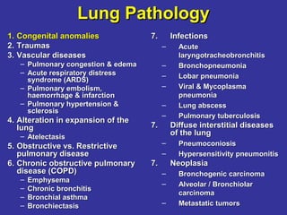
  • 1. Lung PathologyLung Pathology 1.1. Congenital anomaliesCongenital anomalies 2.2. TraumasTraumas 3.3. Vascular diseasesVascular diseases – Pulmonary congestion & edemaPulmonary congestion & edema – Acute respiratory distressAcute respiratory distress syndrome (ARDS)syndrome (ARDS) – Pulmonary embolism,Pulmonary embolism, haemorrhage & infarctionhaemorrhage & infarction – Pulmonary hypertension &Pulmonary hypertension & sclerosissclerosis 4.4. Alteration in expansion of theAlteration in expansion of the lunglung – AtelectasisAtelectasis 5.5. Obstructive vs. RestrictiveObstructive vs. Restrictive pulmonary diseasepulmonary disease 6.6. Chronic obstructive pulmonaryChronic obstructive pulmonary disease (COPD)disease (COPD) – EmphysemaEmphysema – Chronic bronchitisChronic bronchitis – Bronchial asthmaBronchial asthma – BronchiectasisBronchiectasis 7.7. InfectionsInfections – AcuteAcute laryngotracheobronchitislaryngotracheobronchitis – BronchopneumoniaBronchopneumonia – Lobar pneumoniaLobar pneumonia – Viral & MycoplasmaViral & Mycoplasma pneumoniapneumonia – Lung abscessLung abscess – Pulmonary tuberculosisPulmonary tuberculosis 7.7. Diffuse interstitial diseasesDiffuse interstitial diseases of the lungof the lung – PneumoconiosisPneumoconiosis – Hypersensitivity pneumonitisHypersensitivity pneumonitis 7.7. NeoplasiaNeoplasia – Bronchogenic carcinomaBronchogenic carcinoma – Alveolar / BronchiolarAlveolar / Bronchiolar carcinomacarcinoma – Metastatic tumorsMetastatic tumors
  • 2. Congenital anomaliesCongenital anomalies • Agenesis or hypoplasia of both lungs, one lung, or single lobes – Very common, seen in 10% of neonatal autopsies • Tracheal and bronchial anomalies (atresia, stenosis, tracheoesophageal fistula) • Vascular anomalies • Congenital lobar over-inflation (emphysema) • Foregut cysts – Bronchogenic cyst • Congenital pulmonary airway malformation – Harmatoma • Pulmonary sequestrations – Lung tissue without normal connection to airway – Blood supply from aorta or its branches.
  • 3. Cystic Diseases of the LungCystic Diseases of the Lung 3 conditions with large air space in the lung:3 conditions with large air space in the lung: 1.1. Congenital bronchogenic cystic diseaseCongenital bronchogenic cystic disease 2.2. Alveolar cystsAlveolar cysts 3.3. Bullous emphysemaBullous emphysema Only 1 & 2 are congenital diseases.Only 1 & 2 are congenital diseases. Bronchogenic cystsBronchogenic cysts • caused by abnormal formation of bronchioles in fetalcaused by abnormal formation of bronchioles in fetal developmentdevelopment • mechanism is unknownmechanism is unknown • GrossGross:: Delicate thin walled cyst. The lining is smooth andDelicate thin walled cyst. The lining is smooth and similar in color to the normal lungsimilar in color to the normal lung • HistologyHistology:: Ciliated respiratory type epithelium lines cystCiliated respiratory type epithelium lines cyst • Clinical courseClinical course:: may cause dyspnea, atelectasis, ormay cause dyspnea, atelectasis, or hyperinflation with obstructionhyperinflation with obstruction
  • 5. Alveolar CystsAlveolar Cysts • caused by progressive rupture of alveolar wallscaused by progressive rupture of alveolar walls  large air spaceslarge air spaces • GrossGross:: – More often multiple than are bronchogenic cystsMore often multiple than are bronchogenic cysts – Locate more centrally, most often in upper lobesLocate more centrally, most often in upper lobes – Walls are thin, fragile and poorly definedWalls are thin, fragile and poorly defined – Vary in size from minimal to entire lobe (= theVary in size from minimal to entire lobe (= the vanishing lung syndrome)vanishing lung syndrome) • HistologyHistology:: – Linings are compressed preexisting alveolar wallsLinings are compressed preexisting alveolar walls with fibrous trabeculation – Surrounding lung is compressed and atelectaticSurrounding lung is compressed and atelectatic • Clinical courseClinical course:: – Similar to bronchogenic cyst
  • 6. Lung TraumasLung Traumas 1. Mechanical injuries by either blunt or1. Mechanical injuries by either blunt or sharp objectssharp objects - Abrasion- Abrasion - Contusion- Contusion - Laceration- Laceration - Incised wound- Incised wound - Penetrating wound- Penetrating wound 2. Bone injuries2. Bone injuries -- FracturesFractures Most common causes :Most common causes : - Traffic injuries- Traffic injuries - Firearm injuries- Firearm injuries - Suicide- Suicide
  • 7. Lung: Stab wound Gunshot wound • Entrance is small & round • Exit is much larger with ragged edge
  • 8. Lung PathologyLung Pathology 1.1. Congenital anomaliesCongenital anomalies – Cystic diseasesCystic diseases 2.2. TraumasTraumas 3.3. Vascular diseasesVascular diseases – Pulmonary congestion &Pulmonary congestion & edemaedema – Acute respiratory distressAcute respiratory distress syndrome (ARDS)syndrome (ARDS) – Pulmonary embolism,Pulmonary embolism, haemorrhage & infarctionhaemorrhage & infarction – Pulmonary hypertension &Pulmonary hypertension & sclerosissclerosis 4.4. Alteration in expansion ofAlteration in expansion of the lungthe lung – AtelectasisAtelectasis 5.5. Obstructive vs. RestrictiveObstructive vs. Restrictive pulmonary diseasepulmonary disease 6.6. Chronic obstructiveChronic obstructive pulmonary disease (COPD)pulmonary disease (COPD) 7.7. InfectionsInfections – Acute laryngotracheobronchitisAcute laryngotracheobronchitis – BronchopneumoniaBronchopneumonia – Lobar pneumoniaLobar pneumonia – Viral & Mycoplasma pneumoniaViral & Mycoplasma pneumonia – Lung abscessLung abscess – Pulmonary tuberculosisPulmonary tuberculosis 7.7. Diffuse interstitial diseases of theDiffuse interstitial diseases of the lunglung – PneumoconiosisPneumoconiosis – Hypersensitivity pneumonitisHypersensitivity pneumonitis 7.7. NeoplasiaNeoplasia – Bronchogenic carcinomaBronchogenic carcinoma – Alveolar / Bronchiolar carcinomaAlveolar / Bronchiolar carcinoma – Metastatic tumorsMetastatic tumors
  • 9. Pulmonary Congestion & EdemaPulmonary Congestion & Edema EtiologyEtiology:: • Generally associated with left-sided heart failureGenerally associated with left-sided heart failure • Etiology of heart failure includes myocardial damageEtiology of heart failure includes myocardial damage associated with valvular, coronary arterial, or intrinsicassociated with valvular, coronary arterial, or intrinsic myocardial injurymyocardial injury • Other causes include blood loss and peripheralOther causes include blood loss and peripheral vasodilatation (shock)vasodilatation (shock) PathogenesisPathogenesis:: • Increased left atrial pressure leads to increasedIncreased left atrial pressure leads to increased pulmonary venous pressure and then to increasedpulmonary venous pressure and then to increased pulmonary capillary pressure with congestion andpulmonary capillary pressure with congestion and ultimately transudation into the alveolar spaceultimately transudation into the alveolar space (edema)(edema)
  • 10. Classification and Causes of Pulmonary EdemaClassification and Causes of Pulmonary Edema 1.1. Hemodynamic EdemaHemodynamic Edema – Increased hydrostatic pressure (increased pulmonary venousIncreased hydrostatic pressure (increased pulmonary venous pressure)pressure) – Left-sided heart failure (common)Left-sided heart failure (common) – Volume overloadVolume overload – Pulmonary vein obstructionPulmonary vein obstruction – Decreased oncotic pressure (less common)Decreased oncotic pressure (less common) – Lymphatic obstruction (rare)Lymphatic obstruction (rare) 2.2. Edema Due to Microvascular Injury (Alveolar Injury)Edema Due to Microvascular Injury (Alveolar Injury) – Infections: pneumonia, septicemiaInfections: pneumonia, septicemia – Inhaled gases: oxygen, smokeInhaled gases: oxygen, smoke – Liquid aspiration: gastric contents, near-drowningLiquid aspiration: gastric contents, near-drowning – Drugs and chemicals: chemotherapeutic agents (Drugs and chemicals: chemotherapeutic agents (bleomycinbleomycin), other), other medicationsmedications (amphotericin B ),(amphotericin B ), heroin, kerosene, paraquatheroin, kerosene, paraquat – Shock, traumaShock, trauma – RadiationRadiation – Transfusion relatedTransfusion related 3.3. Edema of Undetermined OriginEdema of Undetermined Origin – High altitudeHigh altitude – Neurogenic (central nervous system trauma)Neurogenic (central nervous system trauma) •HypoalbuminHypoalbumin emiaemia •NephroticNephrotic syndromesyndrome •LiverLiver diseasedisease •Protein-Protein- losinglosing enteropathiesenteropathies
  • 11. Pulmonary Congestion & EdemaPulmonary Congestion & Edema GrossGross:: • Heavy wet lungsHeavy wet lungs • Deep red in the congested lower lobesDeep red in the congested lower lobes • Ooze bloody fluid on sectioningOoze bloody fluid on sectioning • When chronic, rusty discoloration from haemosiderinWhen chronic, rusty discoloration from haemosiderin deposit + fibrosis may occurdeposit + fibrosis may occur  firm & brown indurationfirm & brown induration HistologyHistology:: • Dilated blood vessels containing abundant RBC'sDilated blood vessels containing abundant RBC's • Scattered RBC's may be found in the alveoli due toScattered RBC's may be found in the alveoli due to diapedesis which lead to Hemosiderin-laden macrophagesdiapedesis which lead to Hemosiderin-laden macrophages (heart failure cells) appearing within a few days(heart failure cells) appearing within a few days • Protein-rich edema fluid may also be presentProtein-rich edema fluid may also be present
  • 12. Pulmonary CongestionPulmonary Congestion & Edema:& Edema: • granular precipitate in alveolargranular precipitate in alveolar spaces = edematous fluidspaces = edematous fluid • haemosiderin-laden macrophagehaemosiderin-laden macrophage = heart failure cell= heart failure cell
  • 13. Acute Respiratory Distress SyndromeAcute Respiratory Distress Syndrome Etiology:Etiology: • Result of diffuse capillary damage in the lung with capillary leakResult of diffuse capillary damage in the lung with capillary leak Pathogenesis:Pathogenesis: • Endothelial cell damage (direct, secondary to inflammatory cells and mediatorsEndothelial cell damage (direct, secondary to inflammatory cells and mediators especially neutrophils, due to endotoxin)especially neutrophils, due to endotoxin) • Capillary leak into the alveoli with accumulation of edema and proteinaceous,Capillary leak into the alveoli with accumulation of edema and proteinaceous, necrotic material that forms hyaline membrane.necrotic material that forms hyaline membrane. • Edema interferes with gas exchangeEdema interferes with gas exchange GrossGross:: • Heavy lungs which are deep reddish purple and stiffHeavy lungs which are deep reddish purple and stiff HistologyHistology:: • Congestion of the vessels and proteinaceous acellular edema in the alveoliCongestion of the vessels and proteinaceous acellular edema in the alveoli • Accumulation of vividly eosinophilic, acellular hyaline membranes againstAccumulation of vividly eosinophilic, acellular hyaline membranes against alveolar septaealveolar septae • Organize into circular fibrous swirls which over time replace alveoliOrganize into circular fibrous swirls which over time replace alveoli • Occasionally resolve completelyOccasionally resolve completely
  • 14. Conditions Associated with Development ofConditions Associated with Development of Acute Respiratory Distress SyndromeAcute Respiratory Distress Syndrome 1.1. InfectionInfection • Sepsis*Sepsis* • Diffuse pulmonary infections*Diffuse pulmonary infections*  Viral,Viral, MycoplasmaMycoplasma, and, and PneumocystisPneumocystis pneumonia;pneumonia; miliary tuberculosismiliary tuberculosis • Gastric aspiration*Gastric aspiration* 2.2. Physical/InjuryPhysical/Injury • Mechanical trauma, includingMechanical trauma, including head injuries*head injuries* • Pulmonary contusionsPulmonary contusions • Near-drowningNear-drowning • Fractures with fat embolismFractures with fat embolism • BurnsBurns • Ionizing radiationIonizing radiation 3.3. Inhaled IrritantsInhaled Irritants • Oxygen toxicityOxygen toxicity • SmokeSmoke • Irritant gases and chemicalsIrritant gases and chemicals 4.4. Chemical InjuryChemical Injury • Heroin or methadoneHeroin or methadone overdoseoverdose • Acetylsalicylic acidAcetylsalicylic acid • Barbiturate overdoseBarbiturate overdose • ParaquatParaquat 5.5. Hematologic ConditionsHematologic Conditions • Multiple transfusionsMultiple transfusions • DisseminatedDisseminated intravascular coagulationintravascular coagulation 6.6. PancreatitisPancreatitis 7.7. UremiaUremia 8.8. Cardio-pulmonary BypassCardio-pulmonary Bypass 9.9. Hypersensitivity ReactionsHypersensitivity Reactions • Organic solventsOrganic solvents • DrugsDrugs
  • 17. Lung: Hyaline membrane disease from oxygen toxicity
  • 18. Pulmonary EmbolismPulmonary Embolism EtiologyEtiology:: • Most pulmonary emboli are from deep leg vein thrombiMost pulmonary emboli are from deep leg vein thrombi • In situ thromboses are rare and develop in pulmonary HT only PathogenesisPathogenesis:: • Conditions which promote deep vein stasis such as immobility, hypercoagulableConditions which promote deep vein stasis such as immobility, hypercoagulable states, and endothelial damage lead to thrombosisstates, and endothelial damage lead to thrombosis GrossGross:: • Large or medium sized pulmonary artery involvedLarge or medium sized pulmonary artery involved • Deep reddish purple firm material containing some fibrin strands or lines of ZahnDeep reddish purple firm material containing some fibrin strands or lines of Zahn (alternating platelet and red cell layers)(alternating platelet and red cell layers) • May be quite adherent to vessel wall if organization has begunMay be quite adherent to vessel wall if organization has begun • Smaller strands of thrombus may extend into smaller vesselsSmaller strands of thrombus may extend into smaller vessels HistologyHistology:: • Mixture of red blood cells, platelets and fibrinMixture of red blood cells, platelets and fibrin • Over a few days capillaries, smooth muscle cells and fibroblasts grow into theOver a few days capillaries, smooth muscle cells and fibroblasts grow into the embolus from the pulmonary vessel wallembolus from the pulmonary vessel wall • Surface of the embolus will become endothelialisedSurface of the embolus will become endothelialised • Recanalisation may occurRecanalisation may occur
  • 19. Pulmonary EmbolismPulmonary Embolism Clinical CourseClinical Course:: • Large emboli obstructing more than 1/2Large emboli obstructing more than 1/2 pulmonary circulation may cause sudden deathpulmonary circulation may cause sudden death • Smaller emboli may result in nothing moreSmaller emboli may result in nothing more severe than hemorrhage if sufficient bronchialsevere than hemorrhage if sufficient bronchial vascular or collateral supply to distalvascular or collateral supply to distal parenchymaparenchyma • If no other supply to the distal lung orIf no other supply to the distal lung or underlying chronic pulmonary disease infarctunderlying chronic pulmonary disease infarct resultsresults
  • 21. pulmonary thromboembolus in a large pulmonary artery. There are interdigitating areas of pale pink and red that form the "lines of Zahn" representing layers of red cells, platelets, and fibrin which are layed down in the vessel as the thrombus forms The fibrous bands of connective tissue across this branch of pulmonary artery indicate organization of a remote pulmonary thromboembolus. If many pulmonary arteries are involved by this process, pulmonary hypertension could result
  • 22. Lung: Hemorrhagic InfarctLung: Hemorrhagic Infarct
  • 23. Pulmonary Hypertension & SclerosisPulmonary Hypertension & Sclerosis • Primary pulmonary hypertension –Primary pulmonary hypertension – unknownunknown causes, 50% related to mutation of bone morphogeneticcauses, 50% related to mutation of bone morphogenetic protein receptor type 2 (BMPR2) geneprotein receptor type 2 (BMPR2) gene • Secondary pulmonary hypertension –Secondary pulmonary hypertension –knownknown causes for cor pulmonalecauses for cor pulmonale (e.g. COPD, restrictive lung dis,(e.g. COPD, restrictive lung dis, congenital or acquired heart dis, recurrent thrombo-emboli,congenital or acquired heart dis, recurrent thrombo-emboli, auto-immune dis)auto-immune dis) PathogenesisPathogenesis:: • Overactivity of sympathetic autonomic systemOveractivity of sympathetic autonomic system  chronicchronic vasoconstrictionvasoconstriction  pulmonary HTpulmonary HT • Multiple small pulmonary emboliMultiple small pulmonary emboli  thrombosisthrombosis • Some hypersensitivity diseasesSome hypersensitivity diseases  primary vascular lesionprimary vascular lesion associated with hyper-Ig & arthritisassociated with hyper-Ig & arthritis • Dietary & medicinal agentsDietary & medicinal agents  pulmonary HTpulmonary HT
  • 24. Pathogenesis of primary pulmonary hypertensionPathogenesis of primary pulmonary hypertension
  • 25. Pulmonary Hypertension & SclerosisPulmonary Hypertension & Sclerosis GrossGross:: • Atheromatous deposits from the main pulmonary arteriesAtheromatous deposits from the main pulmonary arteries down to arterioles, resembling atherosclerosis in thedown to arterioles, resembling atherosclerosis in the systemic arteries but of milder degreesystemic arteries but of milder degree HistologyHistology:: • Medial hypertrophy of the medium-sized arteriesMedial hypertrophy of the medium-sized arteries • Intemal thickening and fibrosis + adventitial fibrosisIntemal thickening and fibrosis + adventitial fibrosis Clinical courseClinical course:: • Early signs = dyspnea, fatigue & occasional syncopeEarly signs = dyspnea, fatigue & occasional syncope • Chest pain, respiratory distress & cyanosisChest pain, respiratory distress & cyanosis • Right ventricular hypertrophy & cor pulmonale in 2-8 yrsRight ventricular hypertrophy & cor pulmonale in 2-8 yrs
  • 26. Pulmonary Hypertension & SclerosisPulmonary Hypertension & Sclerosis Thickened small peripheralThickened small peripheral pulmonary arteries with thepulmonary arteries with the larger pulmonary arterieslarger pulmonary arteries demonstrate atherosclerosisdemonstrate atherosclerosis with pulmonary hypertensionwith pulmonary hypertension Thickening of the smallThickening of the small arteries along witharteries along with reduplication to form areduplication to form a plexiform lesion.plexiform lesion.
  • 27. AtelectasisAtelectasis Incomplete expansion or collapse of previously inflatedIncomplete expansion or collapse of previously inflated lung, characterised by airless lung parenchymalung, characterised by airless lung parenchyma Applied only for structurally normal alveoli that are re-Applied only for structurally normal alveoli that are re- expandable when removing underlying causeexpandable when removing underlying cause Etiology:Etiology: • Obstruction of airwayObstruction of airway • Compression with fluid/blood/neoplasm/air in pleural spaceCompression with fluid/blood/neoplasm/air in pleural space • Loss of surfactantLoss of surfactant Pathogenesis:Pathogenesis: • Complete obstruction of inflow results in eventual absorptionComplete obstruction of inflow results in eventual absorption of air; partial obstruction results in hyperinflationof air; partial obstruction results in hyperinflation • Compression results from increased intra-thoracic pressureCompression results from increased intra-thoracic pressure by accumulation of material generally in pleural spacesby accumulation of material generally in pleural spaces • Fibrotic changes in the lung or pleuraFibrotic changes in the lung or pleura
  • 28. AtelectasisAtelectasis 1. Resorption atelectasis = complete obstruction of an airway resorption of the oxygen trapped in the dependent alveoli, without impairment of blood flow through the affected alveolar walls. 2. Compression atelectasis results whenever the pleural cavity is partially or completely filled by fluid exudate, tumor, blood, or air. 3. Contraction atelectasis occurs when local or generalized fibrotic changes in the lung or pleura prevent full expansion.
  • 29. AtelectasisAtelectasis GrossGross:: • Dark purple, airless (non-crepitant) tissue as compared toDark purple, airless (non-crepitant) tissue as compared to normal tan aerated lung.normal tan aerated lung. • Hyperinflated regions are tan and pillowyHyperinflated regions are tan and pillowy HistologyHistology:: • Collapse of alveolar septae against each otherCollapse of alveolar septae against each other • May be difficult to separate from artifacts of sectioningMay be difficult to separate from artifacts of sectioning Clinical courseClinical course:: • Reversible process after correcting underlying problemReversible process after correcting underlying problem • Reduction of ventilatory functionReduction of ventilatory function  respiratory distressrespiratory distress  death in massive collapse of both lungsdeath in massive collapse of both lungs
  • 31. COPD: EmphysemaCOPD: Emphysema Abnormal enlargement of the airAbnormal enlargement of the air spaces (over-inflation) distal tospaces (over-inflation) distal to terminal bronchioles, accompaniedterminal bronchioles, accompanied by destruction of their wallsby destruction of their walls 4 types of emphysema:4 types of emphysema: 1.1. Centrilobular (centriacinar) emphysemaCentrilobular (centriacinar) emphysema 2.2. Panlobular (panacinar) emphysemaPanlobular (panacinar) emphysema 3.3. Paraseptal emphysemaParaseptal emphysema 4.4. Irregular emphysemaIrregular emphysema
  • 33. Etiology:Etiology: • Cigarette smoking responsible for vast majority ofCigarette smoking responsible for vast majority of centriacinar emphysemacentriacinar emphysema Pathogenesis:Pathogenesis: • At terminal bronchiole level air flow suddenlyAt terminal bronchiole level air flow suddenly diminishes dropping particulates into adjacentdiminishes dropping particulates into adjacent alveolialveoli • Neutrophil and macrophage elastases turned onNeutrophil and macrophage elastases turned on by particulates or other components of smokeby particulates or other components of smoke • Septal destruction secondary to excess elastaseSeptal destruction secondary to excess elastase and protease activity.and protease activity. COPD: EmphysemaCOPD: Emphysema
  • 35. Centrilobular EmphysemaCentrilobular Emphysema Gross:Gross: • Centrilobular or smoker's emphysema shows air space enlargement mixed withCentrilobular or smoker's emphysema shows air space enlargement mixed with normal airspaces. (Air space enlargement does not involve all alveoli, hence notnormal airspaces. (Air space enlargement does not involve all alveoli, hence not panacinar)panacinar) • Largest spaces representing alveoli which have coalesced with destruction ofLargest spaces representing alveoli which have coalesced with destruction of the septae found in upper portions of all lobes or large bleb consisting of a singlethe septae found in upper portions of all lobes or large bleb consisting of a single air containing space may be seenair containing space may be seen • Black discoloration of walls of spacesBlack discoloration of walls of spaces • Bronchovascular structures stand out from the parenchyma due to loss ofBronchovascular structures stand out from the parenchyma due to loss of parenchymal tissueparenchymal tissue • Pillowy soft lungs that may cover the heartPillowy soft lungs that may cover the heart HistologyHistology:: • Enlarged air spaces with broken septae in the central portion of the acinusEnlarged air spaces with broken septae in the central portion of the acinus around the terminal bronchiolearound the terminal bronchiole • Septal tips have blunt endsSeptal tips have blunt ends • Little fibrosisLittle fibrosis • Many carbon laden macrophagesMany carbon laden macrophages Clinical courseClinical course:: • Patients with pure emphysema develop progressive dyspnea and weight lossPatients with pure emphysema develop progressive dyspnea and weight loss due to loss of oxygen delivery to periphery.due to loss of oxygen delivery to periphery. • "Pink puffer" with slowed forced expirations"Pink puffer" with slowed forced expirations
  • 38. Centrilobular EmphysemaCentrilobular Emphysema The loss of alveolar walls withThe loss of alveolar walls with emphysema is demonstrated.emphysema is demonstrated. Remaining airspaces areRemaining airspaces are dilated.dilated.
  • 39. Panlobular (Panacinar) Emphysema:Panlobular (Panacinar) Emphysema: • The acini are uniformly enlarged from the respiratory bronchioles to alveoliThe acini are uniformly enlarged from the respiratory bronchioles to alveoli • Tend to occur in lower zone & anterior margins of lung, most severe at theTend to occur in lower zone & anterior margins of lung, most severe at the basebase • Associated withAssociated with αα1-antitrypsin deficiency (A1AT1-antitrypsin deficiency (A1AT inhibition of elastolyticinhibition of elastolytic activity of PMN & mactivity of PMN & mφφ in the lung)in the lung) Paraseptal (Distal Acinar) Emphysema:Paraseptal (Distal Acinar) Emphysema: • The peripheral part of acinus (alveolar ducts & sacs) is selectively involvedThe peripheral part of acinus (alveolar ducts & sacs) is selectively involved  striking multiple continuous, enlarged air spaces of 0.5 mm to > 2 cmstriking multiple continuous, enlarged air spaces of 0.5 mm to > 2 cm adjacent to pleuraadjacent to pleura • More severe in the upper lungsMore severe in the upper lungs  spontaneous pneumothoraxspontaneous pneumothorax • Commonly occurs adjacent to areas of fibrosis, scarring or atelectasisCommonly occurs adjacent to areas of fibrosis, scarring or atelectasis Irregular Emphysema:Irregular Emphysema: • The acinus is irregularly involved, most associated with scarringThe acinus is irregularly involved, most associated with scarring  destructive changes of acini adjacent to scarsdestructive changes of acini adjacent to scars • Common results of healing tuberculosis, sarcoidosis or otherCommon results of healing tuberculosis, sarcoidosis or other granulomatous diseases of the lunggranulomatous diseases of the lung • Most are asymptomaticMost are asymptomatic
  • 41. Chronic BronchitisChronic Bronchitis 1. Precipitating causes: – prior acute URI, smoking, – pollution exposure, – hereditary factors. – More common in male > 40 yrs old. 2. Causative agents: mostly normal flora e.g. H. influenzae, S. pneumoniae and/or M. catarrhalis 3. Signs & Symptoms: – Chronic persistent cough with increased sputum production – Progressive deterioration of lung function
  • 42. Chronic BronchitisChronic Bronchitis 1. Diagnosis: – History of persistent cough & excessive mucus production lasting for at least 3 months over a 2-year period. – P.E reveals chest rales and wheezes – X-ray + sputum exam: Not useful 2. Treatment: – Enable the patient to breathe freely, to relieve symptoms and to prevent complications. (Health education + stop smoking) – Bronchodilators + postural drainage. – Do not use expectorant or cough suppressant. – Vaccines against influenza & pneumonia may reduce complications – Coticosteroids therapy by oral or inhaler in moderate or severe cases – Oxygen therapy in severe cases – Naso-tracheal tube or Tracheostomy in end-stage case
  • 43. Chronic BronchitisChronic Bronchitis Persistent cough with sputum production for at least 3Persistent cough with sputum production for at least 3 months in at least 2 consecutive yearsmonths in at least 2 consecutive years Most frequent in middle-aged men, esp. urban adultMost frequent in middle-aged men, esp. urban adult population (10-25%)population (10-25%) Pathogenesis:Pathogenesis: • Chronic irritation by inhaled substances esp. cigaretteChronic irritation by inhaled substances esp. cigarette  excessive mucusexcessive mucus secretion via Vagus nerve stimulation, destroy ciliary action of epitheliumsecretion via Vagus nerve stimulation, destroy ciliary action of epithelium and induce squamous metaplasia & atypical dysplasiaand induce squamous metaplasia & atypical dysplasia • Microbiologic infections, most common areMicrobiologic infections, most common are H. influenzae & D. pneumoniaeH. influenzae & D. pneumoniae Morphology:Morphology: • Hyperemia, swelling & bogginess of mucous membranes of small bronchi &Hyperemia, swelling & bogginess of mucous membranes of small bronchi & bronchioles with excessive mucinous or mucopurulent secretionsbronchioles with excessive mucinous or mucopurulent secretions • Marked thickening (hypertrophy) of the mucous glands with only slightMarked thickening (hypertrophy) of the mucous glands with only slight increase in number of goblet cellsincrease in number of goblet cells  lumen narrowing, mucous plugging &lumen narrowing, mucous plugging & peribronchial inflammatory scarring (most severe =peribronchial inflammatory scarring (most severe = Bronchiolitis fibrosaBronchiolitis fibrosa obliteransobliterans))
  • 44. Chronic Bronchitis showing a bronchus with increasedChronic Bronchitis showing a bronchus with increased numbers of chronic inflammatory cells in the submucosa.numbers of chronic inflammatory cells in the submucosa.
  • 45. Bronchial AsthmaBronchial Asthma Increased irritability of tracheobronchial treeIncreased irritability of tracheobronchial tree  paroxysmal narrowingparoxysmal narrowing of bronchial airways that is reversible spontaneously or by treatmentof bronchial airways that is reversible spontaneously or by treatment Distressing due to unpredictable attacks of severe dyspnea & wheezingDistressing due to unpredictable attacks of severe dyspnea & wheezing from acute bronchospasmmfrom acute bronchospasmm  death indeath in status astmaticusstatus astmaticus Pathogenesis:Pathogenesis: • Extrinsic or allergic/atopic IgE-mediated hypersensitivity reactionExtrinsic or allergic/atopic IgE-mediated hypersensitivity reaction  releaserelease of histamine, SRS-A, ECF-A & PAFof histamine, SRS-A, ECF-A & PAF • Intrinsic (idiosyncratic) reactionIntrinsic (idiosyncratic) reaction Morphology:Morphology: • The lungs are overdistended due to overinflation & small areas of atelectasisThe lungs are overdistended due to overinflation & small areas of atelectasis • Occlusion of bronchi & bronchioles by thick, tenacious, mucous plugs thatOcclusion of bronchi & bronchioles by thick, tenacious, mucous plugs that are shed epithelium (are shed epithelium (Curschmann’ s spiralsCurschmann’ s spirals) + eosinophils with) + eosinophils with Charcot-Charcot- Leyden crystalsLeyden crystals • Thickening of basement membrane of bronchial epithelium + edema &Thickening of basement membrane of bronchial epithelium + edema & inflammatory infiltrate in bronchial walls with hypertrophy of submucosalinflammatory infiltrate in bronchial walls with hypertrophy of submucosal mucous glands and brochial wall musclemucous glands and brochial wall muscle • Emphysematous changes and/or bacterial infections may superveneEmphysematous changes and/or bacterial infections may supervene
  • 46. Lung: Bronchial asthma Note the mucous plug in bronchi & bronchioles Note a submucosa widened by smooth muscle hypertrophy, edema, and inflammation (mainly eosinophils) & mucus in the bronchial lumen.
  • 47. BronchiectasisBronchiectasis Chronic necrotising infection of bronchi & bronchiolesChronic necrotising infection of bronchi & bronchioles leading to or associated with abnormal dilatation of theseleading to or associated with abnormal dilatation of these airwaysairways Etiology:Etiology: • Bronchial obstruction caused by tumors, foreign bodies, mucousBronchial obstruction caused by tumors, foreign bodies, mucous impactionimpaction • Scarring from tuberculosisScarring from tuberculosis • Asthma, chronic bronchitis & emphysemaAsthma, chronic bronchitis & emphysema • Fibrocystic disease of pancreas & lungFibrocystic disease of pancreas & lung • Intra-lobular sequestration of the lung (esp. LLL is not supplied byIntra-lobular sequestration of the lung (esp. LLL is not supplied by pulmonary artery but directly from systemic arteries)pulmonary artery but directly from systemic arteries) Pathogenesis:Pathogenesis: • Bronchial obstructionBronchial obstruction  atelectasisatelectasis  dilatation of the walls ofdilatation of the walls of patent airwayspatent airways • InfectionInfection  bronchial wall inflammation, weakening & dilatation +bronchial wall inflammation, weakening & dilatation + extensive damageextensive damage  endobronchial obliteration & atelectasisendobronchial obliteration & atelectasis  bronchiectasis around atelectasisbronchiectasis around atelectasis
  • 48. Bronchiectasis Morphology: • Marked dilated affected bronchi & bronchioles  long tube-like = cylindroid bronchiectasis or may  fusiform or saccular bronchiectasis • Suppurative, yellow-green or haemorrhagic exudate fill the lumen with necrotic black edematous ulcerated mucosa. Pseudostratified columnar or squamous metaplasia of the remaining epithelium is noted. • Patchy emphysema & atelectasis in lung parenchyma • May progress to lung abscess or extend to pleura  suppurative pleuritis
  • 49. Respiratory Tract Infection (RTI)Respiratory Tract Infection (RTI) • Acute LaryngotracheobronchitisAcute Laryngotracheobronchitis • Acute BronchitisAcute Bronchitis • Chronic BronchitisChronic Bronchitis • Acute Exacerbations of ChronicAcute Exacerbations of Chronic Bronchitis (AECB)Bronchitis (AECB)
  • 50. Acute BronchitisAcute Bronchitis 1.1. Causative agentsCausative agents:: – Mostly viral infectionMostly viral infection – OccasionallyOccasionally M. pneunoniae, C. pneunoniaeM. pneunoniae, C. pneunoniae 2.2. Signs & SymptomsSigns & Symptoms:: – Cough with increased sputum production and mildCough with increased sputum production and mild dyspneadyspnea 3.3. DiagnosisDiagnosis:: – History & P.E. to rule out pneumonia & tuberculosisHistory & P.E. to rule out pneumonia & tuberculosis – X-ray + sputum examX-ray + sputum exam 4.4. TreatmentTreatment:: – Supportive: cough suppressants are the mainstay.Supportive: cough suppressants are the mainstay. – Expectorants & bronchodilators may be helpful.Expectorants & bronchodilators may be helpful. – Antibiotics in cases of definite bacterial infectionAntibiotics in cases of definite bacterial infection
  • 51. AECBAECB • COPD affects 5% of the US populationCOPD affects 5% of the US population • Suffer from cough and daily sputum productionSuffer from cough and daily sputum production for at least 3 consecutive months in 2for at least 3 consecutive months in 2 consecutive yearsconsecutive years • Also have symptoms of acute exacerbationAlso have symptoms of acute exacerbation (increased cough, sputum production, and(increased cough, sputum production, and purulence, dyspnea)purulence, dyspnea) – Type I:Type I: Increased dyspnea, cough or sputumIncreased dyspnea, cough or sputum volume, and purulent sputumvolume, and purulent sputum – Type II:Type II: 2 of the above symptoms2 of the above symptoms – Type III:Type III: 1 of the above symptoms + fever/chill/malaise1 of the above symptoms + fever/chill/malaise Anthonisen NR. Ann Intern Med. 1987;106:196-204.
  • 52. Organisms in AECBOrganisms in AECB • Most common: H. influenzae, S. pneumoniae and Moraxella catarrhalis • Less common: M. pneumoniae, C. pneumoniae and Bordetella pertussis • Rare: Pseudomonas aeruginosa • Some patients may also be implicated by Viruses, allergies and asthma • Fever and a change in the appearance or amount of sputum are signs of bacterial infection. Anthonisen NR. Ann Intern Med. 1987;106:196-204.
  • 53. – Sixth leading cause of death – Leading cause of death due to infectious disease – More than 3 million cases of CAP per year – 500,000 hospitalizations per year – 45,000 deaths per year – Cost: $21 billion Community-AcquiredCommunity-Acquired Pneumonia (CAP)Pneumonia (CAP) Bartlett JG. CID. 2000;31:347-383. File and Tan. Curr Opin Pul Med. 1997;3:89-97. Marston et al. Arch Int Med. 1997;157:1709-1718. EpidemiologyEpidemiology
  • 54. CAPCAP 1. Risk factors: Smoking, Viral infection, especially influenza, Chronic lung disease, Old age, Alcohol/drug abuse, Compromised immune system 2. Causes: • Aspiration of upper respiratory flora into the lungs • Inhalation of airborne droplets • Blood dissemination from another site in the body
  • 55. CAPCAP 3. Causative organisms: • S. pneumoniae (or diplococcal pneumonia) • H. influenzae pneumonia occurs primarily in children & elderly • M. catarrhalis occurs primarily in the elderly • Atypical bacteria e.g. M. pneumonia (in young adults), L. pneumophilia (in elderly) • Special populations: • Pneumocystis carinii pneumonia in patients with HIV • Staphylococcal pneumonia in intravenous drug abusers • Klebsiella pneumoniae almost exclusively in alcoholics • P. aeruginosa pneumonia in patients with lung disease
  • 56. CAPCAP 3. Signs & Symptoms: • Sudden onset of fever • Productive cough with purulent sputum (“rusty sputum”) • In some patients, chest pain. • P.E. shows rales and altered breath sounds indicating pulmonary consolidation, fatigue, myalgia, etc. 3. Diagnosis: • Hx & P.E. • X-ray • Sputum exam (Gram stain & bacterial C/S) • CBC, blood C/S, serology
  • 57. EpidemiologyEpidemiology – Common hospital-acquired infection, second only to UTI.Common hospital-acquired infection, second only to UTI. – Average incidence of 6/1,000 patients.Average incidence of 6/1,000 patients. Risk factorsRisk factors – Surgery, especially chest or abdominalSurgery, especially chest or abdominal – Endotracheal intubation / Mechanical ventilationEndotracheal intubation / Mechanical ventilation – Inability to swallowInability to swallow – Depressed consciousnessDepressed consciousness – Supine positionSupine position – 70+ years70+ years – Chronic lung diseaseChronic lung disease – ICUICU Hospital-Acquired Pneumonia (HAP)Hospital-Acquired Pneumonia (HAP)
  • 58. HAPHAP Causes of nosocomial pneumoniaCauses of nosocomial pneumonia Sources of pathogensSources of pathogens Causative OrganismsCausative Organisms Aspiration ofAspiration of respiratory florarespiratory flora S. pneumoniae, H. influenzaeS. pneumoniae, H. influenzae,, EnterobacterEnterobacter spp. (includingspp. (including MDR strains)MDR strains) AirborneAirborne transmissiontransmission L. pneumophiliaL. pneumophilia Aspiration of stomachAspiration of stomach contentscontents AnaerobesAnaerobes MechanicalMechanical ventilationventilation H. influenzaeH. influenzae,, P. aeuruginosaP. aeuruginosa,, EnterobacterEnterobacter spp. (includingspp. (including MDR strains), MRSAMDR strains), MRSA Catheters and otherCatheters and other invasive instrumentsinvasive instruments S. aureusS. aureus, MRSA, MRSA
  • 59. HAPHAP Signs/Symptoms & DiagnosisSigns/Symptoms & Diagnosis:: • Fever with dyspneaFever with dyspnea • Cough with purulent sputumCough with purulent sputum • None in some cases, just only suspiciousNone in some cases, just only suspicious • Diagnostic procedures are similar to those in CAPDiagnostic procedures are similar to those in CAP TreatmentTreatment:: • Empiric antibiotics to cover nosocomial infectionEmpiric antibiotics to cover nosocomial infection • Change antibiotics according to C/S in caseChange antibiotics according to C/S in case unresponsive to empiric therapyunresponsive to empiric therapy • Supportive treatmentSupportive treatment
  • 60. BronchopneumoniaBronchopneumonia Note pattern of patchy distribution. The consolidatedNote pattern of patchy distribution. The consolidated areas closely match the pattern of lung lobules ("lobular" pneumonia).areas closely match the pattern of lung lobules ("lobular" pneumonia).
  • 61. 07/22/13 Wide-spread fibrinosuppurative consolidation of large areasWide-spread fibrinosuppurative consolidation of large areas to whole lobeto whole lobe 4 stages of inflammatory response as follows:4 stages of inflammatory response as follows: 1.1. CongestionCongestion – 24 hours of developing bacterial infection24 hours of developing bacterial infection – Vascular congestion, intra-alveolar fluid + PMNsVascular congestion, intra-alveolar fluid + PMNs 22.. Red hepatisationRed hepatisation – Massive confluent exudationMassive confluent exudation  obscure pulmonary architectureobscure pulmonary architecture – Extravastion of RBCExtravastion of RBC  red discolourationred discolouration – WBC engulfed bacteriaWBC engulfed bacteria 33.. Gray hepatisationGray hepatisation – Fibrinosuppurative exudateFibrinosuppurative exudate grayish brown dry surfacegrayish brown dry surface 44.. ResolutionResolution – Progressive enzymatic digestionProgressive enzymatic digestion  thin-out alveolar exudatethin-out alveolar exudate Lobar pneumoniaLobar pneumonia
  • 62. Lobar pneumonia in whichLobar pneumonia in which consolidation of the entireconsolidation of the entire left upper lobeleft upper lobe Lobar pneumonia withLobar pneumonia with consolidation of the lowerconsolidation of the lower lobelobe
  • 63. Pneumonia complicated byPneumonia complicated by purulent exudative pleuritispurulent exudative pleuritis Lung abscess cavities that canLung abscess cavities that can be a source for septicemiabe a source for septicemia
  • 64. At the left the alveoli are filled with a neutrophilic exudate (= consolidation seen grossly)At the left the alveoli are filled with a neutrophilic exudate (= consolidation seen grossly) with the bronchopneumonia. More severe pneumonias with destruction of lung tissue,with the bronchopneumonia. More severe pneumonias with destruction of lung tissue, forming lung abscess and hemorrhage.forming lung abscess and hemorrhage.
  • 65. The yellow tan, and veryThe yellow tan, and very firm chronic abscess atfirm chronic abscess at RML, caused by NocardiaRML, caused by Nocardia Chronic abscess = large areas of pink necrotic tissue bordered by granulation tissue. AFB bacilli of Nocardia are seen.
  • 66. 07/22/13 11.. URI / common coldURI / common cold 22.. InfluenzaInfluenza 33.. BronchiolitisBronchiolitis 44.. PneumoniaPneumonia RESPIRATORY VIRAL INFECTIONSRESPIRATORY VIRAL INFECTIONS
  • 67. 07/22/13 Clinical features are nonspecific from mild to severe.Clinical features are nonspecific from mild to severe. (cough, fever, myalgia to dyspnea, toxicity )(cough, fever, myalgia to dyspnea, toxicity ) PathologyPathology 1. Squamous or cuboidal metaplasia of bronchial1. Squamous or cuboidal metaplasia of bronchial epitheliumepithelium 2. Interstitial mononuclear cell infiltrates of alveolar2. Interstitial mononuclear cell infiltrates of alveolar septa & peribronchial tissuessepta & peribronchial tissues 3. Alveolar edema with or without exudate3. Alveolar edema with or without exudate 4. Congestion, emphysema, atelectasis & hemorrhage4. Congestion, emphysema, atelectasis & hemorrhage 5. Hyaline membrane formation in alveoli5. Hyaline membrane formation in alveoli 6. Giant cell formation6. Giant cell formation RESPIRATORY VIRAL INFECTIONSRESPIRATORY VIRAL INFECTIONS
  • 68. Measles pneumoniaMeasles pneumonia Measles & CMVMeasles & CMV Granulomatous reactionGranulomatous reaction Giant cell with inclusionGiant cell with inclusion
  • 69. Respiratory syntytial virus (RSV) pneumonia: Note a typical giant cell with a round, pink intracytoplasmic inclusion. RSV accounts for many cases of pneumonia in children under 2 years, and can be a cause for death in infants 1 to 6 months of age or older
  • 70. TUBERCULOSISTUBERCULOSIS • Jean Antoine Villemin demonstratedJean Antoine Villemin demonstrated transmission of Tbc in rabbit in 1868transmission of Tbc in rabbit in 1868 • Robert Koch first discovered tubercleRobert Koch first discovered tubercle bacilli in 1882bacilli in 1882 MycobacteriumMycobacterium = mould-like pellicular= mould-like pellicular growth of bacilli on liquid medium.growth of bacilli on liquid medium. Characterized by acid fastness due toCharacterized by acid fastness due to complex lipid rich cell wall, no capsule,complex lipid rich cell wall, no capsule, aerobic, non-sporing, non-motile &aerobic, non-sporing, non-motile & divided by binary fission.divided by binary fission.
  • 71. Mycobacterium tuberculosisMycobacterium tuberculosis • Family:Family:MycobacteriaceaeMycobacteriaceae;; Order:Order: Actinomycetales;Actinomycetales; Genus:Genus: Mycobacterium;Mycobacterium; Species:Species: tuberculosistuberculosis complexcomplex • Strictly aerobeStrictly aerobe • acid fastnessacid fastness “Mycolic acid”“Mycolic acid” • virulencevirulence “Cord factor + Sulfolipid“Cord factor + Sulfolipid + Attenuation indicator lipid”+ Attenuation indicator lipid” • DTH responseDTH response “Wax D-mycosides”“Wax D-mycosides”
  • 72. Cell wallCell wall • the most complex,the most complex, • 60% = lipids60% = lipids • several layers of cell walls :several layers of cell walls :
  • 73. InnermostInnermost • Peptidoglycan (Peptidoglycan (mureinmurein) = long) = long polysaccharides X-linked bypolysaccharides X-linked by short peptides -short peptides - M.lepraeM.leprae = L- glycine,= L- glycine, M. tbcM. tbc = L- alanine= L- alanine • Rigidity and shape of cell wallRigidity and shape of cell wall • AdjuvantsAdjuvants (Muramyl dipeptide)(Muramyl dipeptide)
  • 74. ArabinogalactanArabinogalactan layerlayer polysaccharides = Arabinose orpolysaccharides = Arabinose or GalactoseGalactose
  • 75. Mycolic acidMycolic acid • Increase thicknessIncrease thickness • Acid fastness of cell wallAcid fastness of cell wall • Virulence (Virulence (cord factorcord factor)) ((M. leprae = Simple ketomycolic acids)M. leprae = Simple ketomycolic acids)
  • 76. MycosidesMycosides Heterogenous group ofHeterogenous group of • PeptidoglycolipidsPeptidoglycolipids • Phenolic glycolipidsPhenolic glycolipids • Attenuation indicator lipidAttenuation indicator lipid Agglutination serotyping, colonialAgglutination serotyping, colonial morphology, virulence andmorphology, virulence and bacteriophage receptor sitebacteriophage receptor site • Wax D determines DTH responseWax D determines DTH response
  • 77. TransmissionTransmission 1.Droplet nuclei1.Droplet nuclei which are aerosolised bywhich are aerosolised by coughing, sneezing, or speaking.coughing, sneezing, or speaking. The tiny droplets dry rapidly; the smallest (The tiny droplets dry rapidly; the smallest (<<5 to5 to 10 um in diameter) may remain suspended in10 um in diameter) may remain suspended in the air for several hours and may gain directthe air for several hours and may gain direct access to the terminal air passages.access to the terminal air passages. 2. Ingestion of infected raw milk (in the past).2. Ingestion of infected raw milk (in the past). 3. Skin or the placenta, are very rare.3. Skin or the placenta, are very rare.
  • 78. Primary TuberculosisPrimary Tuberculosis ““Ghon lesion”Ghon lesion” = lesion at lower part of= lesion at lower part of upper lobe or upper partupper lobe or upper part of lower lobeof lower lobe 2 Phases2 Phases == Exudative reactionExudative reaction Proliferative reactionProliferative reaction -- Hard tubercleHard tubercle -- Soft tubercleSoft tubercle - common among children up to 4 years of age- common among children up to 4 years of age - usually not transmissible- usually not transmissible
  • 80. Ghon complexGhon complex = Ghon lesion + Hilar L.N.= Ghon lesion + Hilar L.N. CourseCourse :: • Spontaneous resolvedSpontaneous resolved • Progressive 1Progressive 1 oo infectioninfection - Tbc bronchopneumonia- Tbc bronchopneumonia - Miliary Tbc- Miliary Tbc - Isolated organ Tbc- Isolated organ Tbc
  • 81. Secondary TuberculosisSecondary Tuberculosis (Post primary Tbc)(Post primary Tbc) = Reactivation or Reinfection= Reactivation or Reinfection Most common lung lesion =Most common lung lesion = Apical lobeApical lobe Pathology is similar to 1Pathology is similar to 1 oo lesion butlesion but more extensive damagemore extensive damage
  • 82. Acute upper lobeAcute upper lobe tuberculosis withtuberculosis with typical cavitarytypical cavitary infiltrates. Apicalinfiltrates. Apical localization oflocalization of pulmonary Tbcpulmonary Tbc has beenhas been attributed to theattributed to the hyperoxichyperoxic environment ofenvironment of the apices, butthe apices, but another theoryanother theory proposes thatproposes that lymph productionlymph production is deficient at theis deficient at the apices.apices.
  • 83. PulmonaryPulmonary tuberculosistuberculosis Persistence of aPersistence of a right upper loberight upper lobe infiltrate withinfiltrate with areas ofareas of cavitationcavitation..
  • 85. Miliary tuberculosis showing widespread, small, discrete densities. Hematogenous dissemination of infection occurs before development of hypersensitivity
  • 87.
  • 88. Course of diseaseCourse of disease • Arrested TbcArrested Tbc • Progressive pulmonary infectionProgressive pulmonary infection • Pleural invasionPleural invasion  EmpyemaEmpyema • Endotracheal erosionEndotracheal erosion • GI tract invasionGI tract invasion • Miliary TbcMiliary Tbc (Liver, spleen, kidney,(Liver, spleen, kidney, meninges,bone marrow, lymph node, thyroid)meninges,bone marrow, lymph node, thyroid) • Isolated organ TbcIsolated organ Tbc (meninges, kidney,(meninges, kidney, Adrenal gland, bone marrow, fallopian tube orAdrenal gland, bone marrow, fallopian tube or epididymis)epididymis)
  • 89. Other Mycobacteria causingOther Mycobacteria causing tuberculosistuberculosis • Slow growing photochromogens:Slow growing photochromogens: M.M. kansasii, M. marinum, M. simiae, M. asiaticumkansasii, M. marinum, M. simiae, M. asiaticum • Slow growing scotochromogens:Slow growing scotochromogens: M.M. gondonae,gondonae, M. scrofulaceumM. scrofulaceum, M. szulgai, M. szulgai • Slow growing nonchromogens:Slow growing nonchromogens: M. aviumM. avium,, M. intracellulareM. intracellulare, M. ulcarans, M. xenopi, M. ulcarans, M. xenopi • Rapid grower:Rapid grower: – Thermophilic groupThermophilic group: M. phlei, M.smegmatis: M. phlei, M.smegmatis – OthersOthers::M. chelonei, M.fortuitumM. chelonei, M.fortuitum
  • 90. Common OpportunisticCommon Opportunistic Mycobacteria in ManMycobacteria in Man • MAIS complex:MAIS complex: M. aviumM. avium,, M. intracellulareM. intracellulare,, M. scrofulaceumM. scrofulaceum • Others:Others: M. kansasii, M.fortuitum,M. kansasii, M.fortuitum, M. chelonei,M. chelonei,
  • 91. Aspergilloma & Invasive AspergilusAspergilloma & Invasive Aspergilus Increased prevalence in immunocompromised hostIncreased prevalence in immunocompromised host Etiology:Etiology: • Aspergillus spores are widely disseminated in nature.Aspergillus spores are widely disseminated in nature. • Inhalation leads to diseaseInhalation leads to disease Pathogenesis:Pathogenesis: • Organisms will proliferate in a pre-existing cavity in a immunocompetentOrganisms will proliferate in a pre-existing cavity in a immunocompetent host.host. • Organisms will invade in the immunodeficient hostOrganisms will invade in the immunodeficient host Morphology:Morphology: • Preexisting pulmonary cavity (old abscess, infarct etc) filled with brownPreexisting pulmonary cavity (old abscess, infarct etc) filled with brown debrisdebris • Patients with invasive aspergillus will have multiple, hemorrhagic, firmPatients with invasive aspergillus will have multiple, hemorrhagic, firm nodules which may be centered around blood vessels.nodules which may be centered around blood vessels. • Cavity often lined by squamous epithelium with profound cytologic atypiaCavity often lined by squamous epithelium with profound cytologic atypia which may be confused with squamous carcinomawhich may be confused with squamous carcinoma • Septate hyphae with branching at an acute angleSeptate hyphae with branching at an acute angle • Fruiting bodies may be seenFruiting bodies may be seen • In imunocompromised patients hyphae are seen invading vessel wallsIn imunocompromised patients hyphae are seen invading vessel walls surrounded by necrosis and hemorrhagesurrounded by necrosis and hemorrhage
  • 92. This granuloma has an irregular,This granuloma has an irregular, red margin and a firm, tan-orangered margin and a firm, tan-orange center.center. A fungus ball composed of blue-staining hyphal elements of Aspergillus
  • 93. Pneumocystis carinii pneumonia in HIV patient.Pneumocystis carinii pneumonia in HIV patient. • Bronchi are unremarkable • Alveoli are filled with pink, foamy material • Parasites stain with silver stains such as GMS and look helmet shaped or like crushed ping-pong balls, 4-6 µ in diameter • May be congestion and mild inflammation in interstitium
  • 95. Pulmonary Cryptococcosis (C. neoformans) GMS stain CSF, Indian ink stainGMS stain CSF, Indian ink stain
  • 96. Lipid pneumoniaLipid pneumonia Lipid pneumonia in which there is an ill-defined, pale yellow areaLipid pneumonia in which there is an ill-defined, pale yellow area on the left. This yellow appearance explains the term "golden"on the left. This yellow appearance explains the term "golden" pneumonia. There are two main types of lipid pneumonia:pneumonia. There are two main types of lipid pneumonia: endogenous and exogenous.endogenous and exogenous.
  • 97. Lipid pneumoniaLipid pneumonia An endogenous lipid pneumoniaAn endogenous lipid pneumonia with numerous foamy lipid ladenwith numerous foamy lipid laden macrophages in alveolar spaces.macrophages in alveolar spaces. The term endogenous = lipidThe term endogenous = lipid material from breakdown of lungmaterial from breakdown of lung and blood, usually distal to the siteand blood, usually distal to the site of an obstructive process e.g.of an obstructive process e.g. cancercancer An exogenous lipid pneumonia withAn exogenous lipid pneumonia with lipid vacuoles mainly along airways,lipid vacuoles mainly along airways, accompanied by a foreign bodyaccompanied by a foreign body granuloma. The term exogenousgranuloma. The term exogenous refers to the origin of the lipidrefers to the origin of the lipid material outside the body and ismaterial outside the body and is aspirated into the bronchial tree.aspirated into the bronchial tree.

Notas do Editor

  1. Tension pneumothorax is one of the most severe complications of lung injuries  death if unrecognised.
  2. Slide 5
  3. Slide 5
  4. Slide 5
  5. Introduction: AECB Chronic bronchitis is defined as cough and daily sputum production for at least 3 consecutive months in 2 consecutive years. Chronic bronchitis affects an estimated 5% of the U.S. population. People with chronic bronchitis sometimes experience exacerbations in which there is a worsening of their symptoms—increased cough, sputum production and purulence, and shortness of breath. These are the hallmark signs and symptoms of an acute exacerbation of chronic bronchitis (AECB). Reference American Lung Association Website, National Institutes of Health, www.lungusa.org/diseases/lungchronic.html, Nov 23,1999.
  6. Community-Acquired Pneumonia (CAP): Epidemiology Despite the availability of new diagnostic techniques and effective antimicrobials, CAP remains a common and often serious infection. (File and Tan, 1997) The incidence and severity increase with age. Up to 50% of patients with CAP do not have an etiologic diagnosis. (File and Tan, 1997) In recent years a number of pathogens have been recognized as increasingly important, such as L. pneumophila, M. pneumoniae, and C. pneumoniae . S. pneumoniae remains the most-commonly isolated organism and the one most often causing serious illness requiring hospital admission and admission to the ICU. (File and Tan, 1997) When classified according to age, M. pneumoniae and S. pneumoniae are the most common pathogens in younger patients (17 to 44 years old) and S. pneumoniae , followed by C. pneumoniae in patients &gt;55 years old. Annually, more than 3 million cases of CAP result in more than 500,000 hospitalizations and 45,000 deaths. The incidence of CAP requiring hospitalization is estimated to be 267 cases per 100,000 persons in the general population and 1014 cases per 100,000 persons  65 years of age. CAP is the 6th leading cause of death in the U.S., and the leading cause of death due to infectious disease. (Bartlett, 1995)
  7. CAP Management Issues Because no rapid, sensitive, and specific test exists to identify the causative organism of CAP, treatment is largely empiric, usually entailing a broad-spectrum antimicrobial that includes coverage of the pneumococcus and atypicals. The widespread and indiscriminate use of any empiric therapy can, however, contribute to the emergence of drug-resistant strains, a problem that is particularly evident with S. pneumoniae . (Bartlett, 1998) “ Optimally the choice of antimicrobial drug to treat pneumococcal pneumonia should be guided by local or regional prevalence of drug-resistant S. pneumoniae .” (Bartlett, 1998). Other important considerations include the pharmacologic (e.g., AUC:MIC ratio) and microbiologic (e.g., potential to induce resistance) aspects of the drug. For patients requiring hospitalization, it is important to initiate therapy in a timely fashion; an analysis of 14,000 patients showed that a &gt;8h delay from the time of admission to initiation of antibiotic therapy was associated with an increase in mortality. Although 75% of patients can be treated as outpatients, hospitalization may be necessary, especially for the elderly or patients with comorbidities. Indications for admission are based on a constellation of clinical observations. (Bartlett, 1998) Reference Bartlett JG. Practice Guidelines for the Management of CAP in adults. CID 2000;31:347-383.
  8. CAP Management Issues Because no rapid, sensitive, and specific test exists to identify the causative organism of CAP, treatment is largely empiric, usually entailing a broad-spectrum antimicrobial that includes coverage of the pneumococcus and atypicals. The widespread and indiscriminate use of any empiric therapy can, however, contribute to the emergence of drug-resistant strains, a problem that is particularly evident with S. pneumoniae . (Bartlett, 1998) “ Optimally the choice of antimicrobial drug to treat pneumococcal pneumonia should be guided by local or regional prevalence of drug-resistant S. pneumoniae .” (Bartlett, 1998). Other important considerations include the pharmacologic (e.g., AUC:MIC ratio) and microbiologic (e.g., potential to induce resistance) aspects of the drug. For patients requiring hospitalization, it is important to initiate therapy in a timely fashion; an analysis of 14,000 patients showed that a &gt;8h delay from the time of admission to initiation of antibiotic therapy was associated with an increase in mortality. Although 75% of patients can be treated as outpatients, hospitalization may be necessary, especially for the elderly or patients with comorbidities. Indications for admission are based on a constellation of clinical observations. (Bartlett, 1998) Reference Bartlett JG. Practice Guidelines for the Management of CAP in adults. CID 2000;31:347-383.
  9. CAP Management Issues Because no rapid, sensitive, and specific test exists to identify the causative organism of CAP, treatment is largely empiric, usually entailing a broad-spectrum antimicrobial that includes coverage of the pneumococcus and atypicals. The widespread and indiscriminate use of any empiric therapy can, however, contribute to the emergence of drug-resistant strains, a problem that is particularly evident with S. pneumoniae . (Bartlett, 1998) “ Optimally the choice of antimicrobial drug to treat pneumococcal pneumonia should be guided by local or regional prevalence of drug-resistant S. pneumoniae .” (Bartlett, 1998). Other important considerations include the pharmacologic (e.g., AUC:MIC ratio) and microbiologic (e.g., potential to induce resistance) aspects of the drug. For patients requiring hospitalization, it is important to initiate therapy in a timely fashion; an analysis of 14,000 patients showed that a &gt;8h delay from the time of admission to initiation of antibiotic therapy was associated with an increase in mortality. Although 75% of patients can be treated as outpatients, hospitalization may be necessary, especially for the elderly or patients with comorbidities. Indications for admission are based on a constellation of clinical observations. (Bartlett, 1998) Reference Bartlett JG. Practice Guidelines for the Management of CAP in adults. CID 2000;31:347-383.
  10. Community-Acquired Pneumonia (CAP): Epidemiology Despite the availability of new diagnostic techniques and effective antimicrobials, CAP remains a common and often serious infection. (File and Tan, 1997) The incidence and severity increase with age. Up to 50% of patients with CAP do not have an etiologic diagnosis. (File and Tan, 1997) In recent years a number of pathogens have been recognized as increasingly important, such as L. pneumophila, M. pneumoniae, and C. pneumoniae . S. pneumoniae remains the most-commonly isolated organism and the one most often causing serious illness requiring hospital admission and admission to the ICU. (File and Tan, 1997) When classified according to age, M. pneumoniae and S. pneumoniae are the most common pathogens in younger patients (17 to 44 years old) and S. pneumoniae , followed by C. pneumoniae in patients &gt;55 years old. Annually, more than 3 million cases of CAP result in more than 500,000 hospitalizations and 45,000 deaths. The incidence of CAP requiring hospitalization is estimated to be 267 cases per 100,000 persons in the general population and 1014 cases per 100,000 persons  65 years of age. CAP is the 6th leading cause of death in the U.S., and the leading cause of death due to infectious disease. (Bartlett, 1995)
  11. CAP Management Issues Because no rapid, sensitive, and specific test exists to identify the causative organism of CAP, treatment is largely empiric, usually entailing a broad-spectrum antimicrobial that includes coverage of the pneumococcus and atypicals. The widespread and indiscriminate use of any empiric therapy can, however, contribute to the emergence of drug-resistant strains, a problem that is particularly evident with S. pneumoniae . (Bartlett, 1998) “ Optimally the choice of antimicrobial drug to treat pneumococcal pneumonia should be guided by local or regional prevalence of drug-resistant S. pneumoniae .” (Bartlett, 1998). Other important considerations include the pharmacologic (e.g., AUC:MIC ratio) and microbiologic (e.g., potential to induce resistance) aspects of the drug. For patients requiring hospitalization, it is important to initiate therapy in a timely fashion; an analysis of 14,000 patients showed that a &gt;8h delay from the time of admission to initiation of antibiotic therapy was associated with an increase in mortality. Although 75% of patients can be treated as outpatients, hospitalization may be necessary, especially for the elderly or patients with comorbidities. Indications for admission are based on a constellation of clinical observations. (Bartlett, 1998) Reference Bartlett JG. Practice Guidelines for the Management of CAP in adults. CID 2000;31:347-383.
  12. CAP Management Issues Because no rapid, sensitive, and specific test exists to identify the causative organism of CAP, treatment is largely empiric, usually entailing a broad-spectrum antimicrobial that includes coverage of the pneumococcus and atypicals. The widespread and indiscriminate use of any empiric therapy can, however, contribute to the emergence of drug-resistant strains, a problem that is particularly evident with S. pneumoniae . (Bartlett, 1998) “ Optimally the choice of antimicrobial drug to treat pneumococcal pneumonia should be guided by local or regional prevalence of drug-resistant S. pneumoniae .” (Bartlett, 1998). Other important considerations include the pharmacologic (e.g., AUC:MIC ratio) and microbiologic (e.g., potential to induce resistance) aspects of the drug. For patients requiring hospitalization, it is important to initiate therapy in a timely fashion; an analysis of 14,000 patients showed that a &gt;8h delay from the time of admission to initiation of antibiotic therapy was associated with an increase in mortality. Although 75% of patients can be treated as outpatients, hospitalization may be necessary, especially for the elderly or patients with comorbidities. Indications for admission are based on a constellation of clinical observations. (Bartlett, 1998) Reference Bartlett JG. Practice Guidelines for the Management of CAP in adults. CID 2000;31:347-383.
  13. Measles (Rubeola) Acute highly communicable childhood disease Characterized by fever + coryza, conjuctivitis &amp; maculopapular eruptions. Pathology Maculopapular rashes, progressively formed from face to feet. Koplik spot in buccal mucosa Histology : perivascular mononuclear infiltrates. Hyperkeratosis &amp; vacuolar degeneration of epidermis  Hyaline degeneration &amp; necrosis. More necrosis &amp; neutrophil infiltrate + ulceration at Koplik spot . No inclusion body found. Multinucleated giant cell s in lymphoid tissues