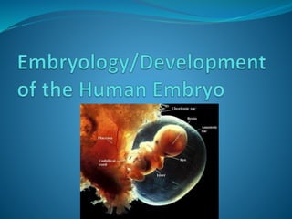
2. What is embryology?
The study of developmental events that
occur during the prenatal stage .
The branch of biology and medicine
concerned with the study of embryos
and their development.
3. Embryonic period vs. Fetal period
Embryonic – first 8 weeks Development of the
three primary germ layers give rise to all structures and
Basic body plan takes shape
Fetal period – remaining 30 weeks. Structures
and organs continue to grow and develop.
8 weeks
5. Stages of Development
1. Fertilization
2. Cleavage
3. Gastrulation Embryogenesis
4.Organogenesis
5.Maturation
6. Fertilization
The process of fusion or union of the
spermatozoon with the mature ovum is known as
conception / fertilizaiton/impregnantation.
Which produced the fertilized single mono-
nucleated cell called the zygote.
8. 2. Gastrulation : is a phase early in the
embryonic development of most animals/human
being, during which the single-layered blastula is
reorganized into a trilaminar ("three-layered")
structure known as the gastrula. These three germ
layers are known as the ectoderm, mesoderm, and
endoderm.
3. Organogenesis: The production and
development of the organs of an animal or plant.
9. How fertilization occurs????
Following ovulation, the ovum, which is about 0.15 mm in
diameter is picked up by the tubal fimbriae and is moved along by
the cilia and by peristaltic movement of the tube.
At the time the cervix under the influence of estrogen, secretes a
flow of alkaline mucus that deposited in the vagina, only
thousands capacitated spermatozoa enter the uterine tube while
300-500 reach the ovum, and remainder are destroyed by the acid
medium of the vagina.
It takes about 1 hour for sperm to reach the site.
10. The sperm release the enzyme, Hylluronidase
which allows penetration of the zona pellucida and
the cell membrane surrounding the ovum.
Many sperm are needed for this to take place but
only one will enter the ovum.
After this the membrane is sealed to prevent entry
of any further sperm and the nucluei of the two
cell fuse.
11. The sperm and ovum contribute half the
complement of chromosomes to make a
total of 46.
The sperm and ovum is known as the male
and female gametes and the fertilized ovum
as the zygote
13. Normal site for Conception
The most common site of conception is the
ampullary part (Ampulla ) of the fallopian
tube which is the widest part located closed
to the ovary
14. Usual Time for Conception
Neither sperm nor ovum can survive longer than 2-3
days and the fertilization is most likely to occur when
intercourse takes place not more than 48 hours before
and 24 hours after ovulation.
So the conception will take place about 14 days before
the next period is due.
The sex of the new individual at the time of
conception is determined by sex chromosomes.
15. Every human cell contains 46 chromosomes,
which are made up of 44 autosome chromosome
and 2 sex chromosomes.
The sex chromosome are X and Y .
Woman have no Y chromosome and male has Y
chromosome (male 44+X+Y) (female 44+X+X).
Therefor e, sex of child is always determined by
father
16.
17. Morula
1. After fertilization, the Zygote divides into 2 cells
(blastomere) (mitosis division)in about 30 hours
after fertilization.
2. The blastomeres continue to divide by binary
division through 4, 8, and 16 cell stage until a
cluster of cells is formed– Morula, resemblibg
a mulberry
18. The morula after spending about 3 days(72 hours)
in the uterine tube enters the uterine cavity
through the narrow uterine ostium (1mm) on the
4th day .
21. 4. Blastocyst
Morula, once entering the uterine cavity, floats
freely(next 2 days) and is covered by endometrial
fluid and mucus.
This fluid is absorbed through the canaliculi of the
zona pellucida and Morula begins to accumulate
fluid and forms a cavity between its cells.
Once cavity appears, it is now called a blastocyst.
23. Blastocyst
The zona pellucida
becomes stretched, thinned
and gradually disappear
soon prior to implantation.
The cell of the outer cell
mass forms the wall of the
blastocyst and is known as
trophoblast.
The inner cell mass is
concerned with the
development of the
embryo.
24. Two Distinct Cell Types
1. Trophoblasts – will form the invading
placenta
2. Inner cell mass – will form the embryo
Trophoblasts
26. hCG is produced
hCG is human chorionic gonadotropin
hormone
It is produced by the trophoblasts starting on day 6
hCG causes endometrium of uterus to grow and
proliferate
hCG prevents the menstrual cycle from occuring
This is why a female misses her periods when she
is pregnant
28. Implantation occurs about 6th day after
fertilization (approx the 20th day of a regular
menstrual cycle).
As the zona pellucida surrounding the blastocyst
disappear, there is increased adhesiveness of the
trophoblastic cells to the endometrium.
The trophoblastic cells invade the stromal cells
lying between the secretary endometrial glands by
histolytic action of the blastocyst bringing about
deeper penetration in the decidua.
29. The syncytial cells ( multinucleate cell) ultimately
penetrate the endothelial coat of the maternal
decidual capillaries establishing communication with
the syncytial lacunar system
Thus maternal blood circulates through the syncytial
lacunae providing nourishment to the blastocyst.
Maternal blood vassels
30. Further penetration is arrested by the maternal
immunological blocking factor. The point of entry of
the blastocyst into the decidual is sealed by a fibrin
coat and later by epithelium
The process of implantation is completed by the 10th
or 11th day of fertilization.
31. This type of deeper penetration of the human
blastocyst is called interstitial implantation and the
blastocyst is covered on all sides by the endometrium
(decidual).
Occasionally, there may be increased blood flow into
the lacunar space and cause bleeding. It is called
implantation bleeding and This corresponds approx to
13th day after fertilization (at about the expected day of
period).
This may produce confusion in determination of the
EDD
32.
33. The outer cell mass of the blastocyst form the
trophoblast layer.
The inner cell mass on the 8th day differentiates into
bilaminar germ disc consisting of a dorsal ectodermal
layer of the tall columnar cells and a ventral
endodermal layer of flat polyhydral cells.
The bilaminar germ disc is connected to the
trophoblast by a mesenchymal connecting stalk called
the body stalk which later forms the umblical cord.
34. Formation of Amniotic cavity and
Yolk sac
Two cavities appear on either side of the bilaminar
embryonic disc.
The dorsal cavity between the ectoderm and
trophoblastic layers lined by mesenchyme(connective
tissue) is called the amniotic cavity.
The cavity on the ventral aspect lined by the primitive
mesenchyme on the outside and the endodermal layer
of the germ cell disc on the inside is called the yolk sac
35. Week 2
Inner cell mass divides into
epiblast and hypoblast
2 fluid filled sacs
Amniotic sac from epiblast within
which the embryo and later the
fetus develop until birth
Yolk sac from hypoblast which is
one of the structures through
which the mother supplies
nutrients to the early embryo
Bilaminar embryonic disc: area
of contact
(gives rise to the whole body)
36. Bilaminar to trilaminar disc
Three primary “germ” layers: all body tissues
develop from these
Ectoderm
Endoderm
Mesoderm
Week 3
37. Formation of the 3 “germ” layers
Primitive streak (groove) on
dorsal surface of epiblast
Grastrulation: invagination
of epiblast cells
Days 14-15: they replace
hypoblast becoming
endoderm
Day 16: mesoderm (a new
third layer) formed
in between
Epiblast cells remaining on
surface: ectoderm
53. Trophoblast
Small projection begins to appear all over
the surface of the blastocyst, becoming most
prolific at the area of contact. These
trophoblastic cells differentiate into 3 layer.
1. The outer syncytiotrophoblast
(syncytium): it erodes the walls of the blood
vessels of the decidua, making the nutrient
in the maternal blood accessible to the
developing fetus.
54. 2. The inner cytotrophoblast: it’s a single
layer cells which produces a hormone HCG. This is
responsible for informing the corpus luteum that a
pregnancy has begun and corpus luteum
continues to produce estrogen and progesterone,
which maintain the integrity of the decidua.
3. In the inner aspect: a layer of mesoderm or
primitive mesenchyme, which consists of loose
connective tissue.
There is similar tissue in the inner cell mass and
the two are continuous at the point where they join
in the body stalk. (fig 3.9 1st picture from book
tuitui)
55.
56.
57. The trophoblast is responsible for the formation of
the placenta and fetal membrane and sub-serve
the important functions of attachment of the fetus
to maternal tissues, providing nutrition,
oxygenation, and clearing of the fetal metabolic
wastes and producing hormones, thus ensuring
growth and development of the fetus.
58. Decidua
The endometrial lining of the uterus is
called decidua during pregnancy and it is
shed after delivery.
Progesterone from the corpus luteum and
placenta maintain decidua during
pregnancy. It is called the decidual reaction.
59. Well developed Decidua consists
of 3 layers.
1. Superficial compact layer: consists of
compact mass of decidual cells, gland, ducts and
dilated capillaries. The greater part of the surface
epithelium is either thinned out or lost.
2. Intermediate spongy layer (cavernous
or functional layer): contains dilated uterine
glands, decidual cells and blood vessesls. Placenta
separation occurs in this layer.
60. 3. The basal layer: containing the basal
portion of the glands and is opposed to the
uterine muscle. Regeneration of the mucous
coat occurs from this layer following
parturition.
After the implantation of the blastocyst into the
compact layer of the decidua, the different
portions of the decidua are renamed as:
61. 1. Decidua basalis or
serotina: The portion of
the decidua in contact
with the base of the
blastocyst.
2. Decidua capsularies or
reflexa: The thin
superficial compact layer
covering the blastocyst.
3. Decidua vera or
parietalis: the rest of
the decidua lining the
uterine cavity outside the
site of implantation.
62. The ovum bulges into the
uterine cavity. The space
between the decidua
capsularies and decidua vera
is called the decidual space.
It progressively diminishes
as the ovum enlarges in
pregnancy
Until at 16 weeks, the space
is obliterate because of the
fusion of the decidua
capsularis with the decidua
vera.
At term this fused layer is
gradually attenuated and its
constituent layers can not
be identified.
The decidua basalis
becomes the maternal
portion of the placenta.
63. Functions of Decidua
1. To provide place for implantation of the
fertilized ovum.
2. To provide nutrition to the growing ovum.
3. Provide a protective action.
4. Provides the basal plate of the placenta.
64. Important Events Following
Fertilization
“O” hour - Fertilization
30 hours - 2 cell stage
40-50 hours - 4 cell stage
72 hours - 12 cell stage
96 hours - 16 cell stage morula enter the
uterine cavity
5th day - blastocyst
6-7th day - zona pellucida disappear.
Interstitial implantation
occurs.
65. 9th day - lacunar period, endometrial
vessels tapped
10-11th day - implantation completed
13th day - primary villi seen
16th day - secondary villi
21st day - tertiary villi
21st -22nd - fetal heart, fetal placental
circulation started
66.
67. The prenatal development of the fetus
can be divided into 3 phases.
1. Ovular period or germinal period (It
lasts from the stage of fertilization upto 2 weeks after
ovulation)
The embryonic period (first 8 weeks
Development of the three primary germ layers give rise
to all structures and Basic body plan takes shape )
Fetal period (remaining 30 weeks. Structures
and organs continue to grow and develop)
68. Fetal Length
In the earlier weeks, it is expressed as the
measurement from the vertex to the coccyx
(crown rump length) while after mid
pregnancy (20 weeks onwards), the
measurement of the fetus is determined
from the vertex to the heel (crown heel
length).
69. Age of the Fetus
The length is more reliable criterion than
the weight to calculate the age of the fetus.
Haase’s rule is employed in calculating the
age of fetus from its length in centimeters,
divided by 5.
70. 1. First lunar month: Fertilization to
2weeks of embryonic growth
Implantation is complete.
Primary chorionic villi forming
Embryo develops into two cell layers (bilaminar
embryonic disks)
Amniotic cavity appears.
Note: lunar month is a period that is measured based on the
movement of the moon. It is roughly 28 days and it constitutes
a month. In that context, pregnancy is 10.
71. 2. Second lunar month: 3 to 6
weeks of embryonic growth
At the end of 6 weeks of growth, the embryo is
approximatey 1.2 cm long.
Arms and leg buds are visible; arm buds are more
developed with finger ridges beginning to appear.
Rudiments of the eyes, ears and nose appear.
Primitive intestinal tract is developing.
Primitive cardiovascular system is functioning.
Neural tube, which forms the brain and spinal cord
closes by the 4th week.
72. 3. Third lunar month: 7 to 10
weeks of growth
The middle of this period (8 weeks)marks the end of the
embryonic period and beginning of the fetal period.
At the end of 10 weeks of growth, the fetus is 6.1 cm from
crown to rump and weigh 14 gm.
Appearance of external genitalia.
By the middle of this month, all major organs, systems have
formed.
The heart has formed four chambers (by 7th week)
The fetus assumes a human appearance
Bone ossification begins
Rudimentary kidney begins to secrete urine.
73. 4. Fourth lunar month: 11 to 14
weeks old fetus
At the end of 14 weeks of growth, the fetus is
12 cm crown to rump length and 110 gm.
Head erect; lower extremities well
developed.
Hard palate and nasal septum have fused.
External genitalia of male and female can
now be differentiated.
Eyelids are sealed
74. 5.Fifth lunar month: 15 to 18
weeks old fetus
At the end of 18 weeks of growth, the fetus is 16 cm
crown rump length and 320 gm.
Ossification of fetal skeleton can be seen on X-ray.
Ears stand out from head.
Fetus makes sucking motions and swallows
amniotic fluid.
Fetal movement may be felt by the mother (end of
month)
75. 6. Sixth lunar month: 19-22 weeks
old fetus
At the end of 22 weeks of
growth, the fetus
is 21 cm crown rump length
and 630 gm.
Vernix caseosa covers the skin.
Head and body hair (lanugo)
visible.
Skin is wrinkled and red.
Brown fat(adipose tissue), an
important site of heat
production, is present in neck
and sternal area.
76. 7. Seventh lunar months: 23-26
weeks old fetus
At the end of 26 weeks of
growth, the fetus is 25 cm
crown to rump length and
1000 gm.
Fingernails present.
Lean body
Eyes partially open:
eyelashes present.
Bronchioles are present:
primitive alveoli (terminal
sacs) are forming
Skin begins to thicken on
hands and feet
Startle reflex present,
grasp reflex is strong.
77. 8. Eighth lunar month: 27-30 weeks
old fetus
At the end of 30 weeks
of growth, the fetus is
28 cm crown rump
length and 1,700 gm.
Eyes open
Ample hair on head:
lanugo begins to fade
Skin slightly wrinkled
Toe nails presents
Testes in inguinal
canal, begin descent to
scrotal sac
Surfactant coat in
much of the alveolar
epithelium
78. 9. Ninth lunar months: 31-34
weeks of old fetus
At the end of 34 weeks of growth, the fetus
is about 32 cm crown –rump length and
2,500 gm
Fingernails reach finger tips
Skin pink and smooth
Testes in scrotal sac
79. 10. Tenth lunar month: 35 to 38
weeks old fetus
End of 38 weeks of growth, fetus is about 36 cm
crown –rump length and 3400 gm.
Ample of subcutaneous fat present
Lanugo is decreasing
Toe nails reach upto toe tips
Testes in scrotum
Vernix caseosa mainly on the back
Breasts are firm.