

Recomendados
Mais conteúdo relacionado
Último
Último (20)
Destaque
Destaque (20)
Sickle cell anemia
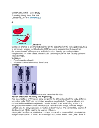
- 1. Sickle Cell Anemia – Case Study Posted by: Daisy Jane, RN, MN October 16, 2010 · Comments (0) 4 Definition Sickle cell anemia is an inherited disorder on the beta chain of the hemoglobin resulting to abnormally shaped red blood cells. RBC’s assume a crescent or C-shape that decreases the cell’s life span and ability to function thereby, producing various complications. In some cases, these sickled cells may block the flow causing pain and organ damage. Incidence Equal male-female ratio Increase incidence in African Americans Etiology Autosomal recessive disorder Review of Related Anatomy and Physiology Red blood cells or erythrocytes carry oxygen to the different parts of the body. Different from other cells, RBC’s do not contain a nucleus (anucleated). These small cells are circular and flattened with depressed centers on both sides resembling to that of a doughnut when viewed under a microscope. Their size and shape provides a large surface area for carrying oxygen in relation to their volume. The normal RBC count is 4- 6 million/mm3. RBC’s has the life span of 120 days. Hemoglobin is a form of protein that contains iron which is responsible for transporting oxygen that is carried in blood. Adult hemoglobin contains a beta chain (HBB) while a
- 2. fetus’ hemoglobin has a gamma chain. Hemoglobin is comprised of four protein (amino acid) components. It has two beta-globin and two alpha-globin. The subunit beta-globin is located inside the RBCs. These amino acids carry an iron-bearing molecule called heme. Heme molecules, which are only found in mature RBC’s, enables the erythrocytes to pick oxygen from the lungs and transport it throughout the body. Once oxygen attaches to hemoglobin it gives the blood its bright red pigment. The more hemoglobin molecules the RBC contain, a higher amount of oxygen will they be able to carry. If the hemoglobin is defective, the erythrocyte will also malfunction. A red blood cell is just a vessel; the one that performs the oxygen transportation is the hemoglobin. Normal hemoglobin is 13-18 grams/100 ml of blood in males and 12-16 grams in females. Pathophysiology Erythrocytes in sickle cell anemia contain abnormal hemoglobin that affects the beta- chain producing hemoglobin S or HbS. In this disorder, the beta-chains (beta-globins) are replaced by Hemoglobin S. Valine (an amino acid) takes the place of the normally appearing glutamic acid in beta-chains. Replacement of glutamic acid with valine causes the polymerization of HbS components to cohere forming long and insoluble particles. These distort the red blood cells, which assumes an inflexible crescent or sickle shape. The abnormally shaped cells become sharp and spiky when the RBCs are discharging oxygen molecules and in cases where the oxygen content in blood is low such as performing vigorous exercise and being in high altitude areas. Typically, a sickled cell’s lifespan is only 20 days. The deformed erythrocytes also rupture easily and they tend to be trapped in the microcirculation, obstructing blood flow and oxygen transport that might lead to painful episodes of ischemic injury. Sickle cell crisis refers to episodes of acute and severe sickling that blocks the circulation posing a threat of extensive organ damage. Severe pain is noted during these incidents caused by occluded vessels in the bone possibly resulting to bone necrosis. The crisis is triggered by hypoxemia, acidosis, or other conditions such as dehydration, infection, vigorous exercise, pregnancy or cold weather. A condition called a sickle cell trait is identified by the presence of a single defective gene, instead of two. Individuals with this trait are essentially normal however, they are carriers. Meaning two sickle cell trait carrier parents can contribute a defective gene to a child that will carry the sickle cell disorder. Diagnosis Prenatal: Chorionic Villi sampling Amniocentesis (blood from the cord) At birth: Newborn Screening Hemoglobin electrophoresis Signs and Symptoms The following manifestations are observed in children with a sickle cell disorder, at about 6 months of age (because fetal hemoglobin contains gamma, not a beta chain): Fever Anemia
- 3. Swelling of the hands and feet (hand-foot syndrome) – caused by blood stasis and infarction Protruding abdomen – due to enlarged spleen and liver secondary to trapping of sickled cells in microcirculation and obstruction of blood flow Icteric sclera – caused by bilirubin release during hemolysis (sickle cell destruction) Priaprism (males) – due to pooling of abnormally shaped erythrocytes in the blood vessels of the penis. Chest syndrome – symptoms same with pneumonia that is the major cause of death in sickle-cell patients Management Conservative Management 1. Pain relief with Acetaminophen (Tylenol) or IV of morphine to reduce metabolic demand of oxygen thereby, terminating cell sickling. 2. Adequate hydration – IV fluids and electrolyte replacement 3. Oxygenation 4. Antibiotics – if the cause of sickling is infection. 5. Blood transfusion with packed RBC’s 6. Hydroxyurea – antineoplastic agent that increases production of fetal hemoglobin in children. 7. Exchange transfusion – replacing sickled with normal cells If the patient does not respond to the usual therapies STEM CELL TRANSPLANTATION is done. Nursing Management 1. Monitor vital signs. Assess for pain. 2. Obtain blood and urine culture, chest x-ray and CBC results if infection is the cause of sickling. 3. Monitor child’s nutritional intake with hydroxyurea. If taken orally, this drug can cause anorexia. 4. Assess for kidney function by noting if the child has urinated or not. (Kidney infarction may occur) 5. Do not administer potassium if the kidney function is not verified. Potassium if not excreted by the kidney may cause arrhythmia. Possible nursing diagnosis 1. Ineffective tissue perfusion R/T decreased hemoglobin concentration in blood 2. Acute pain R/T impaired blood flow due to obstruction of sickled cells image from elev8.com, daviddarling.info Related Nursing Articles 1. Types of Sickle Cell CrisisOverview Sickle cell anemia is an inherited disorder on the beta chain of the hemoglobin resulting to abnormally shaped red blood cells. RBC’s assume a crescent or C-shape that decreases the cell’s life span and... 2. Classifications of AnemiaOverview The main function of a red blood cell or erythrocyte is to carry and transport oxygen to the different parts of the body. The normal RBC count is 4-6 million/mm3. Hemoglobin (Hgb), an iron-bearing... 3. ThalassemiasThalassemias Definition Thalassemia is a group of inherited disorders which is associated with hemoglobin defects. The disorder results in excessive
- 4. destruction of red blood cells leading to anemia. Types of Thalassemia There are two main... 4. ErythropoiesisRed Blood Cells Red Blood Cells (RBC’s) also called erythrocytes, are oxygen carrying cells. It is derived from the Greek words “erythros” meaning “red,” “kytos” meaning “hollow” and “cyte” translated as “cell” in modern language.... 5. Nursing Care Plan – AnemiaAnemia is the reduction in red blood cells (erythrocytes) thus decreasing the oxygen carrying capacity of the blood due to the following (1) excessive blood loss (2) deficiencies and abnormalities of RBC production (3) Excessive... About Daisy Jane, RN, MN Nursing Care Plan – Anemia Posted by: Admin July 6, 2008 · Comments (7) 3 Anemia is the reduction in red blood cells (erythrocytes) thus decreasing the oxygen carrying capacity of the blood due to the following (1) excessive blood loss (2) deficiencies and abnormalities of RBC production (3) Excessive destruction of RBC. *Common Symptoms of Anemia These symptoms appear in most types of anemia: pale skin dizziness fatigue headaches irritability low body temperature numb/cold hands or feet rapid heartbeat shortness of breath weakness chest pain *Types of Anemia Different types of anemia have different causes and symptoms. Iron Deficiency Anemia: Iron deficiency is one of the most common causes of anemia, especially in women. In the U.S. alone, twenty million women suffer from iron deficiency anemia. Iron deficiency causes insufficient hemoglobin production which, in turn, causes anemia and anemia symptoms. Sickle Cell Anemia: Sickle cell anemia, or sickle cell disease, is one of the hereditary causes of anemia. Most often seen in people of African descent, sickle cell anemia is characterized by the production of rigid, sickle-shaped red blood cells. These abnormal sickle cells break down faster then normal red blood cells, resulting in a chronic shortage of red blood cells and anemia symptoms. Sickle cell anemia is a form of hemolytic anemia, which describes types of anemia caused by the rapid destruction of red blood cells. Pernicious Anemia: Certain types of anemia are referred to as megaloblastic, or vitamin deficiency anemia. Pernicious anemia is caused by an inability of the intestines to absorb sufficient amounts of vitamin B12, which is required in the production of red blood cells. Pernicious anemia symptoms include vision problems, memory loss, difficulty walking and numb hands and feet, as well as the more common anemia symptoms. Risk factors for pernicious anemia include a family history of pernicious anemia, Scandinavian or European ancestry or a history of autoimmune endocrine disorders. Vitamin B12 injections are the standard pernicious anemia treatment. Oral vitamin B12 supplements may also be used as pernicious anemia treatment, but they’re not as efficient as B12 injections. Folic Acid Deficiency Anemia: Folic acid deficiency anemia, like pernicious anemia, is a megaloblastic anemia. Folic acid is essential for healthy blood and a healthy nervous system. A folic acid deficiency during pregnancy can cause birth defects.
- 5. Folic acid supplements form the basis of folic acid anemia treatment. Anemia treatment also includes eating a diet high in folic acid, including such foods as: beans and legumes citrus fruits and juices dark green leafy vegetables pork poultry shellfish wheat bran Thalassemia: Causes of anemia often include genetic mutations. Thalassemia is one of the genetically inherited types of anemia. Thalassemia occurs when the body’s genes, which are responsible for proper hemoglobin production, become damaged or mutated. Like sickle cell anemia, thalassemia is one of the hemolytic anemia types. Many possible gene variations can cause thalassemia, and the disease has both mild and severe varieties. Cooley’s anemia is the most common severe thalassemia in the U.S. The disease tends to be present at birth and its symptoms include: dark colored urine deformed facial bones fatigue jaundice poor growth rates a swollen abdomen caused by an enlarged spleen and liver weakness A number of ethnic groups are at higher risk of developing thalassemia, including people of African, Chinese, Filipino, Indian, Italian, Greek and Middle Eastern ethnicity. Treatment for thalassemia depends on the severity of the disease. Mild types of thalassemia may be treated with regular blood transfusions. Severe cases require multiple blood transfusions, iron chelation therapy to remove excess iron from the blood and bone marrow transplants. Without treatment, severe thalassemia often proves fatal in early childhood. Aplastic Anemia: Aplastic anemia is one of the rarer types of anemia, occurring when the body makes insufficient red and white blood cells. Causes of aplastic anemia include: radiation therapy chemotherapy toxic chemicals some medications bone marrow infections