Laboratory diagnosis of visceral leishmaniasis
•
3 gostaram•1,287 visualizações

1. Visceral leishmaniasis (VL), also known as kala-azar, is characterized by fever, spleen and liver enlargement, weight loss, and anemia. It is caused by protozoa of the genus Leishmania and transmitted by sandflies. 2. Laboratory diagnosis of VL can be made by demonstrating the parasite in tissues like spleen, bone marrow or lymph nodes via microscopy, culture, or animal inoculation. It can also be diagnosed by detecting parasite DNA or through immunological tests. 3. While demonstration of the parasite via microscopy of tissues is commonly used but has low sensitivity, splenic aspiration has over 95% sensitivity but risks bleeding. Culture improves sensitivity but is time

Recomendados
Mais conteúdo relacionado
Mais procurados
Mais procurados (20)
Destaque
Semelhante a Laboratory diagnosis of visceral leishmaniasis
Semelhante a Laboratory diagnosis of visceral leishmaniasis (20)
Mais de Aman Ullah
Mais de Aman Ullah (20)
Último
Último (20)
Laboratory diagnosis of visceral leishmaniasis
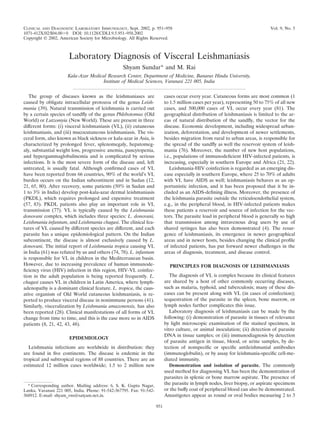
- 1. CLINICAL AND DIAGNOSTIC LABORATORY IMMUNOLOGY, Sept. 2002, p. 951–958 Vol. 9, No. 5 1071-412X/02/$04.00ϩ0 DOI: 10.1128/CDLI.9.5.951–958.2002 Copyright © 2002, American Society for Microbiology. All Rights Reserved. Laboratory Diagnosis of Visceral Leishmaniasis Shyam Sundar* and M. Rai Kala-Azar Medical Research Center, Department of Medicine, Banaras Hindu University, Institute of Medical Sciences, Varanasi 221 005, India The group of diseases known as the leishmaniases are caused by obligate intracellular protozoa of the genus Leish- mania (39). Natural transmission of leishmania is carried out by a certain species of sandfly of the genus Phlebotomus (Old World) or Lutzomyia (New World). These are present in three different forms: (i) visceral leishmaniasis (VL), (ii) cutaneous leishmaniasis, and (iii) mucocutaneous leishmaniasis. The vis- ceral form, also known as black sickness or kala-azar in Asia, is characterized by prolonged fever, splenomegaly, hepatomeg- aly, substantial weight loss, progressive anemia, pancytopenia, and hypergammaglobulinemia and is complicated by serious infections. It is the most severe form of the disease and, left untreated, is usually fatal. Although confirmed cases of VL have been reported from 66 countries, 90% of the world’s VL burden occurs on the Indian subcontinent and in Sudan (12, 21, 65, 80). After recovery, some patients (50% in Sudan and 1 to 3% in India) develop post-kala-azar dermal leishmaniasis (PKDL), which requires prolonged and expensive treatment (57, 83). PKDL patients also play an important role in VL transmission (77). VL is typically caused by the Leishmania donovani complex, which includes three species: L. donovani, Leishmania infantum, and Leishmania chagasi. The clinical fea- tures of VL caused by different species are different, and each parasite has a unique epidemiological pattern. On the Indian subcontinent, the disease is almost exclusively caused by L. donovani. The initial report of Leishmania tropica causing VL in India (61) was refuted by us and others (74, 78). L. infantum is responsible for VL in children in the Mediterranean basin. However, due to increasing prevalence of human immunode- ficiency virus (HIV) infection in this region, HIV-VL coinfec- tion in the adult population is being reported frequently. L. chagasi causes VL in children in Latin America, where lymph- adenopathy is a dominant clinical feature. L. tropica, the caus- ative organism of Old World cutaneous leishmaniasis, is re- ported to produce visceral disease in nonimmune persons (41). Similarly, visceralization by Leishmania amazonensis, has also been reported (28). Clinical manifestations of all forms of VL change from time to time, and this is the case more so in AIDS patients (8, 21, 42, 43, 48). EPIDEMIOLOGY Leishmania infections are worldwide in distribution: they are found in five continents. The disease is endemic in the tropical and subtropical regions of 88 countries. There are an estimated 12 million cases worldwide; 1.5 to 2 million new cases occur every year. Cutaneous forms are most common (1 to 1.5 million cases per year), representing 50 to 75% of all new cases, and 500,000 cases of VL occur every year (81). The geographical distribution of leishmaniasis is limited to the ar- eas of natural distribution of the sandfly, the vector for the disease. Economic development, including widespread urban- ization, deforestation, and development of newer settlements, besides migration from rural to urban areas, is responsible for the spread of the sandfly as well the reservoir system of leish- mania (76). Moreover, the number of new host populations, i.e., populations of immunodeficient HIV-infected patients, is increasing, especially in southern Europe and Africa (21, 22). Leishmania-HIV coinfection is regarded as an emerging dis- ease especially in southern Europe, where 25 to 70% of adults with VL have AIDS as well; leishmaniasis behaves as an op- portunistic infection, and it has been proposed that it be in- cluded as an AIDS-defining illness. Moreover, the presence of the leishmania parasite outside the reticuloendothelial system, e.g., in the peripheral blood, in HIV-infected patients makes these patients a reservoir and source of infection for the vec- tors. The parasite load in peripheral blood is generally so high that transmission among intravenous drug users by use of shared syringes has also been demonstrated (4). The resur- gence of leishmaniasis, its emergence in newer geographical areas and in newer hosts, besides changing the clinical profile of infected patients, has put forward newer challenges in the areas of diagnosis, treatment, and disease control. PRINCIPLES FOR DIAGNOSIS OF LEISHMANIASIS The diagnosis of VL is complex because its clinical features are shared by a host of other commonly occurring diseases, such as malaria, typhoid, and tuberculosis; many of these dis- eases can be present along with VL (in cases of coinfection); sequestration of the parasite in the spleen, bone marrow, or lymph nodes further complicates this issue. Laboratory diagnosis of leishmaniasis can be made by the following: (i) demonstration of parasite in tissues of relevance by light microscopic examination of the stained specimen, in vitro culture, or animal inoculation; (ii) detection of parasite DNA in tissue samples; or (iii) immunodiagnosis by detection of parasite antigen in tissue, blood, or urine samples, by de- tection of nonspecific or specific antileishmanial antibodies (immunoglobulin), or by assay for leishmania-specific cell-me- diated immunity. Demonstration and isolation of parasite. The commonly used method for diagnosing VL has been the demonstration of parasites in splenic or bone marrow aspirate. The presence of the parasite in lymph nodes, liver biopsy, or aspirate specimens or the buffy coat of peripheral blood can also be demonstrated. Amastigotes appear as round or oval bodies measuring 2 to 3 * Corresponding author. Mailing address: 6, S. K. Gupta Nagar, Lanka, Varanasi 221 005, India. Phone: 91-542-367795. Fax: 91-542- 368912. E-mail: shyam_vns@satyam.net.in. 951
- 2. m in length and are found intracellularly in monocytes and macrophages. In preparations stained with Giemsa or Leish- man stain, the cytoplasm appears pale blue, with a relatively large nucleus that stains red. In the same plane as the nucleus, but at a right angle to it, is a deep red or violet rod-like body called a kinetoplast (Fig. 1). After identification, parasite den- sity can be scored microscopically by means of a logarithmic scale ranging from 0 (no parasite per 1,000 oil immersion fields) to ϩ6 (Ͼ100 parasites per field) (19). The sensitivity of the bone marrow smear is about 60 to 85%. Splenic aspirate, though associated with risk of fatal hemorrhage in inexperi- enced hands, is one of the most valuable methods for diagnosis of kala-azar, with a sensitivity exceeding 95%. It requires no special equipment, from the patient’s standpoint is generally preferable to the more painful bone marrow aspirate, and has proven to be safe and relatively easy to perform in experienced hands. For patients suspected to have VL, splenic aspirate can be performed even when spleen is not palpable, after demar- cating the area of splenic dullness by percussion. The only risk of splenic puncture is bleeding from a soft and enlarged spleen. At our treatment center, fatal bleeding has occurred only twice in 9,612 splenic aspirate procedures performed over the last 10 years. To avoid the risk of excessive blood loss, splenic punc- ture should be avoided in patients with a platelet count of less than 40,000 platelets/l and a prothrombin time of more than 5 s over the control. A tissue specimen, e.g., a spleen, liver, or lymph node tissue specimen, may be subjected to imprint cytology by the re- peated pressing of its cut flat surface on microscopic slides. The smear is fixed with absolute alcohol and stained with Giemsa stain. In imprint cytology, a monolayer of cells is formed and amastigotes are easily identifiable. The results are expressed as the number of leishmania per 100 host cell nuclei. Tissue specimens can also be subjected to histology, and the presence of parasites can be demonstrated by standard hema- toxylin and eosin stain. Tissue specimens are usually uneven in thickness; consequently the amastigotes are unevenly distri- buted. Long searches may be required to demonstrate the parasite. The sensitivity of the test can be increased by staining the specimen with fluorescent dye-tagged antibodies to the surface receptors of the parasite. Fluorescein isothiocyanate isomer- or rhodamide B isothiocyanate-conjugated antiserum is usually used for this purpose. Fluorescent dye-conjugated monoclonal antibodies are also used for speciation of the par- asite. Culture of parasite can improve the sensitivity of detection of parasite, but leishmania culture is rarely needed in routine clinical practice. However, cultures are required for (i) obtain- ing a sufficient number of organisms to use an antigen for immunologic diagnosis and speciation, (ii) obtaining parasites to be used in inoculating susceptible experimental animals, (iii) in vitro screening of drugs, and (iv) accurate diagnosis of the infection with the organism (as a supplement to other methods or to provide a diagnosis when routine methods have failed). Leishmania strains can be maintained as promastigotes in ar- tificial culture medium. The culture media used may be monophasic (Schneider’s insect medium, M199, or Grace’s medium) or diphasic (Novy-McNeal Nicolle medium and To- bies medium). We prefer diphasic medium containing modi- fied diphasic rabbit blood agar overlaid with RPMI 1640 (Gibco BRL, Grand Island, N.Y.) (74) for primary isolation, and we prefer M199 medium containing 20% fetal calf serum to amplify parasite numbers (74). Hockmeyer’s medium, which is Schneider’s commercially prepared culture medium supple- mented with 30% heat-inactivated fetal calf serum with 100 IU of penicillin and 100 g of streptomycin, is simple to use and FIG. 1. Microphotograph showing intracellular and extracellular L. donovani bodies in splenic aspirate from a patient with visceral leish- maniasis. 952 MINIREVIEWS CLIN. DIAGN. LAB. IMMUNOL.
- 3. satisfactory for diagnosis of VL, but it is expensive (29). Cul- ture tubes are inoculated with 1 to 2 drops of bone marrow or splenic aspirate and incubated at a temperature between 22 and 28°C. The tubes are examined weekly for the presence of promastigotes by phase-contrast microscopy or by wet mount of culture fluid for 4 weeks before being discarded as negative. If promastigotes are present, they are maintained by weekly passage to fresh medium. Blood can also be used to isolate the parasite, but the method is slow and takes longer. Aseptically collected blood (1 to 2 ml) is diluted with 10 ml of citrated saline, and the cellular deposit obtained after centrifugation is inoculated in culture media. Contamination of the culture me- dia by bacteria or yeast species or other fungi usually compli- cates the culture but can be avoided by use of good sterile techniques and by the addition of penicillin (200 IU/ml) and streptomycin (200 g/ml) to the medium (for bacteria), as well as 5-flucytosine (500 g/ml) (as an antimycotic agent) (64). In vitro culture of the amastigotes is done for chemothera- peutic studies and to study the interrelationship of the amas- tigotes and macrophages. The amastigotes are grown in tissue or macrophage culture. These cell lines are produced from (i) human peripheral blood monocytes, after these are set apart by density sedimentation with lymphocyte separation medium (LSM; Organon-Teknika, Durham, N.C.), in which case a new batch of macrophages must be produced anew (24); (ii) mac- rophage cell lines, e.g., P388D and J774G8 lines from mice; and (iii) dog sarcoma and hamster peritoneal exudates of cell lines, in which case continuous culture can be achieved (64). The parasite can also be demonstrated after inoculation of laboratory animals (such as hamsters, mice or guinea pigs) with infected specimen (42). Animal inoculation is not usually em- ployed as a diagnostic test, since several months may be re- quired to obtain a positive result. Golden hamster is the animal of choice for maintaining L. donovani complex (15). It can be infected via many routes, including across mucous membranes, but intraperitoneal and intrasplenic routes are preferred. Both amastigotes and promastigotes can infect the animal. After inoculation, the animal is examined weekly for signs of infec- tion, such as cutaneous lesions, hepatosplenomegaly, or met- astatic lesions. Amastigotes can be harvested by biopsy from the spleen and the liver of an animal that is under anesthesia and that is allowed to survive following the procedure as a source of infective parasite. In the absence of signs of obvious infection, the animal is generally sacrificed after 4 months, at which point liver and spleen samples are examined for the presence of the parasite. In areas of endemicity, recognition of species of leishmania is rarely required. However, identification of an organism to the species level is helpful epidemiologically and is also impor- tant for the treatment of and prognosis determination for global travelers who are not immune to the parasite and tend to develop unusual manifestations of the disease (41). Identi- fication of species of the L. donovani complex is particularly difficult, because morphologically the species are almost indis- tinguishable from each other. For species-level identification, a large amount of promastigotes is obtained by culture of the organism and the species-specific isoenzyme pattern is ana- lyzed by cellulose acetate electrophoresis (35). Typing of washed live promastigotes by direct agglutination test with species-specific monoclonal antibodies is another highly sensi- tive taxonomic tool frequently utilized for this purpose (33). Species-level identification can also be done by analysis of amplified minicircle kinetoplast DNA (KDNA), by choosing primers from conserved regions of different leishmania species KDNA minicircles (61, 71). Yet another method used for iden- tification of species of leishmania is the analysis of the in vitro promastigotes’ released antigenic factors, which are different for different leishmanial species (32). Although demonstration of even a single amastigote upon microscopic examination of tissue smears or multiple promas- tigotes in cultures is considered sufficient for positive diagnosis of the disease, the sensitivity of the tissue examination, except in the case of splenic aspirate, is low. Moreover, the proce- dure(s) for obtaining tissue specimen(s) is traumatic and asso- ciated with considerable risk. Identification of amastigotes re- quires considerable expertise and training and is subject to the ability of the observer. Besides, culturing parasites is expensive and time consuming and requires expertise and costly equip- ment, severely restricting its use in routine clinical practice. DNA detection method. Due to the limitations inherent in techniques used for detection of parasites, new approaches to the detection of parasites, such as DNA hybridization, have been attempted since the early 1980s. Although these methods had considerable sensitivity (detecting as few as 50 to 100 parasites) (40), their potential use in routine diagnosis is ham- pered by the complex procedure of hybridization. The devel- opment of PCR has provided a powerful approach to the application of molecular biology techniques to the diagnosis of leishmaniasis. Primers designed to amplify conserved se- quences found in minicircles of KDNA of leishmanias of dif- ferent species were tested in various tissues of relevance. Such a target was eminently suitable because the kinetoplast is known to possess thousands of copies of minicircle DNA. In recent years, PCR-based diagnostic methods with a wide range of sensitivities and specificities have been described (1, 5, 51, 54). In a study reported from Sudan, PCR was found to be more sensitive than microscopy for the detection of Leishma- nia parasites in lymph node and bone marrow aspirations. However, its sensitivity for the detection of Leishmania DNA in the blood of parasitologically proven VL cases was only 70% (51). In another study reported from India, in which a species- specific primer for L. donovani (LDI primer) was used, the sensitivity of PCR with whole blood from VL patients was 96% and Leishmania DNA was detected in skin specimens from 45 of 48 patients with PKDL (sensitivity, 93.8%) (54). A PCR–enzyme-linked immunosorbent assay (ELISA) tech- nique using a primer that was able to identify 33 L. infantum strains from 19 different zymodemes has been developed. It has a sensitivity higher than that of other diagnostic tech- niques, e.g., indirect fluorescent-antibody (IFA) test, parasite culture, or microscopy, and was able to detect a minimum of 0.1 promastigote or 1 fg of genomic material. This PCR- ELISA technique can potentially be used for diagnosis of VL from peripheral blood samples (44). PCR done from blood spots on filter paper can also be used as a screening test to identify Leishmania infection in immunocompromised patients with high parasite loads in peripheral blood. The sensitivity of this technique for detecting leishmania (75%) was consider- ably higher than the respective sensitivities of microscopy (26.3%) and blood culture (42.3%) (17). However, PCR assay VOL. 9, 2002 MINIREVIEWS 953
- 4. with buffy coat preparations to detect Leishmania was 10 times more sensitive than that with whole-blood preparations, and particularly good results were obtained when proteinase K- based methods were used. Proteinase K-based PCR was able to detect 10 parasites/ml (37). A fluorescent DNA probe spe- cific for a conserved region of the small subunit rRNA gene of Leishmania and a pair of flanking primers, when used for DNA amplification in one assay, proved to be a highly specific and rapid diagnostic modality to detect infection with Leishmania (82). Using this rapid fluorogenic PCR technique, DNA could be amplified from 27 strains of cultured Leishmania, and the turnaround time from fresh human tissue biopsy to test result was found to be less than 24 h (82). Besides being a highly sensitive and specific tool for diagnosis of both VL and PKDL and a useful method for species identification (46), PCR can also be used to distinguish between relapse and reinfection in treated VL patients. Restriction fragment length polymor- phism analysis of the PCR-amplified minicircle of leishmanial DNA can be utilized for this purpose (47). PCR could also prove to be an important tool in assessing the success of VL treatment: of patients treated for VL who tested negative by PCR with lymph node tissue, none relapsed or developed PKDL, while more than half of patients who tested positive by PCR with lymph node tissue either relapsed or developed PKDL after apparent cure of disease following supervised treatment (50, 52). On the other hand, a substantial number of the patients who tested positive by PCR, after apparent cure, did not relapse or develop PKDL, a result that suggests the limitation of PCR in deciding the end point of treatment. The PCR positivity observed in these patients may be due to non- viable parasite. Similarly, PCR results for healthy endemic controls may be positive (38, 54), which may lead to the erro- neous conclusion that they suffer from VL. In these healthy endemic controls, a combination of direct agglutination test (DAT) (which shows low titers in healthy endemic controls) and PCR may be helpful in defining the status of these pa- tients. Immunodiagnosis. (i) Antigen detection. Antigen detection is more specific than antibody-based immunodiagnostic tests (20, 79). This method is also useful in the diagnosis of disease in cases where there is deficient antibody production (as in AIDS patients). De Colmenares et al. (20) from Spain have reported two polypeptide fractions of 72-75 kDa and 123 kDa in the urine of kala-azar patients. The sensitivities of the 72- 75-kDa fractions were 96%, and the specificities were 100%. Besides, these antigens were not detectable within 3 weeks of anti-kala-azar treatment, suggesting that the test has a very good prognostic value (20). A new latex agglutination test (KATEX) for detecting leish- manial antigen in urine of patients with VL has showed sensi- tivities between 68 and 100% and a specificity of 100% in preliminary trials. The antigen is detected quite early during the infection and the results of animal experiments suggest that the amount of detectable antigen tends to decline rapidly fol- lowing chemotherapy. The test performed better than any of the serological tests when compared to microscopy. Large field trials are under way to evaluate its utility for the diagnosis and prognosis of VL (6). (ii) Antibody detection. For several decades, nonspecific methods, which depend upon raised globulin levels, have been used in the diagnosis of VL. Some of the tests used for detect- ing these nonspecific immunoglobulins are Napier’s formol gel or aldehyde test and the Chopra antimony test. Since these tests depend upon raised globulin levels, results can be positive in a host of conditions (13, 14). Lack of specificity, as well as varying sensitivities, renders them highly unreliable. Several immunodiagnostic methods which are more sensi- tive and specific have been developed. They are useful in iden- tifying specific cases and can be used for community surveil- lance. The human body makes an attempt to fight against VL by producing some of the highest levels of antibodies found in response to any disease, all to no avail. This is due to polyclonal activation of the B cells, resulting in marked elevation of levels (in serum) of immunoglobulin G (IgG) and IgM against vari- ous nonspecific proteins and haptens (23). The consistent pres- ence of high levels of antibodies against parasite antigens can simplify diagnosis of VL. Several serological techniques are based on detection of these antibodies. The specificity of the antibody depends upon the antigen or epitope used in the test, as the parasite stimulates production of a wide array of anti- bodies, including group-, genus-, and species-specific antibod- ies. Therefore, the sensitivity may depend upon the test and its methodology, but the specificity will depend on the antigen rather than the serological procedure used. In most serological tests, the sensitivity and specificity data are compared against demonstration of parasites in various tissues. Conventional methods for antibody detection included gel diffusion, complement fixation test, indirect hemagglutination test, IFA test, and countercurrent immunoelectrophoresis (2, 13, 14, 25, 30, 69, 80). However, aside from practical difficulties at peripheral laboratories, the sensitivities and specificities of most of the above tests have been the limiting factors. Except for the IFA test, which is used on a limited scale, these tests are rarely used for routine diagnosis of VL. In 1988, a modified DAT was reported to be useful in kala-azar and is being used in several countries of endemicity (27). In this test, the trypsinized whole promastigotes are formalin fixed and stained with Coomasie brilliant blue; serum from the patient is then incubated with the antigen, and agglutination is observed the next day. Use of an 0.8% concentration of 0.1 M 2-mercapto- ethanol in the sample diluent further improves its performance (66). DAT in various studies has shown to be 91 to 100% sensitive and 72 to 100% specific (45, 67, 72, 84, 85). In Sudan, in specially set up field laboratories, Medecins Sans Frontieres uses DAT for diagnosis of VL; patients with high titers receive treatment, and a confirmatory parasitic diagnosis is done in those with low titers (10). From India, several laboratories reported satisfactory sensitivity and specificity levels for this test (72, 79). Although DAT showed a high degree of repeat- ability within the centers, its reproducibility across the centers was quite weak (11). Moreover, difficult field conditions, the fragility of aqueous antigen, the lack of cold chain, and batch- to-batch variations in the antigen, along with the nonstandard- ization of test readings, have severely limited its widespread applicability in regions of endemicity. Freeze-dried antigens developed in Belgian and Dutch laboratories are likely to over- come some of these handicaps (10, 49). Unless this improved antigen is produced indigenously to make it affordable and DAT is made user friendly with one-step dilution and reduced incubation time, its field use is unlikely in countries of ende- 954 MINIREVIEWS CLIN. DIAGN. LAB. IMMUNOL.
- 5. micity like India. Like most antibody-based tests, DAT may yield positive results for a long time after complete cure and thus has not proved to be of much prognostic value (27). ELISA has been used as a potential serodiagnostic tool for almost all infectious diseases, including leishmaniasis. The technique is highly sensitive, but its specificity depends upon the antigen used. Several antigens have been tried. The com- monly used antigen is a crude soluble antigen (CSA). It is prepared by repeated freezing and thawing (four to six cycles) of a suspension of promastigotes in phosphate-buffered saline, followed by cold centrifugation at 10,000 to 20,000 ϫ g. The supernatant is used as soluble antigen and is used to coat ELISA plates after estimation of protein content (100 to 5,000 ng/ml). The sensitivity of ELISA using these concentrations of CSA is reported to range from 80 to 100%, but cross-reactions with sera from patients with trypanosomiasis, tuberculosis, and toxoplasmosis have been recorded (13, 14, 18, 36, 67, 70). On the other hand, when various selective antigenic masses (116 kDa, 72 kDa, and 66 kDa) were used, a specificity of 100% could be achieved, but only at the cost of sensitivity, which went down to as low as 37.5% (20, 79). Palatnik-de-Souza et al. (53) described the use of fucose-mannose ligand as the anti- genic molecule. It is a 36-kDa glycoprotein present throughout the life cycle of leishmania (amastigote and promastigote stag- es). Its use in ELISA has been found to result in 100% sensi- tivity and 96% specificity (53). In a recent study, it was found that the sensitivity and specificity of ELISA in diagnosing VL could also be increased by the use of soluble antigens derived from promastigotes cultivated in a protein-free medium. One study, done with 129 VL and 143 cutaneous leishmaniasis patients, showed a sensitivity of 95% (56, 60). A recombinant antigen, rK39, has been shown to be specific for antibodies in patients with VL caused by members of the L. donovani complex (7, 9, 16). This antigen, which is conserved in the kinesin region, is highly sensitive and predictive of the onset of acute disease. The antigen is derived from L. chagasi, which in the United States is used for veterinary purposes, though it is not approved for human use. High antibody titers in immunocompetent patients with VL have been demon- strated. This antigen has been reported to be 100% sensitive and 100% specific in the diagnosis of VL and PKDL by ELISA (36, 55, 67). Another important facet of anti-rK39 antibody is that the titer correlates directly with the disease activity, indi- cating its potential for use in predicting response to chemo- therapy. It was previously shown that anti-rK39 antibody titers were 59-fold higher than those of antibody against CSA at the time of diagnosis, and with successful therapy, it fell sharply at the end of treatment and fell further during follow-up moni- toring. In patients who experience disease relapse, the titer rose steeply again (36). The diagnostic and prognostic utility of rK39 for HIV-infected patients has also been demonstrated (31). Because of the conditions prevailing in areas of endemicity, any sophisticated method cannot be employed on a wider scale. There is a need for a simple rapid and accurate test with good sensitivity and specificity, which can be used without any specific expertise. A promising ready-to-use immunochromato- graphic strip test based on rK39 antigen has been developed as a rapid test for use in difficult field conditions. The recombi- nant antigen is immobilized on a small rectangular piece of nitrocellulose membrane in a band form, and goat anti-protein A is attached to the membrane above the antigen band. After the finger is pricked, half a drop of blood is smeared at the tip of the strip, and the lower end of the strip is allowed to soak in 4 to 5 drops of phosphate-buffered saline, placed on a clean glass slide or tube. If the antibody is present, it will react with the conjugate (protein A colloidal gold) that is predried on the assay strip. The mixture moves along the strip by capillary action and reacts with rK39 antigen on the strip, yielding a pink band. In the strip of patients who are infected, two pinkish lines appear in the middle of the nitrocellulose membrane (the upper pinkish band serves as a procedural control). In the first extensive field trial in 323 patients, we found the strip test to be 100% sensitive (confidence interval, 98 to 100%) and 98% specific (confidence interval, 95 to 100%) (75). Several studies from the Indian subcontinent reported the test to be 100% sensitive (9, 73, 75). However, when evaluated in Sudan, the sensitivity of the test was only 67%. In the Sudan study, all the parasitologically confirmed VL patients who tested negative by the rK39 strip test showed IgG against rK39 by micro-ELISA (though at lower titers) (86). In a study done in southern Europe, the rK39 strip test results were positive in only 71.4% of the cases of VL (34). These differences in sensitivity may be due to differences in the antibody responses observed in dif- ferent ethnic groups (67). When tested for PKDL, the test had a 91% sensitivity (63). High levels of specificity (97 to 100%) have been reported uniformly for this test; however, with a later version of the rK39-treated strips, some (12.5%) healthy endemic control subjects also tested positive (73). While such reactions might be considered to be false positive, these prob- ably represent subclinical infections: PCR assay for L. dono- vani was positive in a few of these cases (62, 73). Anti-rK39 IgG may be present in serum for an extended period after success- ful treatment for VL; thus, patients with suspected relapse of VL with a past history of infection would not be candidates for diagnosis by strip testing. Another drawback of this format is that an individual with a positive rK39 strip test result may suffer from an illness(es) (malaria, typhoid fever, or tubercu- losis) with clinical features similar to those of VL yet be mis- diagnosed as suffering from VL. Notwithstanding these limita- tions, the rK39 immunochromatographic strip test has proved to be versatile in predicting acute infection, and it is the only available format for diagnosis of VL with acceptable sensitivity and specificity levels which is also inexpensive (ϳ1 to 1.5 U.S. dollars) and simple and can be performed even by paramedics in prevailing difficult field conditions. Specific antibodies can also be detected by Western blotting. For this type of testing, promastigotes of L. donovani are grown to log phase and lysed and the soluble protein is run on sodium dodecyl sulfate-polyacrylamide gel electrophoresis gels. The separated proteins are electroblotted onto a nitro- cellulose membrane and probed with serum from the patient. The sensitivity of this technique can be enhanced using the chemiluminescent antibody probes. Using Western blotting, one can find even minor antigenic differences among various organisms and thus detect cross-reactive antigens. However, the process is time consuming, technically cumbersome, and expensive (68). (iii) Skin testing. Delayed type hypersensitivity (DTH) or T-cell-mediated immunity is a group-specific immune re- VOL. 9, 2002 MINIREVIEWS 955
- 6. sponse. The Montenegro skin test (leishmanin skin test) is a test for DTH specific to leishmaniasis, but its role is limited (26, 43, 80). In this method, 0.5 ml of phenol-killed whole parasites (5 ϫ 107 promastigotes) is injected on the volar aspect of the forearm of the patient. After 48 to 72 h, the size of induration is measured and compared with the size of in- duration produced by injection of a phenol-saline control in the other forearm. Presently, there is no available standardized leishmanin reagent. All leishmanins are said to be alike and nonspecific. The test is negative in acute cases of VL due to the absence of DTH and is positive only in cases where kala-azar has been cured (26, 42). HIV-LEISHMANIA COINFECTION Atypical clinical presentations of VL in HIV-infected pa- tients pose a considerable diagnostic challenge. In fact, the clinical triad of fever, splenomegaly, and hepatomegaly is found in less than half of such patients, though more so in patients with low CD4 counts (Ͻ50 CD4 cells/mm3 ) (3, 4, 59). In these patients, leishmaniasis can present with gastrointesti- nal involvement (stomach, duodenum, or colon); ascites; pleu- ral or pericardial effusion; involvement of lungs, tonsils, and skin; and even as widely disseminated disease (4, 58). The diagnostic principles remain essentially the same as those for non-HIV-infected patients. The presence of amastigotes may be demonstrated in buffy coat preparation. Sometimes the presence of amastigotes in unusual sites may be demonstrated (e.g., amastigotes may be present in specimens from bron- choalveolar lavage, pleural fluid, or biopsy specimens from the gastrointestinal tract). For HIV patients, the sensitivity of an- tibody-based immunologic tests like the IFA test and ELISA is low (3, 4). Since the parasite load is quite heavy in these patients, the presence of leishmania amastigotes in the bone marrow can often be demonstrated, but there are well-de- scribed instances in the literature where amastigotes were not demonstrable on bone marrow, though they were found at unexpected locations like the stomach, the colon, or the lungs. PCR analysis of the whole blood or its buffy coat preparation may prove a useful screening test for these patients, obviating the need for traumatic procedures. CONCLUSIONS Various noninvasive tests, with various specificities and sen- sitivities, are available for the diagnosis of leishmaniasis (Table 1); however, none have become popular in areas of endemicity. Very few are commercially available; generally speaking, they also are expensive, require skilled personnel, expensive equip- ment, and electricity, and are technically demanding. Parasite diagnosis by splenic, marrow, or skin lesion remains the “gold standard,” with its usual limitations. DAT can be performed only in a few centralized laboratories that are equipped for the purpose (and have trained personnel); cost, multiple steps, incubation, and antigenic variations are limiting factors. The rK39 strip test has the potential to be used for diagnosis of VL under field conditions. Other tests, which are likely candidates for diagnosis and prognosis of leishmaniasis in the future, are KATEX and a field-adaptable version of PCR, which would be simple, inexpensive, and easily available. ACKNOWLEDGMENTS This work was supported by the UNDP/World Bank/WHO Special Programme for Research and Training in Tropical Diseases (TDR ID no. 990106). We are grateful to Kalpana Pai and M. Sahu for the review of the manuscript. REFERENCES 1. Adhya, S., M. Chatterjee, M. Q. Hassan, S. Mukherjee, and S. Sen. 1995. Detection of leishmania in blood of early kala-azar patients with the aid of polymerase chain reaction. Trans. R. Soc. Trop. Med. Hyg. 89:622–624. 2. Aikat, B. K., S. Sehgal, R. C. Mahajan, A. G. Pathania, P. K. Bhattacharya, S. Sahaya, A. B. Choudhury, N. Pasricha, and L. S. Prasad. 1979. The role of counter immunoelectrophoresis as a diagnostic tool for kala-azar. Indian J. Med. Res. 70:592–597. 3. Albrecht, H. 1998. Leishmaniasis: new perspectives on an underappreciated opportunistic infection. AIDS 12:2225–2226. 4. Alvar, J., C. Can˜avate, B. Gutie´rrez-Solar, M. Jime´nez, F. Laguna, R. Lo´pez- Ve´lez, R. Molina, and J. Moreno. 1997. Leishmania and human immunode- ficiency virus coinfection: the first 10 years. Clin. Microbiol. Rev. 10:298–319. 5. Andresen, K., S. Gasim, A. M. El. Hassan, E. A. Khalil, D. C. Barker, T. G. Theander, and A. Kharazmi. 1997. Diagnosis of visceral leishmaniasis by polymerase chain reaction using blood, bone marrow and lymphnode sam- ples from patients from the Sudan. Trop. Med. Int. Health 2:440–444. 6. Attar, Z. J., M. L. Chance, S. el-Safi, J. Carney, A. Azazy, M. El-Hadi, C. Dourado, and M. Hommel. 2001. Latex agglutination test for the detection of urinary antigens in visceral leishmaniasis. Acta Trop. 78:11–16. 7. Badaro, R., D. Benson, M. C. Eulalio, M. Freire, S. Cunha, E. M. Netto, D. Pedral-Sampaio, C. Madureira, J. M. Burns, R. L. Houghton, J. R. David, and S. G. Reed. 1996. rK39: a cloned antigen for Leishmania chagasi that predicts active visceral leishmaniasis. J. Infect. Dis. 173:758–761. 8. Berhe, N., A. Hailu, D. Wolday, Y. Negessey, P. Cenini, and D. Frommel. 1995. Ethiopian visceral leishmaniasis patients co-infection with human im- munodeficiency virus. Trans. R. Soc. Trop. Med. Hyg. 89:205–207. 9. Bern, C., S. N. Jha, A. B. Joshi, G. D. Thakur, and M. B. Bista. 2000. Use of the recombinant k39 dipstick test and the direct agglutination test in a setting TABLE 1. Sensitivities and specificities of various methods used for diagnosis of visceral leishmaniasis Method and test or tissue useda Sensitivity (%) Specificity (%) Immunodiagnosis by: Antibody detection (13, 14, 25, 54, 80) Complement fixation test 70–80 60–73 Immunodiffusion test 70–75 90–95 CCIEP 80–90 50–70 Indirect hemagglutination 73–75 80–90 IFA test 55–70 70–89 DAT (67, 72, 84, 85) 91–100 72–95 ELISA with CSA (18) 80–100 84–95 ELISA with fucose-mannose ligand (53) 63–100 90–95 ELISA with rK39 antigen (36, 67) 100 100 Rapid strip test with rK39 100b 88–98b Rapid strip test with rK39 67–71c 97–100c Antigen detection KATEX (6) 68–100 Under evaluation DNA detection by: PCR with LDI primer (62) Blood 96 Under evaluation Bone marrow 100 Skin 93.8 a References are cited in parentheses. CCIEP, countercurrent immunoelec- trophoresis. b Values reported from the Indian subcontinent (73, 75). c Values reported from outside the Indian subcontinent (34, 84). 956 MINIREVIEWS CLIN. DIAGN. LAB. IMMUNOL.
- 7. endemic for visceral leishmaniasis in Nepal. Am. J. Trop. Med. Hyg. 63:153– 157. 10. Boelaert, M., L. Lynen, P. Desjeux, and P. Van der Stuyft. 1999. Cost- effectiveness of competing diagnostic-therapeutic strategies for visceral leish- maniasis. Bull. W. H. O. 77:667–674. 11. Boelaert, M., S. El Safi, H. Mousa, J. Githure, P. Mbati, V. Gurubacharya, J. Shrestha, D. Jacquet, A. De Muynck, D. Le Ray, and P. Vander Stuyft. 1999. Multicenter evaluation of repeatability and reproducibility of the direct agglutination test for visceral leishmaniasis. Trop. Med. Int. Health 4:31–37. 12. Bora, D. 1999. Epidemiology of visceral leishmaniasis in India. Natl. Med. J. India 12:62–68. 13. Bray, R. S. 1976. Immunodiagnosis of leishmaniasis, p. 65–76. In S. Cohen and E. H. Sadun (ed.), Immunology of parasitic infections. Blackwell Scien- tific Publications, Oxford, United Kingdom. 14. Bray, R. S. 1985. Immunodiagnosis of leishmaniasis, p. 177–181. In K. P. Chang and R. S. Bray (ed.), Leishmaniasis. Elsevier Science Publishers, Amsterdam, The Netherlands. 15. Bray, R. S. Experimental leishmaniasis of mammals, p. 425–464. In W. Peters and R. Killick-Kendrick (ed.), The leishmaniasis in biology and medicine, vol. 1. Academic Press, New York, N.Y. 16. Burns, J. M, Jr., W. G. Shreffler, D. R. Benson, H. W. Ghalib, R. Badaro, and S. G. Reed. 1993. Molecular characterization of a kinesin-related antigen of Leishmania chagasi that detects specific antibody in both African and Amer- ican visceral leishmaniasis. Proc. Natl. Acad. Sci. USA 90:775–790. 17. Campino, L., S. Cortes, R. Pires, L. Oskam, and P. Abranches. 2000. De- tection of leishmania in immunocompromised patients using peripheral blood spot on filter paper and the polymerase chain reaction. Eur. J. Clin. Microbiol. Infect. Dis. 19:396–398. 18. Choudhry, A., P. Y. Guru, R. P. Saxena, A. Tandon, and K. C. Saxena. 1990. Enzyme-linked immunosorbent assay in the diagnosis of kala-azar in Bhadohi (Varanasi), India. Trans. R. Soc. Trop. Med. Hyg. 84:363–366. 19. Chulay, J. D., and A. D. Bryceson. 1983. Quantitation of amastigotes of Leishmania donovani in smears of spleenic aspirates from patients with visceral leishmaniasis. Am. J. Trop. Med. Hyg. 32:475–479. 20. De Colmenares, M., M. Portus, C. Riera, M. Gallego, M. J. Aisa, S. Torras, and C. Munoz. 1995. Detection of 72–75 kD and 123 kD fractions of leish- mania antigen in urine of patients with visceral leishmaniasis. Am. J. Trop. Med. Hyg. 52:427–428. 21. Desjeux, P. 1999. Global control and Leishmania HIV co-infection. Clin. Dermatol. 17:317–325. 22. Desjeux, P. 2001. The increase in risk factors for leishmaniasis worldwide. Trans. R. Soc. Trop. Med. Hyg. 95:239–243. 23. Galvao-Castro, B., J. A. Sa Ferreira, K. F. Marzochi, M. C. Marzochi, S. G. Countinho, and P. H. Lambert. 1984. Polyclonal B-cell activation, circulating immune complexes and autoimmunity in human visceral leishmaniasis. Clin. Exp. Immunol. 56:58–66. 24. Grogl, M., J. L. Daugirda, D. L. Hoover, A. J. Magill, and J. D. Berman. 1993. Survivability and infectivity of viscerotropic Leishmania tropica from operation desert storm participants in human blood product maintained under blood bank conditions. Am. J. Trop. Med. Hyg. 49:308–315. 25. Haldar, J. P., K. C. Saha, and A. C. Ghose. 1981. Serologic profiles in Indian post kala-azar dermal leishmaniasis. Trans. R. Soc. Trop. Med. Hyg. 75:514– 517. 26. Haldar, J. P., S. Ghose, K. C. Saha, and A. C. Ghose. 1983. Cell-mediated immune response in Indian kala-azar and post kala-azar dermal leishmani- asis. Infect. Immun. 42:702–707. 27. Harith, A. E., A. H. Kolk, J. Leewenburg, R. Muigai, E. Huigen, T. Jelsma, and P. A. Kagar. 1988. Improvement of a direct agglutination test for field studies of visceral leishmaniasis. J. Clin. Microbiol. 26:1321–1325. 28. Herwaldt, B. L. 2001. Leishmaniasis, p. 1213–1218. In E. Braunwald, A. S. Fauci, D. L. Kasper, S. L. Hauser, D. L. Longo, and J. L. Jameson (ed.), Harrison’s principles of internal medicine. McGraw Hill Companies, Inc., New York, N.Y. 29. Hockmeyer, W. T., P. A. Kager, P. H. Rees, and L. D. Hendricks. 1981. The culture of Leishmania donovani in Schneider’s insect medium: its value in the diagnosis and management of patients with visceral leishmaniasis. Trans. R. Soc. Trop. Med. Hyg. 75:861–863. 30. Hockmeyer, W. T., B. T. Wellde, C. L. Sabwa, D. H. Smith, P. H. Rees, and P. A. Kager. 1984. A complement fixation test for visceral leishmaniasis using homologous parasite antigen I. Ann. Trop. Med. Parasitol. 78:489–493. 31. Houghton, R. L., M. Petrescu, D. R. Benson, Y. A. Skeiky, A. Scalone, R. Badaro, S. G. Reed, and L. Gradoni. 1998. A cloned antigen (recombinant K39) of Leishmania chagasi diagnostic for visceral leishmaniasis in human immunodeficiency virus type 1 patients and a prognostic indicator for mon- itoring patients undergoing drug therapy. J. Infect. Dis. 177:1339–1344. 32. Ilg, T., Y. D. Stierhof, M. Wiese, M. J. McConville, and P. Overath. 1994. Characterization of phosphoglycan containing secretory products of leish- mania. Parasitology 108:563–571. 33. Jaffe, C. L., and R. Sarfstein. 1987. Species-specific antibodies to Leishmania tropica (minor) recognize somatic antigens and exometabolites. J. Immunol. 139:1310–1319. 34. Jelinek, T., S. Eichenlaub, and T. Loscher. 1999. Sensitivity and specificity of a rapid immunochromatographic test for diagnosis of visceral leishmaniasis. Eur. J. Clin. Microbiol. Infect. Dis. 18:669–670. 35. Kreutzer, R. D., M. Grogl, F. A. Neva, D. J. Fryauff, A. J. Magill, and M. M. Aleman-Munoz. 1993. Identification and genetic comparison of leishmanial parasites causing viscerotropic and cutaneous disease in soldiers returning from Operation Desert Storm. Am. J. Trop. Med. Hyg. 49:357–363. 36. Kumar, R., K. Pai, K. Pathak, and S. Sundar. 2001. Enzyme-linked immu- nosorbent assay for recombinant K39 antigen in diagnosis and prognosis of Indian visceral leishmaniasis. Clin. Diagn. Lab. Immunol. 8:1220–1224. 37. Lachaud, L., E. Chabbert, P. Dubessay, J. Reynes, J. Lamothe, and P. Bastein. 2001. Comparison of various sample preparation methods of PCR diagnosis of visceral leishmaniasis using peripheral blood. J. Clin. Microbiol. 38:613–617. 38. Le Fichoux, Y., J. F. Quaranta, J. P. Aufeuvre, A. Lelievre, P. Marty, I. Suffia, D. Rousseau, and J. Kubar. 1999. Occurrence of Leishmania infantum par- asitemia in asymptomatic blood donors living in an area of endemicity in southern France. J. Clin. Microbiol. 37:1953–1957. 39. Leishman, W. B. 1903. On the possibility of occurrence of trypanosomiasis in India. Br. Med. J. 30:1252. 40. Lopez, M., Y. Montoya, M. Arana, F. Cruzalegui, J. Braga, L. Llanos- Cuentas, G. Romero, and J. Arvalo. 1988. The use of non-radioactive DNA probes for the characterization of leishmania isolates from Peru. Am. J. Trop. Med. Hyg. 38:308–314. 41. Magill, A. J., M. Grogl, R. A. Gasser, S. Wellington, and C. N. Oster. 1993. Viscerotropic leishmaniasis caused by Leishmania tropica in soldiers return- ing from Operation Desert Storm. N. Engl. J. Med. 328:1383–1387. 42. Marsden, P. D. 1986. Mucosal leishmaniasis (espundia Escomel 1911). Trans. R. Soc. Trop. Med. Hyg. 80:859–876. 43. Marsden, P. D., and T. C. Jones. 1985. Clinical manifestations, diagnosis and treatment of leishmaniasis, p. 183–198. In K. P. Chang and R. S. Bray (ed.), Leishmaniasis. Elsevier Science Publishers, Amsterdam, The Netherlands. 44. Martin Sanchez, J., M. C. Lopez-Lopez, C. Acedo-Sanchez, J. J. Castro- Fajardo, J. A. Pineda, and F. Morillas-Marquez. 2001. Diagnosis of infection with Leishmania infantum using PCR-ELISA. Parasitology 122:607–615. 45. Mengistu, G., R. Klessling, and H. Akuffo. 1990. The value of a direct agglutination test in the diagnosis of cutaneous and visceral leishmaniasis in Ethiopia. Trans. R. Soc. Trop. Med. Hyg. 84:359–362. 46. Minodier, P., R. Piarroux, F. Gambrelli, C. Joblet, and J. Dumon. 1997. Rapid identification of causative species in patients with Old World leish- maniasis. J. Clin. Microbiol. 35:2551–2555. 47. Morales, M. A., C. Chicharro, M. Ares, C. Canavate, D. C. Barker, and J. Alvar. 2001. Molecular tracking of infections by Leishmania infantum. Trans. R. Soc. Trop. Med. Hyg. 95:104–107. 48. Munoz-Rodriguez, P. J., S. Padro, P. Pastor, D. Rosa-Re, M. E. Valls, J. M. Miro, and J. M. Gatell. 1997. Pleural and peritoneal leishmaniasis in an AIDS patient. Eur. J. Clin. Microbiol. Infect. Dis. 16:246–248. 49. Oskam, L., J. L. Nieuwenhuijs, and A. Hailu. 1999. Evaluation of the direct agglutination test (DAT) using freeze-dried antigen for the detection of anti-Leishmania antibodies in stored sera from various patient groups in Ethiopia. Trans. R. Soc. Trop. Med. Hyg. 93:275–277. 50. Osman, O. F., L. Oskam, E. E. Zijlstra, A. M. el-Hassan, D. A. el-Naeim, and P. A. Kager. 1998. Use of polymerase chain reaction to assess the success of visceral leishmaniasis treatment. Trans. R. Soc. Trop. Med. Hyg. 92:397–400. 51. Osman, O. F., L. Oskam, E. E. Zijlstra, N. C. M. Kroon, G. J. Schoone, E.-T. A. G. Khalil, A. M. El-Hassan, and P. A. Kager. 1997. Evaluation of PCR for diagnosis of visceral leishmaniasis. J. Clin. Microbiol. 35:2454–2457. 52. Osman, O. F., P. A. Kager, E. E. Zijlstra, A. M. el-Hassan, and L. Oskam. 1997. Use of PCR on lymph node samples as test of cure of visceral leish- maniasis. Ann. Trop. Med. Parasitol. 92:845–850. 53. Palatnik-de-Sousa, C. B., E. M. Gomes, E. Paraguail-de-Souza, M. Palatnik, K. Leez, and R. Borojevic. 1995. Leishmania donovani: titration of the anti- bodies to the fucose mannose ligand as an aid in the diagnosis and prognosis of visceral leishmaniasis. Trans. R. Soc. Trop. Med. Hyg. 89:390–393. 54. Piarroux, R., F. Gamberelli, H. Dumon, M. Fontes, S. Dunan, C. Mary, B. Toga, and M. Quilici. 1994. Comparison of PCR with direct examination of bone marrow aspiration, myeloculture, and serology for diagnosis of visceral leishmaniasis in immunocompromised patients. J. Clin. Microbiol. 32:746– 749. 55. Qu, J. Q., L. Zhong, M. Masoom-Yasinzai, M. Abdur-Rab, H. S. Aksu, S. G. Reed, K. P. Chang, and A. Gilman-Sachs. 1994. Serodiagnosis of Asian leishmaniasis with a recombinant antigen from the repetitive domain of Leishmania kinesin. Trans. R. Soc. Trop. Med. Hyg. 88:543–545. 56. Rajasekariah, G. H., J. R. Rykan, S. R. Hillier, L. P. Yi, J. M. Stiteler, L. Cui, A. M. Smithyman, and S. K. Martin. 2001. Optimisation of an ELISA for the serodiagnosis of visceral leishmaniasis using in vitro derived promastigote antigens. J. Immunol. Methods 252:105–119. 57. Ramesh, V., and A. Mukherjee. 1995. Post kala-azar dermal leishmaniasis. Int. J. Dermatol. 34:85–91. 58. Rosenthal, E., P. Marty, P. del Giudice, C. Pradier, C. Ceppi, J. A. Gastaut, Y. L. Fichoux, and J. P. Cassuto. 2000. HIV and leishmania coinfection: a review of 91 cases with focus on atypical locations of leishmania. Clin. Infect. Dis. 31:1093–1095. VOL. 9, 2002 MINIREVIEWS 957
- 8. 59. Rosenthal, E., P. Marty, I. Poizot-Martin, et al. 1995. Visceral leishmaniasis and HIV co-infection in Southern France. Trans. R. Soc. Trop. Med. Hyg. 89:159–162. 60. Ryan, J. R., A. M. Smithyman, G. H. Rajasekariah, L. Hochberg, J. M. Stiteler, and S. K. Martin. 2002. Enzyme-linked immunosorbent assay based on soluble promastigote antigen detects immunoglobulin M (IgM) and IgG antibodies in sera from cases of visceral and cutaneous leishmaniasis. J. Clin. Microbiol. 40:1037–1043. 61. Sacks, D. L., R. T. Kenny, R. D. Kreutzer, C. L. Jaffe, A. K. Gupta, M. C. Sharma, S. P. Sinha, F. V. Neua, and R. Saran. 1995. Indian kala-azar caused by Leishmania tropica. Lancet 345:959–961. 62. Salotra, P., G. Sreenivas, G. P. Pogue, L. Nancy, H. L. Nakhasi, V. Ramesh, and N. S. Negi. 2001. Development of a species-specific PCR assay for detection of Leishmania donovani in clinical samples from patients with kala-azar and post-kala-azar dermal leishmaniasis. J. Clin. Microbiol. 39: 849–854. 63. Salotra, P., G. Sreenivas, V. Ramesh, and S. Sundar. 2001. A simple and sensitive test for field diagnosis of post kala-azar dermal leishmaniasis. Br. J. Dermatol. 145:630–632. 64. Schur, L. F., and R. L. Jacobson. Parasitological techniques, p. 500–541. In W. Peters and R. Killick-Kendrick (ed.), Leishmaniasis in biology and med- icine, vol. 1. Academic Press, New York, N.Y. 65. Seaman, J., A. J. Mercer, H. E. Sondorp, and B. L. Herwaldt. 1996. Epidemic visceral leishmaniasis in Southern Sudan: treatment of severely debilitated patients under wartime conditions with limited resources. Ann. Int. Med. 124:664–672. 66. Shiddo, S. A., H. O. Akuffo, A. A. Mohamed, G. Huldt, L. A. Nilsson, O. Oochterlony, and R. Jhorstensson. 1995. Visceral leishmaniasis in Somalia, prevalence of leishmanin positive and seropositive inhabitants in an endemic area. Trans. R. Soc. Trop. Med. Hyg. 89:21–24. 67. Singh, S., A. Gilman-Sachs, K. P. Chang, and S. G. Reed. 1995. Diagnostic and prognostic value of K39 recombinant antigen in Indian leishmaniasis. J. Parasitol. 81:1000–1003. 68. Singh, S. 1996. Recent advances in the laboratory diagnosis of leishmaniasis, p. 39–56. In S. Sundar (ed.), Indian kala-azar. Khandelwal Offsets, Varanasi, India. 69. Sinha, R., and S. Sehgal. 1994. Comparative evaluation of serological tests in Indian kala-azar. J. Trop. Med. Hyg. 97:333–340. 70. Smrkovski, L. L., and C. L. Larson. 1977. Antigenic cross reactivity between Mycobacterium bovis (BCG) and Leishmania donovani. Infect. Immun. 18: 561–562. 71. Smyth, A. J., A. Gosh, M. Q. Hassan, D. Basu, M. H. De Bruijn, S. Adhya, K. K. Mallik, and D. C. Barker. 1992. Rapid and sensitive detection of leishmania kinetoplast DNA from spleen and blood samples of kala-azar patients. Parasitology 105:183–192. 72. Sundar, S., G. S. Singh, V. P. Singh, N. Singla, K. Kumar, and V. K. Vinayak. 1996. Comparative evaluation of DAT, IFAT and micro-ELISA in the se- rodiagnosis of Indian kala-azar. J. Parasitic Dis. 20:41–43. 73. Sundar, S., K. Pai, M. Sahu, V. Kumar, and H. W. Murray. 2002. Immuno- chromatogaphic strip test detection of anti k39 antibody in Indian visceral leishmaniasis. Ann. Trop. Med. Parasitol. 96:19–23. 74. Sundar, S., K. Pai, R. Kumar, K. P. Tripathi, A. A. Gam, M. Roy, and R. T. Kenny. 2001. Resistance to treatment in kala-azar: speciation of isolates from Northeast India. Am. J. Trop. Med. Hyg. 65:193–196. 75. Sundar, S., S. G. Reed, V. P. Singh, P. C. K. Kumar, and H. W. Murray. 1998. Rapid accurate field diagnosis of visceral leishmaniasis. Lancet 351: 563–565. 76. Thakur, C. P. 2000. Socioeconomics of visceral leishmaniasis in Bihar (In- dia). Tran. R. Soc. Trop. Med. Hyg. 94:156–157. 77. Thakur, C. P., and K. Kumar. 1992. Post kala-azar dermal leishmaniasis: a neglected aspect of kala-azar control programmes. Ann. Trop. Med. Para- sitol. 86:355–359. 78. Thakur, C. P., J. P. Dedet, S. Narain, and F. Pratlong. 2001. Leishmania species, drug unresponsiveness and visceral leishmaniasis in Bihar, India. Trans. R. Soc. Trop. Med. Hyg. 95:187–189. 79. Vinayak, V. K., D. Mahajan, R. C. Sobti, N. Singla, and S. Sunder. 1994. Anti-66 kDa anti-leishmanial antibodies as specific immunodiagnostic probe for visceral leishmaniasis. Indian J. Med. Res. 99:109–114. 80. World Health Organization. 1995. Bridging the gap. World Health Report. World Health Organization, Geneva, Switzerland. 81. World Health Organization. 1998. Life in twenty first century: a vision for all. World Health Report. World Health Organization, Geneva, Switzerland. 82. Wortmann, G., C. Sweeny, H. S. Houng, N. Aronson, J. Stiteler, J. Jackson, and C. Ockenhouse. 2001. Rapid diagnosis of leishmaniasis by fluorogenic polymerase chain reaction. Am. J. Trop. Med. Hyg. 65:583–587. 83. Zijlstra, E. E., A. M. el Hassan, and A. Ismael. 1995. Endemic kala-azar in eastern Sudan: post kala-azar dermal leishmaniasis. Am. J. Trop. Med. Hyg. 52:299–305. 84. Zijlstra, E. E., M. S. Ali, A. M. el-Hassan, I. A. el-Toum, M. Satti, H. W. Ghalib, and P. A. Kager. 1991. Direct agglutination test for diagnosis and seroepidemiological survey of kala-azar in the Sudan. Trans. R. Soc. Trop. Med. Hyg. 85:474–476. 85. Zijlstra, E. E., M. S. Ali, A. M. el-Hassan, I. A. el-Toum, M. Satti, H. W. Ghalib, and P. A. Kager. 1992. Kala-azar: a comparative study of parasito- logical methods and the direct agglutination test in diagnosis. Trans. R. Soc. Trop. Med. Hyg. 86:505–507. 86. Zijlstra, E. E., Y. Nur, P. Desjeux, E. A. Khalil, A. M. el-Hassan, and J. Groen. 2001. Diagnosing visceral leishmaniasis with the recombinant K39 strip test: experience from the Sudan. Trop. Med. Int. Health 6:108–113. 958 MINIREVIEWS CLIN. DIAGN. LAB. IMMUNOL.