SlideShare uma empresa Scribd logo
1 de 50
The nucleus
Presented By: Group 1
Afsha Zia,
Alizay,
Ayesha Khalid,
Noor-ul-naeem,
Tehreem Ishtiaq,
Tehreem Tariq
CONTENTS
 Introduction
 Nuclear Envelope
 Transport Through Nucleus
 Internal Organization of Nucleus
 Assembly of RNAs
THE NUCLEUS
 When you look at a eukaryotic cell in a light microscope nucleus is the
largest visible compartment.
 The presence of a nucleus distinguishes eukaryotic cells from
prokaryotic cells.
 The nucleus houses all of the eukaryotic cell’s genome and acts as a
center for controlling cellular activities.
 Processes such as DNA replication, transcription, and RNA processing
all takes place within the nucleus.
DISCOVERY
 Antony van Leeuwenhoek (1632–
1723) was probably the first to
observe nucleus in the blood cells of
birds and amphibians.
 But Felice Fontana (1730–1805) was
the actual discoverer of nucleus by
observing epidermal cells of eel.
 The Scottish botanist, Robert Brown
(1773–1858) observed the nucleus in
plant cells and was the first to call
these structures ‘nuclei’.
STRUCTURE
 A double membrane called nuclear envelope encloses the nucleus.
 The lumen separates the two membranes and is continuous with the Endoplasmic
Reticulum.
 Macromolecules pass between the nucleus and cytoplasm through the Nuclear Pore
complexes (NPCs) that are channels spanning the envelope.
 The nucleus has non-enveloped sub-compartments with specialized function.
 Nucleolus is the most clearly visible structure.
 Regions other than the nucleolus are referred to as the nucleoplasm.
 Other sub-compartments include speckles, cajal bodies and PML bodies etc.
 Inside the nucleus the DNA can be found in the compacted and highly stained form,
heterochromatin or in the less densely compacted form the euchromatin.
Structure
ADVANTAGES TO EUKARYOTES IN HAVING A NUCLEUS
 By separating the genome from the
cytoplasm, the nuclear envelope allows
gene expression to be regulated by
mechanisms that are unique to
eukaryotes.
 In prokaryotes mRNAs are translated
before their transcription is completed,
eukaryotic mRNAs are produced from
precursor RNAs through complete
processes before being transported
from the nucleus to the cytoplasm.
 When DNA is replicating during
interphase the nucleus protects the
relaxed DNA from breakage by force of
cytoskeleton.
NUCLEI VARY IN NUMBER AND APPEARANCE
 Nuclei can vary in size
according to the amount of
DNA they contain. The single
celled organism
Saccharomyces cerevisiae
has a nucleus of 1µ in
diameter.
 Many multicellular organisms
have a nucleus of size 5-10µ
in diameter.
 The percentage of volume
occupied by nuclei in
different cells varies.
 Volume occupied by nucleus
in yeasts cells is 1%-2%,
10% in somatic cells and
40%-60% in cells that do not
have many cytoplasmic
functions.
 In most cells the nucleus is
oblong or oval shaped to
minimize surface area of
enclosure. We can identify
different cells from the shape of
their nucleus.
 Example: During leukemia an
excess of white blood cells are
produced which have different
shapes of nuclei at different
stages of development. The type
of leukemia can be diagnosed by
studying the morphology of
nuclei of the cells produced in
excess.
 Most cells are mononucleate, some
are multi nucleate and some are
anucleate.
 Example: Multinucleate cells include
those of Drosophila melanogaster in
embryonic stages.
 Myocytes formed by fusion of
myoblasts are also multinucleate.
 Cells like the mammalian red blood
cells and cells of lens of vertebrate
eye lack nucleus.
Figure:
A Drosophila embryo at the
multinucleate stage
Nuclear envelope
NUCLEAR MEMBRANE
 Nuclear Membrane is a fence between
nucleus and cytoplasm to stave off free
transmission of molecules.
 It provides nucleus an identity of separate
biochemical compounds.
 The nuclear membrane consists of:
1. Outer nuclear membrane
2. Inner nuclear membrane
3. Perinuclear space
4. Nuclear pores
5. Nuclear lamina
OUTER NUCLEAR MEMBRANE
 The outer nuclear membrane is
continuous with endoplasmic reticulum,
therefore the lumen of nuclear membrane
is directly connected with lumen of ER.
 The outer nuclear membrane is
functionally homologous to ER
membrane.
 The cytoplasmic surface of outer nuclear
membrane has ribosomes that are
different in composition of protein and
these ribosomes are enriched in
membrane proteins (for cytoskeleton
binding).
PERINUCLEAR SPACE
 Space is present between ONM
and INM and is called Perinuclear
space or lumen of envelope.
 The thickness of each nuclear
membrane is 7-8nm thick while
perinuclear space is 20-40nm
thick.
INNER NUCLEAR MEMBRANE
 Proteins that are specific to nucleus are present in
INM such as those that bind the nuclear lamina.
 Including Lamin B receptor (LBR), lamina-
associated polypeptide (LAP) 1, LAP2, emerin,
MAN1 and nurim.
 Most of these proteins interact with lamins and
chromatin.
 Mutations in emerin and nuclear lamins have been
associated with muscular dystrophies and
lipodystrophy.
 Integral proteins of the inner nuclear membrane are
synthesized on the rough ER and reach the inner
nuclear membrane by lateral diffusion in the
connected ER and nuclear envelope membranes.
NUCLEAR PORE CHANNEL (NPC)
 The Phospholipid bilayer is only
permeable for non-polar
micromolecules.
 The only channel through which
transmission of polar
micromolecules and
macromolecules occurs is through
Nuclear Pore Complex.
 NPCS are the points where lNM
and ONM are continuous.
Figure:
The inner and outer membranes of the
nuclear envelope are fused at the NPC.
NUCLEAR LAMINA
 In multicellular eukaryotes, a fibrous mesh work supports
the inner nuclear membrane called Nuclear Lamina
 The nuclear lamina is present inside the nuclear
envelope.
 Lamins are 60-80 kilo Dalton fibrous proteins that
makeup the nuclear lamina
 Some associated proteins are also present.
 Lamins belong to a class of intermediate filament
proteins.
 Nuclear Lamina disease:
1. Emery-Dreifuss muscular dystrophy
2. Hutchinson-Gilford progeria syndrome
MODEL OF LAMIN ASSEMBLY
• Association of lamins with each other occurs to form higher order structure called nuclear
lamina.
• Attachment of lamins to the nuclear envelope is mediated by prenylation and binding to
specific inner nuclear membrane proteins such as emerin and Lamin B receptor.
DURING MITOSIS THE NUCLEAR ENVELOPE DISASSEMBLES
Onset
 The nuclear membrane disassembles and
the nuclear lamina depolymerizes due to
phosphorylation of nuclear lamins by Cyclin-
dependant protein kinase.
 The nuclear envelope membrane proteins
also dissemble by phosphorylation leading
to disassembling of NPCs.
 NPC proteins become bound to nuclear
import receptors, which play an important
part in the reassembly of NPCs at the end of
mitosis.
 Motor proteins on microtubules also help in
tearing down the nuclear membrane.
Offset
 The nuclear envelope forms around
chromatin. Chromatin is surrounded by a
shroud of Ran-GTP.
 The assembly of NPCs is started by
nuclear import receptors from NPC
proteins displaced by the Ran-GTP cloud.
 Alongside the nuclear envelope
membrane proteins with
dephosphorylated lamins start attaching
to chromatin again.
 Membranes from ER fuse over the
chromatin until a complete Nuclear
envelope is formed.
Nuclear Pore:
transport through nucleus
WHAT ARE NUCLEAR PORES?
 Small polar molecules, ions and macro-molecules can only move in
between the nucleus and cytoplasm through channels.
 The large circles with diameter of 120nm and molecular mass of ̴125
million Dalton and 30X the size of ribosomes are pores that are
collectively called as Nuclear Pore complex.
 They are composed of several proteins of 30 different types.
 Those specialized proteins are named as Nucleoporins.
VARIATION IN NUMBER OF NPCS IN
MEMBRANES
 Requirement of nuclear transmission
of a cell decides the number of NPCs.
 150-250 NPCs are present in the much
smaller yeast cell's nuclear envelope.
 Some cells contain millions of NPCs
(e.g. Xenopus) because they are very
active transcriptionally and
translationally.
FIGURE The surface of the nuclear envelope
of a Xenopus laevis oocyte is covered with
NPCs.
MODEL OF THE NUCLEAR
PORE COMPLEX
 The complex consists of an
assembly of eight spokes attached
to rings on the cytoplasmic and
nuclear sides of the nuclear
envelope.
 The spoke-ring assembly surrounds
a central channel containing the
central transporter.
 Cytoplasmic filaments extend from
the cytoplasmic ring, and filaments
forming the nuclear basket extend
from the nuclear ring.
FUNCTIONS OF NUCLEAR PORE
 Nuclear pores play an important role in physiology of eukaryotic cells
by controlling the traffic of molecules between nucleus and cytoplasm.
 RNAs that are synthesized in nucleus are carried out through the
nuclear pores in order to synthesize proteins in the cytoplasm.
 Conversely, proteins required for nuclear functions (e.g., transcription
factors) must be transported to the nucleus from their sites of synthesis
in the cytoplasm.
 Many proteins shuttle continuously in between nucleus and cytoplasm
which is also a very specialized function of nuclear pore.
MOLECULAR TRAFFIC THROUGH NUCLEAR PORE
COMPLEXES
 Small molecules are able to pass
rapidly through open channels in
the nuclear pore complex by passive
diffusion.
 In contrast, macromolecules are
transported by a selective, energy-
dependent mechanism that acts
predominantly to import proteins to
the nucleus and export RNAs to the
cytoplasm.
NUCLEAR LOCALIZATION SIGNALS
Figure:
The nuclear localization signal of
nucleoplasm is bipartite, consisting
of a Lys-Arg sequence, followed by a
Lys-Lys-Lys-Lys sequence located
ten amino acids farther downstream.
 These proteins are targeted to the
nucleus by specific amino acid
sequences called nuclear localization
signals.
 These localization signals are detected
by nuclear transport receptors
(Importins) as they carry proteins into
the nucleus.
 They direct protein transport through
nuclear pore complex.
 Nuclear localization signals have yet
been observed in many proteins, and
amino acids that are involved in these
localization signals are found close to
each other but not immediately
adjacent to each other.
TRANSPORT OF RNAs
 Since proteins are synthesized in the cytoplasm, the export of mRNAs, rRNAs, tRNAs,
and microRNAs (miRNAs) is a critical step in gene expression in eukaryotic cells.
 As proteins are selectively transported from cytoplasm to nucleus in the same way
RNAs are transported from nucleus to cytoplasm to synthesize these proteins.
 Like protein import, the export of all RNAs through the nuclear pore complex is an
active, energy-dependent process requiring the transport receptors to interact with the
nuclear pore complex.
 Karyopherin, importins and exportins transport most tRNAs, rRNAs, miRNAs and small
nuclear RNAs.
 In contrast to mRNAs, tRNAs, and rRNAs, which function in the cytoplasm, many small
RNAs (snRNAs and snoRNAs) function within the nucleus as components of the RNA
processing machinery.
TRANSPORT OF SnRNAs BETWEEN NUCLEUS AND
CYTOPLASM
 snRNAs firstly get transported into the
cytoplasm from nucleus.
 Then they associate with proteins to form
functional snRNPs.
 And then go back to the nucleus.
 Crm1 and other transport receptor proteins that
bind to the 5' 7-methylguanosine caps of
snRNAs are involved in the export of the
snRNAs to the cytoplasm. Figure: Small nuclear RNAs are initially exported
from the nucleus to the cytoplasm, where they
associate with proteins to form snRNPs.
The assembled snRNPs are then transported back
into the nucleus.
ACTIVE TRANSPORT OF MACROMOLECULES
 Uncharged molecules including
water, can diffuse freely through
phospholipid bilayers.
 Other macromolecules that are
transported across the nuclear
envelope move through NPCs.
 The process of moving through
the NPC is called translocation.
Internal organization of nucleus
INTERNAL ORGANIZATION OF NUCLEUS
 A loosely organized matrix of
nuclear lamins extends from
nuclear lamina into the interior of
nucleus in animal cell, which
serves as sites of chromatin
attachment and bind other proteins
into the nuclear bodies.
 Chromatin is organized into large
loops of DNA and regions of these
loops are bound to the lamin
matrix by lamin binding proteins.
 Many other nuclear proteins form
Lamin-dependent complex
CHROMOSOMES AND HIGHER ORDER CHROMATIN
STRUCTURES
 During interphase the heterochromatin remains highly condensed and
inactive and euchromatin is de-condensed and distributed throughout
the nucleus, a heterochromatin is of two types:
 Constitutive heterochromatin remains permanently in heterochromatin
stage, having DNA sequences that are not transcribed.
 Facultative heterochromatin consist of euchromatin that takes on the
staining and compactness characteristics of heterochromatin during
some phase of development, having DNA sequences that are
transcribed in cell which we are studying but are transcribed in other
cells.
 Although interphase chromatin
appears to be uniformly
distributed, the chromosomes are
actually arranged in an organized
fashion and divided into discrete
functional domains that play an
important role in regulating gene
expression.
Heterochromatin in interphase nuclei
The euchromatin is distributed throughout the nucleus. The
heterochromatin is indicated by arrowheads and the
nucleolus by an arrow.
SUB-COMPARTMENTS WITHIN THE NUCLEUS
 The internal organization of nucleus is
the result of localization of nuclear
processes to specific regions of
nucleus.
 Many enzymes and proteins of nucleus
are organized to discrete sub-nuclear
bodies. The nature and function of
these nuclear bodies are not clear.
 Replication of multiple DNA molecules
takes place on the cluster site of nuclei
in mammalian cell.
Sub-compartments within the nucleolus,
as
 Actively transcribed genes appears to be distributed
throughout the nucleus.
 Components of mRNA splicing machinery are concerted
in nuclear speckles. Immunoflourescent staining
showed that rather than being distributed uniformly
throughout the nucleus the components of RNA splicing
apparatus are concerted in these 20-50 discrete
structures:
 Speckles: storage sites of splicing components where
pre mRNA processing occurs. In addition to speckles
nuclei also contain PML and cajal bodies.
 PML bodies: transcription factors & chromatin-modifying
enzymes localize here.
 Cajal bodies/coiled body: involved in snRNP biogenesis,
histone mRNA processing & telomere maintenance.
 Gemini Bodies: are not found in all cells, and some of
their components are also found in Cajal bodies,
suggesting they may not perform distinct functions.
Cajal bodies and Gemini bodies
can be detected by using specific
antibodies and indirect immuno-
fluorescence.
CHROMOSOMES OCCUPY DISTINCT TERRITORIES
 Although the nucleus lacks
internal membranes, nuclei are
highly organized and contain
many sub-compartments.
 Each chromosome occupies a
distinct region or territory, which
prevents chromosomes from
becoming entangled with one
another.
 The nucleus contains both
chromosome domains and inter-
chromosomal regions.
Individual chromosomes occupy distinct areas
of the nucleus called chromosome territories.
NUCLEAR SUB COMPARTMENTS ARE NOT
MEMBRANE-BOUNDED
 Nuclear sub compartments are not membrane-bounded.
 rRNA is synthesized and ribosomal subunits are assembled in the
nucleolus.
 Genes that encode runs are present on multiple chromosomes that
cluster together to form nucleoli sub-compartments, mRNA splicing
factors are stored in nuclear speckles and move to sites of transcription
where they function.
 Other nuclear bodies have been identified using antibodies; some of
these bodies are believed to concentrate specific nuclear proteins, but
the functions of most nuclear bodies are unknown.
nucleolus
THE NUCLEOLUS
 The most prominent nuclear body is the nucleolus.
 It is the site of rRNA transcription and processing as well as aspects
of ribosome assembly.
 The nucleolus is a ribosome production factory, designed to fulfill
the need for regulated and efficient production of rRNAs and
assembly of the ribosomal subunits.
 Actively growing mammalian cells, for example, contain 5 million to
10 million ribosomes that must be synthesized each time the cell
divides.
 Recent evidence suggests that nucleoli also have a more general
role in RNA modification and that several types of RNA move in and
out of the nucleolus at specific stages during their processing.
Nucleoli in amphibian
oocytes The amplified rRNA
genes of Xenopus oocytes
are clustered in multiple
nucleoli (darkly stained
spots).
RIBOSOMAL RNA GENES AND THE ORGANIZATION OF THE
NUCLEOLUS
 The nucleolus is associated with the chromosomal regions that contain
the genes for the 5.8S, 18S, and 28S rRNAs.
 Ribosomes of higher eukaryotes contain four types of RNA designated
the 5S, 5.8S, 18S, and 28S rRNAs.
 The 5.8S, 18S, and 28S rRNAs are transcribed as a single unit within the
nucleolus by RNA polymerase I, yielding a 45S ribosomal precursor
RNA.
 Transcription of the 5S rRNA, which is also found in the 60S ribosomal
subunit, takes place outside the nucleolus in higher eukaryotes and is
catalyzed by RNA polymerase III.
rRNA Genes
 The human genome, for example, contains about 200 copies
of the gene that encodes the 5.8S, 18S, and 28S rRNAs and
approximately 2000 copies of the gene that encodes 5S RNA.
 The genes for 5.8S, 18S, and 28S rRNAs are clustered in
tandem arrays on five different human chromosomes
(chromosomes 13, 14, 15, 21, and 22)
 The 5S rRNA genes are present in a single tandem array on
chromosome 1.
RIBOSOMAL RNA GENES
Each rRNA gene is a single transcription unit containing the 185, 5.85, and 285
rRNAs as well as transcribed spacer sequences. The rRNA genes are organized
in tandem arrays, separated by non-transcribed spacer DNA
IMPORTANCE OF RIBOSOME PRODUCTION
 The importance of ribosome production is particularly evident in
oocytes in which the rRNA genes are amplified to support the synthesis
of the large numbers of ribosomes required for early embryonic
development.
 In Xenopus oocytes, the rRNA genes are amplified approximately two-
thousand-fold, resulting in about one million copies per cell.
 These amplified rRNA genes are distributed to thousands of nucleoli,
which support the accumulation of nearly 1012 ribosomes per oocyte.
 Recently, it has been shown that ribosome biogenesis is intimately
linked to multiple cellular signaling pathways and that defects in
ribosome production can lead to a wide variety of human diseases.
TRANSCRIPTION AND PROCESSING OF rRNA
 Each nucleolar organizing region contains a cluster of tandemly
repeated rRNA genes separated from each other by non-transcribed
spacer DNA.
 These genes are very actively transcribed by RNA polymerase I,
allowing their transcription to be readily visualized by electron
microscopy.
 In such electron micrographs, each of the tandemly arrayed rRNA
genes is surrounded by densely packed growing RNA chains forming a
structure that looks like a Christmas tree.
 The high density of growing RNA chains reflects that of RNA polymerase
molecules, which are present at a maximal density of approximately one
polymerase per hundred base pairs of template DNA.
RIBOSOME ASSEMBLY
 Early in ribosome assembly, the processing of the two nascent
ribosomal subunits occur separately
 Processing of the smaller subunit, which contains only the 18S rRNA, is
simpler and involves only four endonuclease cleavages.
 In higher eukaryotes, this is completed within the nucleus but in yeast
the final cleavage to the mature 18S rRNA actually occurs after export of
the 40S subunit to the cytosol.
 Processing of the larger subunit, which contains the 28S, 5.8and 5S
rRNAs, involves extensive nuclease cleavages and is completed within
the nucleolus. Consequently, most of the pre-ribosomal particles in the
nucleolus represent precursors to the large (60S) subunit.
 The final stages of ribosomal subunit maturation follow the export of
pre-ribosomal particles to the cytoplasm, forming the active 40S and
60S subunits of eukaryotic ribosomes.
Ribosomal proteins
are imported to the
nucleolus from the
cytoplasm and begin
to assemble on pre-
rRNA prior to its
cleavage.
As the pre-rRNA is
processed, additional
ribosomal proteins and
the 55
rRNA (which is
synthesized elsewhere
in the nucleus)
assemble to form
preribosomal
particles. The final
steps of maturation
follow the export of
preribosomal
particles to the
cytoplasm, yielding the
RIBOSOME ASSEMBLY
THANK
YOU
REFERENCES
 Pelava, A., Schneider, C., & Watkins, N. J. (2016). The importance of ribosome production, and the 5S
RNP-MDM2 pathway, in health and disease. Biochemical Society transactions, 44(4), 1086-90.
 Holmer, L. & Worman, H. CMLS, Cell. Mol. Life Sci. (2001) 58: 1741.
https://doi.org/10.1007/PL00000813
 Plopper, G. (n.d.). Lewins cells (3rd ed.). Burlington, MA;Jones & Bartlett Learning;2015.
 Cooper, G. M. (2007). Cell: A molecular approach (4rth ed.). Place of publication not identified: Sinauer
Associates.
 Alberts, B. (2008). Molecular biology of the cell (5th ed.). New York: Garland Science.
 Google Images

Mais conteúdo relacionado

Mais procurados

Ribosomes structure & function
Ribosomes structure & functionRibosomes structure & function
Ribosomes structure & functionAbu Raihan
 
Golgi complex structure and functions
Golgi complex structure and functionsGolgi complex structure and functions
Golgi complex structure and functionsbikashsingh2510
 
THE CYTOSKELETON
THE CYTOSKELETONTHE CYTOSKELETON
THE CYTOSKELETONMariya Raju
 
Structure and functions of endoplasmic reticulum
Structure and functions of endoplasmic reticulumStructure and functions of endoplasmic reticulum
Structure and functions of endoplasmic reticulumICHHA PURAK
 
Intermediate filaments
Intermediate filamentsIntermediate filaments
Intermediate filamentsSarita Nanda
 
Nucleus structure and nuclear pore complex
Nucleus structure and nuclear pore complexNucleus structure and nuclear pore complex
Nucleus structure and nuclear pore complexHARINATHA REDDY ASWARTHA
 
Chloroplast: Structure & functions
Chloroplast: Structure & functionsChloroplast: Structure & functions
Chloroplast: Structure & functionsDeepaThanage
 
Centrosome and centrioles
Centrosome and centriolesCentrosome and centrioles
Centrosome and centriolesSAKEEL AHMED
 
Watson and crick model of dna
Watson and crick model of dnaWatson and crick model of dna
Watson and crick model of dnaBangaluru
 
Mitochondrial biogenesis.pptx
Mitochondrial biogenesis.pptxMitochondrial biogenesis.pptx
Mitochondrial biogenesis.pptxBangaluru
 
Mitochondria ppt
Mitochondria pptMitochondria ppt
Mitochondria pptdevadevi666
 

Mais procurados (20)

Ribosomes structure & function
Ribosomes structure & functionRibosomes structure & function
Ribosomes structure & function
 
Golgi complex structure and functions
Golgi complex structure and functionsGolgi complex structure and functions
Golgi complex structure and functions
 
Nucleus
NucleusNucleus
Nucleus
 
Cytoskeleton
CytoskeletonCytoskeleton
Cytoskeleton
 
THE CYTOSKELETON
THE CYTOSKELETONTHE CYTOSKELETON
THE CYTOSKELETON
 
Nucleosomes
NucleosomesNucleosomes
Nucleosomes
 
Structure and functions of endoplasmic reticulum
Structure and functions of endoplasmic reticulumStructure and functions of endoplasmic reticulum
Structure and functions of endoplasmic reticulum
 
Lysosome
Lysosome Lysosome
Lysosome
 
Nuclear pores
Nuclear poresNuclear pores
Nuclear pores
 
Vacuoles
VacuolesVacuoles
Vacuoles
 
Microtubules
MicrotubulesMicrotubules
Microtubules
 
Structure of model membrane
Structure of model membrane Structure of model membrane
Structure of model membrane
 
Intermediate filaments
Intermediate filamentsIntermediate filaments
Intermediate filaments
 
Nucleus structure and nuclear pore complex
Nucleus structure and nuclear pore complexNucleus structure and nuclear pore complex
Nucleus structure and nuclear pore complex
 
Chloroplast: Structure & functions
Chloroplast: Structure & functionsChloroplast: Structure & functions
Chloroplast: Structure & functions
 
Centrosome and centrioles
Centrosome and centriolesCentrosome and centrioles
Centrosome and centrioles
 
Plasma membrane
Plasma membranePlasma membrane
Plasma membrane
 
Watson and crick model of dna
Watson and crick model of dnaWatson and crick model of dna
Watson and crick model of dna
 
Mitochondrial biogenesis.pptx
Mitochondrial biogenesis.pptxMitochondrial biogenesis.pptx
Mitochondrial biogenesis.pptx
 
Mitochondria ppt
Mitochondria pptMitochondria ppt
Mitochondria ppt
 

Semelhante a The Nucleus

Semelhante a The Nucleus (20)

the_nucleus.pptx
the_nucleus.pptxthe_nucleus.pptx
the_nucleus.pptx
 
Nucleus sivaraj
Nucleus sivarajNucleus sivaraj
Nucleus sivaraj
 
Nucleus report
Nucleus reportNucleus report
Nucleus report
 
Chromosome & nuclear structure.pdf
Chromosome & nuclear structure.pdfChromosome & nuclear structure.pdf
Chromosome & nuclear structure.pdf
 
Cellular organelles nucleus, mitochondria & plastids
Cellular organelles nucleus, mitochondria & plastidsCellular organelles nucleus, mitochondria & plastids
Cellular organelles nucleus, mitochondria & plastids
 
Nucleus
NucleusNucleus
Nucleus
 
Nucleus.pptx
Nucleus.pptxNucleus.pptx
Nucleus.pptx
 
The cell nucleus
The cell nucleusThe cell nucleus
The cell nucleus
 
easylearningwithned.blogspot.com-What is Nucleus Explained its structure and...
easylearningwithned.blogspot.com-What is Nucleus  Explained its structure and...easylearningwithned.blogspot.com-What is Nucleus  Explained its structure and...
easylearningwithned.blogspot.com-What is Nucleus Explained its structure and...
 
C &m ppt [autosaved]
C &m ppt [autosaved]C &m ppt [autosaved]
C &m ppt [autosaved]
 
Molecular Cell Biology the nucleus of cell
Molecular Cell Biology  the nucleus of cellMolecular Cell Biology  the nucleus of cell
Molecular Cell Biology the nucleus of cell
 
Nucleus
NucleusNucleus
Nucleus
 
DNA and its DISCOVERY.pdf
DNA and its DISCOVERY.pdfDNA and its DISCOVERY.pdf
DNA and its DISCOVERY.pdf
 
Nucleus & Endoplasmic reticulum
Nucleus & Endoplasmic reticulumNucleus & Endoplasmic reticulum
Nucleus & Endoplasmic reticulum
 
The cell Nucleus
The cell NucleusThe cell Nucleus
The cell Nucleus
 
Membrane bounded organelles
Membrane bounded organellesMembrane bounded organelles
Membrane bounded organelles
 
Cell nucleus
Cell nucleus Cell nucleus
Cell nucleus
 
Nucleus
NucleusNucleus
Nucleus
 
nucleus
nucleusnucleus
nucleus
 
Nucleus
NucleusNucleus
Nucleus
 

Mais de Alizay Shahid

In Vitro Studies of Persister Cells: A review
In Vitro Studies of Persister Cells: A reviewIn Vitro Studies of Persister Cells: A review
In Vitro Studies of Persister Cells: A reviewAlizay Shahid
 
Microbial Ecology of Soil
Microbial Ecology of SoilMicrobial Ecology of Soil
Microbial Ecology of SoilAlizay Shahid
 
Sustainable Development Goals
Sustainable Development GoalsSustainable Development Goals
Sustainable Development GoalsAlizay Shahid
 
My Favourite Myths,legends & folktales
My Favourite Myths,legends & folktalesMy Favourite Myths,legends & folktales
My Favourite Myths,legends & folktalesAlizay Shahid
 

Mais de Alizay Shahid (9)

Osteoimmunology
OsteoimmunologyOsteoimmunology
Osteoimmunology
 
In Vitro Studies of Persister Cells: A review
In Vitro Studies of Persister Cells: A reviewIn Vitro Studies of Persister Cells: A review
In Vitro Studies of Persister Cells: A review
 
Microbial Ecology of Soil
Microbial Ecology of SoilMicrobial Ecology of Soil
Microbial Ecology of Soil
 
Quorum Sensing
Quorum SensingQuorum Sensing
Quorum Sensing
 
Gene Sequencing
Gene SequencingGene Sequencing
Gene Sequencing
 
Sustainable Development Goals
Sustainable Development GoalsSustainable Development Goals
Sustainable Development Goals
 
My Favourite Myths,legends & folktales
My Favourite Myths,legends & folktalesMy Favourite Myths,legends & folktales
My Favourite Myths,legends & folktales
 
Prebiotic evolution
Prebiotic evolutionPrebiotic evolution
Prebiotic evolution
 
Cytochrome
CytochromeCytochrome
Cytochrome
 

Último

Hubble Asteroid Hunter III. Physical properties of newly found asteroids
Hubble Asteroid Hunter III. Physical properties of newly found asteroidsHubble Asteroid Hunter III. Physical properties of newly found asteroids
Hubble Asteroid Hunter III. Physical properties of newly found asteroidsSérgio Sacani
 
Asymmetry in the atmosphere of the ultra-hot Jupiter WASP-76 b
Asymmetry in the atmosphere of the ultra-hot Jupiter WASP-76 bAsymmetry in the atmosphere of the ultra-hot Jupiter WASP-76 b
Asymmetry in the atmosphere of the ultra-hot Jupiter WASP-76 bSérgio Sacani
 
❤Jammu Kashmir Call Girls 8617697112 Personal Whatsapp Number 💦✅.
❤Jammu Kashmir Call Girls 8617697112 Personal Whatsapp Number 💦✅.❤Jammu Kashmir Call Girls 8617697112 Personal Whatsapp Number 💦✅.
❤Jammu Kashmir Call Girls 8617697112 Personal Whatsapp Number 💦✅.Nitya salvi
 
GUIDELINES ON SIMILAR BIOLOGICS Regulatory Requirements for Marketing Authori...
GUIDELINES ON SIMILAR BIOLOGICS Regulatory Requirements for Marketing Authori...GUIDELINES ON SIMILAR BIOLOGICS Regulatory Requirements for Marketing Authori...
GUIDELINES ON SIMILAR BIOLOGICS Regulatory Requirements for Marketing Authori...Lokesh Kothari
 
TEST BANK For Radiologic Science for Technologists, 12th Edition by Stewart C...
TEST BANK For Radiologic Science for Technologists, 12th Edition by Stewart C...TEST BANK For Radiologic Science for Technologists, 12th Edition by Stewart C...
TEST BANK For Radiologic Science for Technologists, 12th Edition by Stewart C...ssifa0344
 
Biogenic Sulfur Gases as Biosignatures on Temperate Sub-Neptune Waterworlds
Biogenic Sulfur Gases as Biosignatures on Temperate Sub-Neptune WaterworldsBiogenic Sulfur Gases as Biosignatures on Temperate Sub-Neptune Waterworlds
Biogenic Sulfur Gases as Biosignatures on Temperate Sub-Neptune WaterworldsSérgio Sacani
 
Bacterial Identification and Classifications
Bacterial Identification and ClassificationsBacterial Identification and Classifications
Bacterial Identification and ClassificationsAreesha Ahmad
 
Biopesticide (2).pptx .This slides helps to know the different types of biop...
Biopesticide (2).pptx  .This slides helps to know the different types of biop...Biopesticide (2).pptx  .This slides helps to know the different types of biop...
Biopesticide (2).pptx .This slides helps to know the different types of biop...RohitNehra6
 
COST ESTIMATION FOR A RESEARCH PROJECT.pptx
COST ESTIMATION FOR A RESEARCH PROJECT.pptxCOST ESTIMATION FOR A RESEARCH PROJECT.pptx
COST ESTIMATION FOR A RESEARCH PROJECT.pptxFarihaAbdulRasheed
 
Vip profile Call Girls In Lonavala 9748763073 For Genuine Sex Service At Just...
Vip profile Call Girls In Lonavala 9748763073 For Genuine Sex Service At Just...Vip profile Call Girls In Lonavala 9748763073 For Genuine Sex Service At Just...
Vip profile Call Girls In Lonavala 9748763073 For Genuine Sex Service At Just...Monika Rani
 
Chemistry 4th semester series (krishna).pdf
Chemistry 4th semester series (krishna).pdfChemistry 4th semester series (krishna).pdf
Chemistry 4th semester series (krishna).pdfSumit Kumar yadav
 
Labelling Requirements and Label Claims for Dietary Supplements and Recommend...
Labelling Requirements and Label Claims for Dietary Supplements and Recommend...Labelling Requirements and Label Claims for Dietary Supplements and Recommend...
Labelling Requirements and Label Claims for Dietary Supplements and Recommend...Lokesh Kothari
 
Formation of low mass protostars and their circumstellar disks
Formation of low mass protostars and their circumstellar disksFormation of low mass protostars and their circumstellar disks
Formation of low mass protostars and their circumstellar disksSérgio Sacani
 
Animal Communication- Auditory and Visual.pptx
Animal Communication- Auditory and Visual.pptxAnimal Communication- Auditory and Visual.pptx
Animal Communication- Auditory and Visual.pptxUmerFayaz5
 
Zoology 4th semester series (krishna).pdf
Zoology 4th semester series (krishna).pdfZoology 4th semester series (krishna).pdf
Zoology 4th semester series (krishna).pdfSumit Kumar yadav
 
GBSN - Biochemistry (Unit 1)
GBSN - Biochemistry (Unit 1)GBSN - Biochemistry (Unit 1)
GBSN - Biochemistry (Unit 1)Areesha Ahmad
 
Stunning ➥8448380779▻ Call Girls In Panchshil Enclave Delhi NCR
Stunning ➥8448380779▻ Call Girls In Panchshil Enclave Delhi NCRStunning ➥8448380779▻ Call Girls In Panchshil Enclave Delhi NCR
Stunning ➥8448380779▻ Call Girls In Panchshil Enclave Delhi NCRDelhi Call girls
 
Discovery of an Accretion Streamer and a Slow Wide-angle Outflow around FUOri...
Discovery of an Accretion Streamer and a Slow Wide-angle Outflow around FUOri...Discovery of an Accretion Streamer and a Slow Wide-angle Outflow around FUOri...
Discovery of an Accretion Streamer and a Slow Wide-angle Outflow around FUOri...Sérgio Sacani
 
Green chemistry and Sustainable development.pptx
Green chemistry  and Sustainable development.pptxGreen chemistry  and Sustainable development.pptx
Green chemistry and Sustainable development.pptxRajatChauhan518211
 

Último (20)

Hubble Asteroid Hunter III. Physical properties of newly found asteroids
Hubble Asteroid Hunter III. Physical properties of newly found asteroidsHubble Asteroid Hunter III. Physical properties of newly found asteroids
Hubble Asteroid Hunter III. Physical properties of newly found asteroids
 
Asymmetry in the atmosphere of the ultra-hot Jupiter WASP-76 b
Asymmetry in the atmosphere of the ultra-hot Jupiter WASP-76 bAsymmetry in the atmosphere of the ultra-hot Jupiter WASP-76 b
Asymmetry in the atmosphere of the ultra-hot Jupiter WASP-76 b
 
❤Jammu Kashmir Call Girls 8617697112 Personal Whatsapp Number 💦✅.
❤Jammu Kashmir Call Girls 8617697112 Personal Whatsapp Number 💦✅.❤Jammu Kashmir Call Girls 8617697112 Personal Whatsapp Number 💦✅.
❤Jammu Kashmir Call Girls 8617697112 Personal Whatsapp Number 💦✅.
 
GUIDELINES ON SIMILAR BIOLOGICS Regulatory Requirements for Marketing Authori...
GUIDELINES ON SIMILAR BIOLOGICS Regulatory Requirements for Marketing Authori...GUIDELINES ON SIMILAR BIOLOGICS Regulatory Requirements for Marketing Authori...
GUIDELINES ON SIMILAR BIOLOGICS Regulatory Requirements for Marketing Authori...
 
TEST BANK For Radiologic Science for Technologists, 12th Edition by Stewart C...
TEST BANK For Radiologic Science for Technologists, 12th Edition by Stewart C...TEST BANK For Radiologic Science for Technologists, 12th Edition by Stewart C...
TEST BANK For Radiologic Science for Technologists, 12th Edition by Stewart C...
 
Biogenic Sulfur Gases as Biosignatures on Temperate Sub-Neptune Waterworlds
Biogenic Sulfur Gases as Biosignatures on Temperate Sub-Neptune WaterworldsBiogenic Sulfur Gases as Biosignatures on Temperate Sub-Neptune Waterworlds
Biogenic Sulfur Gases as Biosignatures on Temperate Sub-Neptune Waterworlds
 
Bacterial Identification and Classifications
Bacterial Identification and ClassificationsBacterial Identification and Classifications
Bacterial Identification and Classifications
 
Biopesticide (2).pptx .This slides helps to know the different types of biop...
Biopesticide (2).pptx  .This slides helps to know the different types of biop...Biopesticide (2).pptx  .This slides helps to know the different types of biop...
Biopesticide (2).pptx .This slides helps to know the different types of biop...
 
COST ESTIMATION FOR A RESEARCH PROJECT.pptx
COST ESTIMATION FOR A RESEARCH PROJECT.pptxCOST ESTIMATION FOR A RESEARCH PROJECT.pptx
COST ESTIMATION FOR A RESEARCH PROJECT.pptx
 
Vip profile Call Girls In Lonavala 9748763073 For Genuine Sex Service At Just...
Vip profile Call Girls In Lonavala 9748763073 For Genuine Sex Service At Just...Vip profile Call Girls In Lonavala 9748763073 For Genuine Sex Service At Just...
Vip profile Call Girls In Lonavala 9748763073 For Genuine Sex Service At Just...
 
Chemistry 4th semester series (krishna).pdf
Chemistry 4th semester series (krishna).pdfChemistry 4th semester series (krishna).pdf
Chemistry 4th semester series (krishna).pdf
 
Labelling Requirements and Label Claims for Dietary Supplements and Recommend...
Labelling Requirements and Label Claims for Dietary Supplements and Recommend...Labelling Requirements and Label Claims for Dietary Supplements and Recommend...
Labelling Requirements and Label Claims for Dietary Supplements and Recommend...
 
Formation of low mass protostars and their circumstellar disks
Formation of low mass protostars and their circumstellar disksFormation of low mass protostars and their circumstellar disks
Formation of low mass protostars and their circumstellar disks
 
Animal Communication- Auditory and Visual.pptx
Animal Communication- Auditory and Visual.pptxAnimal Communication- Auditory and Visual.pptx
Animal Communication- Auditory and Visual.pptx
 
Zoology 4th semester series (krishna).pdf
Zoology 4th semester series (krishna).pdfZoology 4th semester series (krishna).pdf
Zoology 4th semester series (krishna).pdf
 
GBSN - Biochemistry (Unit 1)
GBSN - Biochemistry (Unit 1)GBSN - Biochemistry (Unit 1)
GBSN - Biochemistry (Unit 1)
 
CELL -Structural and Functional unit of life.pdf
CELL -Structural and Functional unit of life.pdfCELL -Structural and Functional unit of life.pdf
CELL -Structural and Functional unit of life.pdf
 
Stunning ➥8448380779▻ Call Girls In Panchshil Enclave Delhi NCR
Stunning ➥8448380779▻ Call Girls In Panchshil Enclave Delhi NCRStunning ➥8448380779▻ Call Girls In Panchshil Enclave Delhi NCR
Stunning ➥8448380779▻ Call Girls In Panchshil Enclave Delhi NCR
 
Discovery of an Accretion Streamer and a Slow Wide-angle Outflow around FUOri...
Discovery of an Accretion Streamer and a Slow Wide-angle Outflow around FUOri...Discovery of an Accretion Streamer and a Slow Wide-angle Outflow around FUOri...
Discovery of an Accretion Streamer and a Slow Wide-angle Outflow around FUOri...
 
Green chemistry and Sustainable development.pptx
Green chemistry  and Sustainable development.pptxGreen chemistry  and Sustainable development.pptx
Green chemistry and Sustainable development.pptx
 

The Nucleus

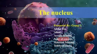
  • 1. The nucleus Presented By: Group 1 Afsha Zia, Alizay, Ayesha Khalid, Noor-ul-naeem, Tehreem Ishtiaq, Tehreem Tariq
  • 2. CONTENTS  Introduction  Nuclear Envelope  Transport Through Nucleus  Internal Organization of Nucleus  Assembly of RNAs
  • 3. THE NUCLEUS  When you look at a eukaryotic cell in a light microscope nucleus is the largest visible compartment.  The presence of a nucleus distinguishes eukaryotic cells from prokaryotic cells.  The nucleus houses all of the eukaryotic cell’s genome and acts as a center for controlling cellular activities.  Processes such as DNA replication, transcription, and RNA processing all takes place within the nucleus.
  • 4.
  • 5. DISCOVERY  Antony van Leeuwenhoek (1632– 1723) was probably the first to observe nucleus in the blood cells of birds and amphibians.  But Felice Fontana (1730–1805) was the actual discoverer of nucleus by observing epidermal cells of eel.  The Scottish botanist, Robert Brown (1773–1858) observed the nucleus in plant cells and was the first to call these structures ‘nuclei’.
  • 6. STRUCTURE  A double membrane called nuclear envelope encloses the nucleus.  The lumen separates the two membranes and is continuous with the Endoplasmic Reticulum.  Macromolecules pass between the nucleus and cytoplasm through the Nuclear Pore complexes (NPCs) that are channels spanning the envelope.  The nucleus has non-enveloped sub-compartments with specialized function.  Nucleolus is the most clearly visible structure.  Regions other than the nucleolus are referred to as the nucleoplasm.  Other sub-compartments include speckles, cajal bodies and PML bodies etc.  Inside the nucleus the DNA can be found in the compacted and highly stained form, heterochromatin or in the less densely compacted form the euchromatin.
  • 8. ADVANTAGES TO EUKARYOTES IN HAVING A NUCLEUS  By separating the genome from the cytoplasm, the nuclear envelope allows gene expression to be regulated by mechanisms that are unique to eukaryotes.  In prokaryotes mRNAs are translated before their transcription is completed, eukaryotic mRNAs are produced from precursor RNAs through complete processes before being transported from the nucleus to the cytoplasm.  When DNA is replicating during interphase the nucleus protects the relaxed DNA from breakage by force of cytoskeleton.
  • 9. NUCLEI VARY IN NUMBER AND APPEARANCE  Nuclei can vary in size according to the amount of DNA they contain. The single celled organism Saccharomyces cerevisiae has a nucleus of 1µ in diameter.  Many multicellular organisms have a nucleus of size 5-10µ in diameter.  The percentage of volume occupied by nuclei in different cells varies.  Volume occupied by nucleus in yeasts cells is 1%-2%, 10% in somatic cells and 40%-60% in cells that do not have many cytoplasmic functions.
  • 10.  In most cells the nucleus is oblong or oval shaped to minimize surface area of enclosure. We can identify different cells from the shape of their nucleus.  Example: During leukemia an excess of white blood cells are produced which have different shapes of nuclei at different stages of development. The type of leukemia can be diagnosed by studying the morphology of nuclei of the cells produced in excess.  Most cells are mononucleate, some are multi nucleate and some are anucleate.  Example: Multinucleate cells include those of Drosophila melanogaster in embryonic stages.  Myocytes formed by fusion of myoblasts are also multinucleate.  Cells like the mammalian red blood cells and cells of lens of vertebrate eye lack nucleus. Figure: A Drosophila embryo at the multinucleate stage
  • 12. NUCLEAR MEMBRANE  Nuclear Membrane is a fence between nucleus and cytoplasm to stave off free transmission of molecules.  It provides nucleus an identity of separate biochemical compounds.  The nuclear membrane consists of: 1. Outer nuclear membrane 2. Inner nuclear membrane 3. Perinuclear space 4. Nuclear pores 5. Nuclear lamina
  • 13. OUTER NUCLEAR MEMBRANE  The outer nuclear membrane is continuous with endoplasmic reticulum, therefore the lumen of nuclear membrane is directly connected with lumen of ER.  The outer nuclear membrane is functionally homologous to ER membrane.  The cytoplasmic surface of outer nuclear membrane has ribosomes that are different in composition of protein and these ribosomes are enriched in membrane proteins (for cytoskeleton binding).
  • 14. PERINUCLEAR SPACE  Space is present between ONM and INM and is called Perinuclear space or lumen of envelope.  The thickness of each nuclear membrane is 7-8nm thick while perinuclear space is 20-40nm thick.
  • 15. INNER NUCLEAR MEMBRANE  Proteins that are specific to nucleus are present in INM such as those that bind the nuclear lamina.  Including Lamin B receptor (LBR), lamina- associated polypeptide (LAP) 1, LAP2, emerin, MAN1 and nurim.  Most of these proteins interact with lamins and chromatin.  Mutations in emerin and nuclear lamins have been associated with muscular dystrophies and lipodystrophy.  Integral proteins of the inner nuclear membrane are synthesized on the rough ER and reach the inner nuclear membrane by lateral diffusion in the connected ER and nuclear envelope membranes.
  • 16. NUCLEAR PORE CHANNEL (NPC)  The Phospholipid bilayer is only permeable for non-polar micromolecules.  The only channel through which transmission of polar micromolecules and macromolecules occurs is through Nuclear Pore Complex.  NPCS are the points where lNM and ONM are continuous. Figure: The inner and outer membranes of the nuclear envelope are fused at the NPC.
  • 17. NUCLEAR LAMINA  In multicellular eukaryotes, a fibrous mesh work supports the inner nuclear membrane called Nuclear Lamina  The nuclear lamina is present inside the nuclear envelope.  Lamins are 60-80 kilo Dalton fibrous proteins that makeup the nuclear lamina  Some associated proteins are also present.  Lamins belong to a class of intermediate filament proteins.  Nuclear Lamina disease: 1. Emery-Dreifuss muscular dystrophy 2. Hutchinson-Gilford progeria syndrome
  • 18. MODEL OF LAMIN ASSEMBLY • Association of lamins with each other occurs to form higher order structure called nuclear lamina. • Attachment of lamins to the nuclear envelope is mediated by prenylation and binding to specific inner nuclear membrane proteins such as emerin and Lamin B receptor.
  • 19. DURING MITOSIS THE NUCLEAR ENVELOPE DISASSEMBLES Onset  The nuclear membrane disassembles and the nuclear lamina depolymerizes due to phosphorylation of nuclear lamins by Cyclin- dependant protein kinase.  The nuclear envelope membrane proteins also dissemble by phosphorylation leading to disassembling of NPCs.  NPC proteins become bound to nuclear import receptors, which play an important part in the reassembly of NPCs at the end of mitosis.  Motor proteins on microtubules also help in tearing down the nuclear membrane. Offset  The nuclear envelope forms around chromatin. Chromatin is surrounded by a shroud of Ran-GTP.  The assembly of NPCs is started by nuclear import receptors from NPC proteins displaced by the Ran-GTP cloud.  Alongside the nuclear envelope membrane proteins with dephosphorylated lamins start attaching to chromatin again.  Membranes from ER fuse over the chromatin until a complete Nuclear envelope is formed.
  • 20.
  • 22. WHAT ARE NUCLEAR PORES?  Small polar molecules, ions and macro-molecules can only move in between the nucleus and cytoplasm through channels.  The large circles with diameter of 120nm and molecular mass of ̴125 million Dalton and 30X the size of ribosomes are pores that are collectively called as Nuclear Pore complex.  They are composed of several proteins of 30 different types.  Those specialized proteins are named as Nucleoporins.
  • 23. VARIATION IN NUMBER OF NPCS IN MEMBRANES  Requirement of nuclear transmission of a cell decides the number of NPCs.  150-250 NPCs are present in the much smaller yeast cell's nuclear envelope.  Some cells contain millions of NPCs (e.g. Xenopus) because they are very active transcriptionally and translationally. FIGURE The surface of the nuclear envelope of a Xenopus laevis oocyte is covered with NPCs.
  • 24. MODEL OF THE NUCLEAR PORE COMPLEX  The complex consists of an assembly of eight spokes attached to rings on the cytoplasmic and nuclear sides of the nuclear envelope.  The spoke-ring assembly surrounds a central channel containing the central transporter.  Cytoplasmic filaments extend from the cytoplasmic ring, and filaments forming the nuclear basket extend from the nuclear ring.
  • 25. FUNCTIONS OF NUCLEAR PORE  Nuclear pores play an important role in physiology of eukaryotic cells by controlling the traffic of molecules between nucleus and cytoplasm.  RNAs that are synthesized in nucleus are carried out through the nuclear pores in order to synthesize proteins in the cytoplasm.  Conversely, proteins required for nuclear functions (e.g., transcription factors) must be transported to the nucleus from their sites of synthesis in the cytoplasm.  Many proteins shuttle continuously in between nucleus and cytoplasm which is also a very specialized function of nuclear pore.
  • 26. MOLECULAR TRAFFIC THROUGH NUCLEAR PORE COMPLEXES  Small molecules are able to pass rapidly through open channels in the nuclear pore complex by passive diffusion.  In contrast, macromolecules are transported by a selective, energy- dependent mechanism that acts predominantly to import proteins to the nucleus and export RNAs to the cytoplasm.
  • 27. NUCLEAR LOCALIZATION SIGNALS Figure: The nuclear localization signal of nucleoplasm is bipartite, consisting of a Lys-Arg sequence, followed by a Lys-Lys-Lys-Lys sequence located ten amino acids farther downstream.  These proteins are targeted to the nucleus by specific amino acid sequences called nuclear localization signals.  These localization signals are detected by nuclear transport receptors (Importins) as they carry proteins into the nucleus.  They direct protein transport through nuclear pore complex.  Nuclear localization signals have yet been observed in many proteins, and amino acids that are involved in these localization signals are found close to each other but not immediately adjacent to each other.
  • 28. TRANSPORT OF RNAs  Since proteins are synthesized in the cytoplasm, the export of mRNAs, rRNAs, tRNAs, and microRNAs (miRNAs) is a critical step in gene expression in eukaryotic cells.  As proteins are selectively transported from cytoplasm to nucleus in the same way RNAs are transported from nucleus to cytoplasm to synthesize these proteins.  Like protein import, the export of all RNAs through the nuclear pore complex is an active, energy-dependent process requiring the transport receptors to interact with the nuclear pore complex.  Karyopherin, importins and exportins transport most tRNAs, rRNAs, miRNAs and small nuclear RNAs.  In contrast to mRNAs, tRNAs, and rRNAs, which function in the cytoplasm, many small RNAs (snRNAs and snoRNAs) function within the nucleus as components of the RNA processing machinery.
  • 29. TRANSPORT OF SnRNAs BETWEEN NUCLEUS AND CYTOPLASM  snRNAs firstly get transported into the cytoplasm from nucleus.  Then they associate with proteins to form functional snRNPs.  And then go back to the nucleus.  Crm1 and other transport receptor proteins that bind to the 5' 7-methylguanosine caps of snRNAs are involved in the export of the snRNAs to the cytoplasm. Figure: Small nuclear RNAs are initially exported from the nucleus to the cytoplasm, where they associate with proteins to form snRNPs. The assembled snRNPs are then transported back into the nucleus.
  • 30. ACTIVE TRANSPORT OF MACROMOLECULES  Uncharged molecules including water, can diffuse freely through phospholipid bilayers.  Other macromolecules that are transported across the nuclear envelope move through NPCs.  The process of moving through the NPC is called translocation.
  • 32. INTERNAL ORGANIZATION OF NUCLEUS  A loosely organized matrix of nuclear lamins extends from nuclear lamina into the interior of nucleus in animal cell, which serves as sites of chromatin attachment and bind other proteins into the nuclear bodies.  Chromatin is organized into large loops of DNA and regions of these loops are bound to the lamin matrix by lamin binding proteins.  Many other nuclear proteins form Lamin-dependent complex
  • 33. CHROMOSOMES AND HIGHER ORDER CHROMATIN STRUCTURES  During interphase the heterochromatin remains highly condensed and inactive and euchromatin is de-condensed and distributed throughout the nucleus, a heterochromatin is of two types:  Constitutive heterochromatin remains permanently in heterochromatin stage, having DNA sequences that are not transcribed.  Facultative heterochromatin consist of euchromatin that takes on the staining and compactness characteristics of heterochromatin during some phase of development, having DNA sequences that are transcribed in cell which we are studying but are transcribed in other cells.
  • 34.  Although interphase chromatin appears to be uniformly distributed, the chromosomes are actually arranged in an organized fashion and divided into discrete functional domains that play an important role in regulating gene expression. Heterochromatin in interphase nuclei The euchromatin is distributed throughout the nucleus. The heterochromatin is indicated by arrowheads and the nucleolus by an arrow.
  • 35. SUB-COMPARTMENTS WITHIN THE NUCLEUS  The internal organization of nucleus is the result of localization of nuclear processes to specific regions of nucleus.  Many enzymes and proteins of nucleus are organized to discrete sub-nuclear bodies. The nature and function of these nuclear bodies are not clear.  Replication of multiple DNA molecules takes place on the cluster site of nuclei in mammalian cell. Sub-compartments within the nucleolus, as
  • 36.  Actively transcribed genes appears to be distributed throughout the nucleus.  Components of mRNA splicing machinery are concerted in nuclear speckles. Immunoflourescent staining showed that rather than being distributed uniformly throughout the nucleus the components of RNA splicing apparatus are concerted in these 20-50 discrete structures:  Speckles: storage sites of splicing components where pre mRNA processing occurs. In addition to speckles nuclei also contain PML and cajal bodies.  PML bodies: transcription factors & chromatin-modifying enzymes localize here.  Cajal bodies/coiled body: involved in snRNP biogenesis, histone mRNA processing & telomere maintenance.  Gemini Bodies: are not found in all cells, and some of their components are also found in Cajal bodies, suggesting they may not perform distinct functions. Cajal bodies and Gemini bodies can be detected by using specific antibodies and indirect immuno- fluorescence.
  • 37. CHROMOSOMES OCCUPY DISTINCT TERRITORIES  Although the nucleus lacks internal membranes, nuclei are highly organized and contain many sub-compartments.  Each chromosome occupies a distinct region or territory, which prevents chromosomes from becoming entangled with one another.  The nucleus contains both chromosome domains and inter- chromosomal regions. Individual chromosomes occupy distinct areas of the nucleus called chromosome territories.
  • 38. NUCLEAR SUB COMPARTMENTS ARE NOT MEMBRANE-BOUNDED  Nuclear sub compartments are not membrane-bounded.  rRNA is synthesized and ribosomal subunits are assembled in the nucleolus.  Genes that encode runs are present on multiple chromosomes that cluster together to form nucleoli sub-compartments, mRNA splicing factors are stored in nuclear speckles and move to sites of transcription where they function.  Other nuclear bodies have been identified using antibodies; some of these bodies are believed to concentrate specific nuclear proteins, but the functions of most nuclear bodies are unknown.
  • 40. THE NUCLEOLUS  The most prominent nuclear body is the nucleolus.  It is the site of rRNA transcription and processing as well as aspects of ribosome assembly.  The nucleolus is a ribosome production factory, designed to fulfill the need for regulated and efficient production of rRNAs and assembly of the ribosomal subunits.  Actively growing mammalian cells, for example, contain 5 million to 10 million ribosomes that must be synthesized each time the cell divides.  Recent evidence suggests that nucleoli also have a more general role in RNA modification and that several types of RNA move in and out of the nucleolus at specific stages during their processing. Nucleoli in amphibian oocytes The amplified rRNA genes of Xenopus oocytes are clustered in multiple nucleoli (darkly stained spots).
  • 41. RIBOSOMAL RNA GENES AND THE ORGANIZATION OF THE NUCLEOLUS  The nucleolus is associated with the chromosomal regions that contain the genes for the 5.8S, 18S, and 28S rRNAs.  Ribosomes of higher eukaryotes contain four types of RNA designated the 5S, 5.8S, 18S, and 28S rRNAs.  The 5.8S, 18S, and 28S rRNAs are transcribed as a single unit within the nucleolus by RNA polymerase I, yielding a 45S ribosomal precursor RNA.  Transcription of the 5S rRNA, which is also found in the 60S ribosomal subunit, takes place outside the nucleolus in higher eukaryotes and is catalyzed by RNA polymerase III.
  • 42. rRNA Genes  The human genome, for example, contains about 200 copies of the gene that encodes the 5.8S, 18S, and 28S rRNAs and approximately 2000 copies of the gene that encodes 5S RNA.  The genes for 5.8S, 18S, and 28S rRNAs are clustered in tandem arrays on five different human chromosomes (chromosomes 13, 14, 15, 21, and 22)  The 5S rRNA genes are present in a single tandem array on chromosome 1.
  • 43. RIBOSOMAL RNA GENES Each rRNA gene is a single transcription unit containing the 185, 5.85, and 285 rRNAs as well as transcribed spacer sequences. The rRNA genes are organized in tandem arrays, separated by non-transcribed spacer DNA
  • 44. IMPORTANCE OF RIBOSOME PRODUCTION  The importance of ribosome production is particularly evident in oocytes in which the rRNA genes are amplified to support the synthesis of the large numbers of ribosomes required for early embryonic development.  In Xenopus oocytes, the rRNA genes are amplified approximately two- thousand-fold, resulting in about one million copies per cell.  These amplified rRNA genes are distributed to thousands of nucleoli, which support the accumulation of nearly 1012 ribosomes per oocyte.  Recently, it has been shown that ribosome biogenesis is intimately linked to multiple cellular signaling pathways and that defects in ribosome production can lead to a wide variety of human diseases.
  • 45. TRANSCRIPTION AND PROCESSING OF rRNA  Each nucleolar organizing region contains a cluster of tandemly repeated rRNA genes separated from each other by non-transcribed spacer DNA.  These genes are very actively transcribed by RNA polymerase I, allowing their transcription to be readily visualized by electron microscopy.  In such electron micrographs, each of the tandemly arrayed rRNA genes is surrounded by densely packed growing RNA chains forming a structure that looks like a Christmas tree.  The high density of growing RNA chains reflects that of RNA polymerase molecules, which are present at a maximal density of approximately one polymerase per hundred base pairs of template DNA.
  • 46. RIBOSOME ASSEMBLY  Early in ribosome assembly, the processing of the two nascent ribosomal subunits occur separately  Processing of the smaller subunit, which contains only the 18S rRNA, is simpler and involves only four endonuclease cleavages.  In higher eukaryotes, this is completed within the nucleus but in yeast the final cleavage to the mature 18S rRNA actually occurs after export of the 40S subunit to the cytosol.  Processing of the larger subunit, which contains the 28S, 5.8and 5S rRNAs, involves extensive nuclease cleavages and is completed within the nucleolus. Consequently, most of the pre-ribosomal particles in the nucleolus represent precursors to the large (60S) subunit.  The final stages of ribosomal subunit maturation follow the export of pre-ribosomal particles to the cytoplasm, forming the active 40S and 60S subunits of eukaryotic ribosomes.
  • 47.
  • 48. Ribosomal proteins are imported to the nucleolus from the cytoplasm and begin to assemble on pre- rRNA prior to its cleavage. As the pre-rRNA is processed, additional ribosomal proteins and the 55 rRNA (which is synthesized elsewhere in the nucleus) assemble to form preribosomal particles. The final steps of maturation follow the export of preribosomal particles to the cytoplasm, yielding the RIBOSOME ASSEMBLY
  • 50. REFERENCES  Pelava, A., Schneider, C., & Watkins, N. J. (2016). The importance of ribosome production, and the 5S RNP-MDM2 pathway, in health and disease. Biochemical Society transactions, 44(4), 1086-90.  Holmer, L. & Worman, H. CMLS, Cell. Mol. Life Sci. (2001) 58: 1741. https://doi.org/10.1007/PL00000813  Plopper, G. (n.d.). Lewins cells (3rd ed.). Burlington, MA;Jones & Bartlett Learning;2015.  Cooper, G. M. (2007). Cell: A molecular approach (4rth ed.). Place of publication not identified: Sinauer Associates.  Alberts, B. (2008). Molecular biology of the cell (5th ed.). New York: Garland Science.  Google Images