
Recomendados
Mais conteúdo relacionado
Mais procurados
Mais procurados (20)
Semelhante a Diabetic foot
Semelhante a Diabetic foot (20)
Mais de ajayyadav753
Mais de ajayyadav753 (20)
Último
Último (20)
Diabetic foot
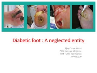
- 1. Diabetic foot : A neglected entity Ajay Kumar Yadav PGY2,Internal Medicine IOM-TUTH, Kathmandu 2074/12/20
- 2. Why is it a neglected entity and still important ? Individuals who develop a DFU are at greater risk of premature death, myocardial infarction and fatal stroke than those without a history of DFU. Around 25% of people with diabetes will develop a DFU during their lifetime. In most developed countries, the annual incidence of foot ulceration amongst people with diabetes is about 2%. Increased economic burden : In England, foot complications account for 20% of the total National Health Service spend on diabetes care, which equates to around £650 million per year
- 3. In these countries, diabetes is the most common cause of non-traumatic amputation; approximately 1% of people with diabetes suffer alower-limb amputation. It has been estimated that every 20 seconds a lower limb is amputated due to complications of diabetes. Mortality following amputation ranges from 50–68% at five years, which is comparable or worse than for most malignancies It has been suggested that up to 85% of amputations can be avoided when an effective care plan is adopted IDF Clinical Practice Recommendations on the Diabetic Foot 2017
- 5. Diabetic foot : risk category
- 6. Aetiology of DFU Neuropathic Ischaemic Neuroischaemic • Loss of protective sensation is a major component of nearly all DFU. It is a/w seven–fold increase in risk of ulceration. • People with diabetes are 4-5 times as likely to have PAD as those without diabetes. It is also a key risk factor for lower extremity amputation. • Even in the absence of a poor arterial supply, microangiopathy (small vessel dysfunction) contributes to poor ulcer healing in neuroischaemic DFU
- 8. Mechanism for Diabetic foot BMJ 2017;359:Supp 1
- 9. Bony/structural deformities Results from neuropathic changes, stiffening of the joints (termed cheiroarthropathy), altered biomechanics or previous surgeries. Motor neuropathy : intrinsic muscle atrophy contracted digits and a displaced fat pad from the metatarsal heads to just below the toesmetatarsal heads become prominent and close to the skin surface increased pressure and a potential ulceration site Clawed toes Hammer toes Hallux limitus : Limitation in the range of motion of the first MTP joint Hallux rigidus : complete immobility
- 12. Classification of diabetic wound
- 13. Wagner classification of diabetic wound Grade Characteristics of wound 0 Intact Skin 1 Superficial ulcer of skin or subcutaneous tissue 2 Ulcers extend into tendon, bone, or capsule 3 Deep ulcer with osteomyelitis, or abscess 4 Gangrene of toes or forefoot 5 Midfoot or hindfoot gangrene
- 15. Management of Diabetic foot Successful diagnosis and treatment of pts with DFUs involves a holistic approach that includes • Optimal diabetes control • Effective local wound care • Infection control • Pressure relieving strategies • Restoring pulsatile blood flow.
- 16. ABCDEFS Scheme for management of DM A – A1c : HbA1C < 7% B – Blood pressure : BP target < 130/80 mmHg. C – Cholesterol : LDL target of 2.0 mmol/L or less. D – Drugs : Protect against heart attack and stroke with appropriate medication. E – Exercise : Regular physical activity. F – Footcare S – Smoking and stress : Stop smoking and manage stress effectively.
- 20. Assessing DFUs • EXAMINATION OF THE ULCER o A physical examination should determine: Is the wound predominantly neuropathic, ischaemic or neuroischaemic? If ischaemic, is there critical limb ischaemia? Are there any musculoskeletal deformities? What is the size/depth/location of the wound? What is the colour/status of the wound bed? : Black (necrosis) : Yellow, red, pink Is there any exposed bone? Is there any necrosis or gangrene? Is the wound infected? If so, are there systemic signs and symptoms of infection (such as fevers, chills, rigors, metabolic instability and confusion)? Is there any malodour? Is there local pain? Is there any exudate? What is the level of production (high, moderate, low, none),colour and consistency of exudate, and is it purulent? What is the status of the wound edge callus, maceration, erythema, oedema, undermining)?
- 22. • Documenting ulcer characteristics Size, depth, appearance and location Digitally photographing DFUs at the first consultation and periodically thereafter to document progress is helpful
- 23. Testing for loss of sensation Two simple and effective tests for peripheral neuropathy are commonly used: 10g (Semmes-Weinstein) monofilament • A positive result is the inability to feel the monofilament when it is pressed against the foot with enough force to bend it • Do not test for neuropathy in areas of callus as this can mask feeling from any of the neuropathy testing devices and may give a false- positive result. Standard 128Hz tuning fork.
- 25. Testing for vascular status Assessment of the femoral, popliteal and pedal (dorsalis pedis and posterior tibial) pulses. The dorsalis pedis pulse is reported to be absent in 8.1% of healthy individuals, and the posterior tibial pulse is absent in 2.0%. If the leg and ankle arterial pulses are normal and auscultation reveals no femoral arterial bruit, PAD can be excluded with specificity and negative predictive value as high as 98.3% and 94.9%, respectively. Grading of pulse : • Strong arterial pulse (0, non-ischemic) • Palpable but slightly diminished (1, mild) • Thready and scarcely palpable (2, moderate) • Non-palpable pulses (3,severe).
- 26. • ABPI (Ankle brachial pressure index ) The sensitivity of ABI is 95%, and the specificity 99%. ABI normal reference value is 1.00 -1.30, 0.91 to 0.99 for borderline PAD. ABI > 1.30 or higher usually means vascular calcification, and impaired arterial elasticity. ABI is not sensitive enough in detecting PAD in the early stage -- calcinosis of the arterial wall in diabetes, can falsely elevate ABI If ABI > 1.30, toe brachial index (TBI) may be measured.
- 27. • Ischemia is defined as an ABI < 0.9 and TBI < 0.75. ABI 0.71 to 0.90 : mild PAD, ABI 0.41 to 0.70 : moderate and ≤ 0.40 : severe PAD or critical limb ischaemia A pt with acute limb ischaemia characterised by the six ‘Ps’ (pulselessness, pain, pallor [mottled colouration], perishing cold, paraesthesia and paralysis)
- 28. Optimising local wound care • TIME framework Tissue debridement Inflammation and infection control Moisture balance (optimal dressing selection) Epithelial edge advancement.
- 29. Factors that may Impact Skin Integrity and Wound Healing Glycemic level : delayed wound healing and compromised chemotaxis and phagocytosis Activity Smoking : compromised blood flow Trauma : prone to injury/re-injury. Footwear: ill-fitting and inappropriate footwear Neuropathy Bony deformity : areas of high pressure and skin breakdown PAD History of wounds: healed wounds are more vulnerable to re-injury due to less resiliency and elasticity Amputation : abnormal mechanics and ill-fitting prosthetics may cause pressure leading to tissue injury and prevent closure of existing wounds Age
- 30. Tissue debridement Surgical/sharp Larval Autolytic Hydrosurgery and Ultrasonic
- 31. Sharp debridement • Gold standard technique • Regular, local, sharp debridement using a scalpel, scissors and/or forceps • Vascular status must always be determined prior to sharp debridement. Patients needing revascularisation should not undergo extensive sharp debridement because of the risk of trauma to vascularly compromised tissues • Benefits Removes necrotic/sloughy tissue and callus Reduces pressure Allows full inspection of the underlying tissues Helps drainage of secretions or pus Helps optimise the effectiveness of topical preparations Stimulates healing.
- 32. • Other debridement methods • While sharp debridement is the gold standard technique, other methods may be appropriate in certain situations: As an interim measure For patients for whom sharp debridement is contraindicated or unacceptably painful When the clinical decision is that another debridement technique may be more beneficial for the patient For patients who have expressed another preference.
- 33. • Larval therapy The larvae of the greenbottle fly can achieve relatively rapid, atraumatic removal of moist, slimy slough, and can ingest pathogenic organisms present in the wound It is not recommended as the sole method of debridement for neuropathic DFUs as the larvae cannot remove callus • Hydrosurgical debridement Alternative method of wound debridement : forces water or saline into a nozzle to create a high-energy cutting beam. Enables precise visualisation and removal of devitalised tissue in the wound bed
- 34. • Autolytic debridement Natural process that uses a moist wound dressing to soften and remove devitalised tissue. Not recommended in the presence of ischaemia and/or dry gangrene
- 35. INFLAMMATION AND INFECTION CONTROL
- 36. Identifying infections Around 56% of DFUs become infected and overall about 20% of patients with an infected foot wound will undergo a lower extremity amputation. • Risk factors for infection A positive probe-to-bone test DFU present for more than 30 days A history of recurrent DFUs A traumatic foot wound The presence of PAD in the affected limb A previous lower extremity amputation Loss of protective sensation The presence of renal insufficiency A history of walking barefoot.
- 37. • Subtle 'secondary'signs s/o infection Friable granulation tissue Wound undermining Malodour or wound exudate • Signs of spreading infection Spreading, intense erythema Increasing induration Lymphangitis Regional lymphadenitis Hypotension, tachypnoea, tachycardia Rigors
- 39. Classification of diabetic foot infections
- 40. Diabetic foot osteomyelitis Osteomyelitis s/b suspected when ulcers lie over a bony prominence and fail to heal in spite of offloading or have associated soft tissue induration (“sausage toe “appearance). A definitive diagnosis of diabetic foot osteomyelitis requires both histologic evidence of bone infection and isolation of a bacterial pathogen from a bone sample. The probe to bone (PTB) test is a useful clinical tool when employed correctly for the diagnosis of osteomyelitis. Plain X-rays have sensitivity and specificity of 0.54 and 0.68 respectively.
- 41. Blood tests for inflammatory markers such as CRP and ESR are often used but are not specific to the type of infection : An ESR elevated to more than 70 mm/hr increases the likelihood of osteomyelitis. ESR drops more slowly with treatment, it may serve as a marker for treatment response MRI : gold standard for diagnosing osteomyelitis. When MRI is contraindicated, or not available, a white-blood-cell labelled radionuclide scan, computed tomography or positron emission tomography may beconsidered
- 42. Antibiotic selection • Acute diabetic foot infections without prior antibiotic treatment caused by aerobic Gram-positive organisms and most commonly by Staphylococcus aureus including MRSA and B-hemolytic streptococcus • Chronic infections Wound becomes polymicrobial and includes anaerobes Cultures should not be taken from clinically non-infected wounds as all ulcers will be contaminated; microbiological sampling cannot discriminate colonisation from infection Obtaining specimens for culture is recommended only for clinically infected wounds prior to initiation of antibiotic therapy. • The specimen for culture should be obtained following wound cleansing and debridement. • If bone is involved, the recommendation is to obtain a bone biopsy
- 44. • Duration of antibiotic treatment The duration of therapy depends on the severity of infection, the involvement of bone and the patient’s response to treatment. In general, for mild soft tissue infections, two weeks of oral treatment is recommended. For more severe soft-tissue infection or for larger necrotic wounds, a longer course may be required. For osteomyelitis, usually four to six weeks of intravenous treatment is recommended (this may be followed by oral antibiotic course).
- 45. • If after two weeks: There is improvement in the wound, but continuing signs of infection : continue the chosen treatment The wound has improved and the signs and symptoms of wound infection are no longer present : antimicrobial s/b discontinued and a non-antimicrobial dressing applied to cover the open wound No improvement : discontinue treatment + re-culture wound + reassess the need for surgical therapy or revascularisation.
- 46. • Superficial DFUs with skin infection (mild infection) Start empiric oral antibiotic therapy targeted at Staphylococcus aureus and ß- haemolytic Streptococcus Change to an alternate antibiotic if the culture results indicate a more appropriate antibiotic Obtain another optimum specimen for culture if the wound does not respond to treatment.
- 47. • Role of topical antimicrobials Advantage of not driving resistance. Provide high local concentrations, but do not penetrate intact skin or into deeper soft tissue • Beneficial in certain situations: Where there are concerns regarding reduced antibiotic tissue penetration — for example, where the patient has a poor vascular supply In non-healing wounds where the classic signs and symptoms of infection are absent, but where there is a clinical suspicion of increased bacterial bioburden. • If there are clinical signs of infection at dressing change, systemic antibiotic therapy should be started • Patients may also require debridement to remove infected material. In addition, infected wounds should be cleansed at each dressing change with saline or an appropriate antiseptic wound cleansing agent.
- 48. • Common topical antimicrobial agents used Silver : dressings containing silver (elemental, inorganic compound or organic complex) or silver sulphadiazine cream/ dressings Polyhexamethylene biguanide (PHMB) : solution, gel or impregnated dressings Iodine : povidone iodine (impregnated dressing) or cadexomer iodine (ointment, beads or impregnated dressings) Medical-grade honey : gel, ointment or impregnated dressings
- 49. • Deep tissue infection (moderate to severe infection) • For treating deep tissue infection (cellulitis, lymphangitis, septic arthritis, fasciitis): Start patients quickly on broad-spectrum antibiotics, commensurate with the clinical history and according to local protocols where possible Take deep tissue specimens or aspirates of purulent secretions for cultures at the start of treatment to identify specific organisms in the wound, but do not wait for results before initiating therapy Change to an alternate antibiotic if: — indicated by microbiology results — the signs of inflammation are not improving Administer antibiotics parenterally for all severe and some moderate infections and switch to the oral route when the patient is systemically well and culture results are available
- 50. Continue antibiotic therapy until the infection resolves, but not through to complete healing. In most cases 1–3 weeks of therapy is sufficient for soft tissue infections Consider giving empiric therapy directed against MRSA: — in patients with a prior history of MRSA infection — when the local prevalence of MRSA colonisation or infection is high — if the infection is clinically severe. Infection in a neuroischaemic foot is often more serious than in a neuropathic foot (which has a good blood supply ) Infections accompanied by a deep abscess, extensive bone or joint involvement, crepitus, substantial necrosis or gangrene, or necrotising fasciitis, need prompt surgical intervention along with appropriate antibiotic therapy, to reduce the risk of major amputation
- 51. • Biofilms and chronic persistent infection Biofilms are complex polymicrobial communities that develop on the surface of chronic wounds, which may lack the overt clinical signs of infection. They are not visible to the naked eye and cannot be detected by routine cultures This matrix acts as a thick, slimy protective barrier, making it very difficult for antimicrobial agents to penetrate it Treatment should aim to : -- Disrupt the biofilm burden through regular, repeated debridement and vigorous wound cleansing -- Prevent reformation and attachment of the biofilm by using antimicrobial dressings. Appropriate wound bed preparation remains the gold standard for biofilm removal
- 54. Moisture balance: optimal dressing selection • Factors to consider include: Location of the wound Extent (size/depth) of the wound Amount and type of exudate The predominant tissue type on the wound surface Condition of the periwound skin Compatibility with other therapies (eg contact casts) Wound bioburden and risk of infection Avoidance of pain and trauma at dressing changes Quality of life and patient wellbeing.
- 56. Types of wound dressing
- 58. • When applying dressings: Avoid bandaging over toes as this may cause a tourniquet effect (instead, layer gauze over the toes and secure with a bandage from the metatarsal heads to a suitable point on foot) Use appropriate techniques (eg avoiding creases and being too bulky) and take care when dressing weight-bearing areas Avoid strong adhesive tapes on fragile skin Avoid tight bandaging at the fifth toe and the fifth metatarsal head (trim the bandage back) Ensure wound dead space is eliminated (eg use a dressing that conforms to the contours of the wound bed) Remember that footwear needs to accommodate any dressing. Wounds should be cleansed at each dressing change and after debridement with a wound cleansing solution or saline. Cleansing can help remove devitalised tissue, re-balance the bioburden and reduce exudate to help prepare the wound bed for healing98. It may also help to remove biofilms
- 59. Epithelial edge advancement • It is important to debride the edges of the ulcer to remove potential physical barriers to the growth of the epithelium across the ulcer bed. • Conversely, ‘die-back’ is an abnormal response to over-aggressive sharp debridement. It involves necrosis at the wound edge and extends through previously healthy tissue
- 60. Pressure offloading • Offload at-risk areas of the foot in order to redistribute pressures evenly. • Inadequate offloading leads to tissue damage and ulceration. • The gold standard is the total contact cast (TCC) : This is a wellmoulded, minimally padded foot and lower leg cast that distributes pressures evenly over the entire plantar surface of the foot.
- 61. • Disadvantages of TCCs include: Must be applied by fully trained and experienced practitioners May cause skin irritation and further ulcers if applied inappropriately Prevents daily inspection (signs of spreading infection may go unnoticed) May disturb sleep Makes bathing difficult Patient may not tolerate it (especially in warm climates) May prevent patient's ability to work Relatively high cost/low availability. Contraindicated in patients with ischaemia because of the risk of inducing further DFUs
- 62. • Removable devices may also be more pragmatic choices for less motivated patients because they allow patients to bathe and sleep more comfortably
- 64. Amputation • Indications Ischaemic rest pain that cannot be managed by analgesia or revascularisation A life-threatening foot infection that cannot be managed by other measures A non-healing ulcer • Around half of patients who undergo an amputation will develop a further DFU on the contralateral limb within 18 months of amputation. • The three–year mortality rate after a first amputation is 20–50%. • In a six-year follow-up study, almost 50% of patients developed critical limb ischemia in the contralateral limb
- 65. • Minor amputations: Amputation of digits Partial foot amputation Ankle disarticulation • Major amputations: Below-knee amputation, aka BKA Knee disarticulation Above-knee amputation, aka AKA (transfemoral) Van Nes rotation/rotationplasty (foot turned around and reattached to allow the ankle joint to be used as a knee) Hip disarticulation Hemipelvectomy/hindquarter amputation
- 66. Charcot osteoarthropathy Progressive condition characterized by pathological fractures, joint dislocation and destruction of the pedal architecture. Predisposing factors • Peripheral neuropathy • Increases in local blood flow • Excessive osteoclastic activity • Unrecognized injury and continued repetitive stress Once the condition is triggered, the uncontrolled inflammation process leads to osteolysis. This is directly responsible for joint and bone destruction
- 67. • Acute Charcot metabolic changes : bony reabsorption and multiple spontaneous fractures. • Increased warmth(8 to 15 F difference compared with a mirror image) is the first indicator of inflammation in an insensate foot and may be the first sign of acute Charcot foot.
- 68. Stage of charcot osteoarthropathy Stage Description 0 (prodromal) Skin flush/redness and increased skin temperature, with or without local edema and bounding pulses. Evidence of instability of the foot. X-ray evidence may or may not be seen. 1(developmental, acute) Acute destructive period that is induced by minor trauma resulting in fragmentation of bone and joint dislocation and subluxation. The most important stage 2(coalescence, subacute) Lessening of edema and healing of fractures 3(reconstruction, chronic) Healing of bone and remodelling on X-ray, plus evidence of deformity
- 69. Management of charcot osteoarthropathy
- 71. Tips on foot care for people with diabetes Inspect both feet daily, including the area between the toes. Wash the feet daily with water at room temperature, with careful drying, especially between the toes. Use lubricating oils or creams for dry skin, but not between the toes. Cut nails straight across. Do not remove corns and calluses using a chemical agent or plaster. They should not be excised at home and must be managed by trained staff. Always wear socks with shoes and check inside shoes for foreign objects before wearing them. Avoid walking barefoot at all times. Ensure a qualified healthcare provider examines your feet regularly. Notify the healthcare provider at once if a blister, cut, scratch,or sore develops.
- 72. Footcare
- 73. Advanced therapies • Negative pressure wound therapy (NPWT) The only data to support the use of NPWT is as a post-surgical intervention. There is insufficient evidence to support the use of NPWT for the routine management of neuropathic foot ulcers • Hyperbaric oxygen therapy (HBOT) No clinical evidence in ulcers related to neuropathy However, in the presence of diminished arterial flow, hyperbaric oxygen therapy may be of benefit • Biologically active dressings
- 74. Reference • International Best Practice Guidelines: Wound Management in Diabetic Foot Ulcers. Wounds International,2013. • 2018 Canadian Association of Wound Care • IDF Clinical Practice Recommendations on the Diabetic Foot – 2017 • BMJ 2017 : Diabetic foot
- 75. Thank you for bearing with me!!