Rotation investigation
•
1 gostou•125 visualizações

Photo-montage in order to conceal medical information, to frustrate medical forensic inbvestigation. Proofing is by image analysis and reconstruction of situation.

Recomendados
Recomendados
Mais conteúdo relacionado
Semelhante a Rotation investigation
Semelhante a Rotation investigation (20)
Mais de siegfried van hoek
Mais de siegfried van hoek (20)
Último
Último (20)
Rotation investigation
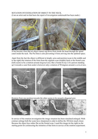
- 1. 1 ROTATION INVESTIGATION OF OBJECT IN THE NECK. (I am an artist and on that basis the report of investigation underneath has been made.) It has become clear that an object cannot sag down from inside the head through the spinal canal inside the neck. The Pia Dura is also preventing it from arriving into the epidural space. Apart from the fact the object is different in height, seize and position (scan in the middle and to the right|) the rotation of the form from the sagittale scan (middle) back to the frontal scan (left) seems to be a rotation around diagonal axis. But a frontal X-ray (‘of a person standing up’) towards a scan from aside is however only a rotation of 90 degrees around a vertical axis. Rotation from one position towards the other seems to be around a diagonal axis: In service of the rotation investigation the image situation has been simulated enlarged. With contrast settings both the scans have sharpened in order to define the 3D-form much closer. Because the object lays rather flat on the frontal scan, I used this image (at the right) as the starting point for constructing the imitation of that form in service of the rotation experiment. RIBS
- 2. 2 In this setup mainly attention has been put at the outer contour lines of the shape regarding direction, seize and position in order to approach the image on the scan as close as possible. The position of the head of the imitated object is true enough a little bit different, but for the rotation experiment in broad outline, this makes no difference at all for the total result. Photo of test set-up based upon / starting from respectively a frontal or a sagittal scan. For making a matching photo in imitation of the object at the starting point of the sagittal scan I had to turn over the camera a little and place it a little bit higher (then perpendicular for the frontal scan). That displacement (in height etc) can be seen as a rotation too (of almost 30o ), but then around a horizontal axis in a vertical field. The experiment however is about the result of rotation of an object with an angle of 90 o around a vertical axis in a horizontal field. The making-process of the imitation of the object, for the simulation of a rotation around a vertical axis of 90 degrees (of a person upright standing) with sight between front towards aside to the left.
- 3. 3 Summary of findings from the first rotation-experiment: Starting from the images at the sagittal scan and the frontal scan both situations I have imitated enlarged in view of the object, and then simulated the situation of rotation (90 degrees around vertical axis). In both situations of test set-up the result after a rotation under an angle of 90o degrees a difference is to be found between the image on the original X-ray- photo and the ultimate result of imitation of rotations. In result thus it became clear that the object-images at the initial Frontal and Sagittal scan- images are not corresponding in rotation. The rotation between the two positions certainly is not at all 90 degrees perpendicular around a vertical axis. - Both shapes at the scans appeared to be imitable with a different position of camera only. So both the positions appeared not to be put on photo perpendicular in order to get a resembling image at the initial corresponding scan already. Note: An X-ray- photo however is being made with an X-ray projector which is beaming perpendicular at the X-ray-negative, with the subject placed in between; and if the client is standing upright, then the rotation is only around a vertical axis. - The imitation on photo has photographic perspective, scan-images don’t, so that minor distortion we take for granted, and we forget about the difference in inclination angle of the camera in both situations, but then still there is a difference in rotation around a vertical axis between the two images. The angle of rotation around a vertical axis (difference between the both visual displays) is not 90 degrees but about 60 degrees. Frontal photo is perpendicular with respect to the protractor / object placed similar for comparison with the object on the Frontal scan (left). For the imitation of the Sagittal however the camera-setup is placed oblique from above and aside scan with the object on it (right). Additional to this little research both situations can be imitated once more in a ‘shadow-play’ (compare that situation with Wajang puppets play), where only contour lines of the object become visible (equal to metal is blocking radiation and likewise in consequence is separating a shape. (The eye's position still has to be corrected somewhat for in respect to the legs.) In proving the incongruity under a rotation of 90 degrees in connection with both the positions such an extra research however is not adding anything more. A report worked out further regarding aspect of forming of this experiment follows below. 60 degrees rotation
- 4. 4 Rotation of the frontal image under 90 degrees doesn’t look as the image of the Sagittal scan. Sagittal image is not matching entirely; the rotation of also does not fit with the Frontal scan. First conclusion in experiment on rotation of the object (with camera perpendicularly at the image) in frontal position rotating clockwise towards the sagittal position (at left side): both positions of the object at the Sagittal-RX respectively the Frontal-RX scan do not correspond together in result of a rotation of 90 degrees of the object around a vertical axis. <= Imitation of the situation starting from the Frontal scan. The camera in this scan-photo investigation is placed with perpendicular sight at the object parallel to the 360 degree surface (with 15 degree subdision ) and the object in countour is placed in a similar position. After a rotation of 90 degrees clockwise this however results into a different position then the related image on the initial Sagittale scan showing. Reverse control: Simulation aproximately of the situation at the Sagittale scan. A rotation of 90 degrees counter-clockwise around avertical axis also results into a different display of the object (with also a difference of about 30 degrees). Added to this the simulation of this image at the Sagittal scan did not resemble precisely when making photo’s of the object perpendiculary. (F) (F) L L L F F F (L) (L)
- 5. 5 (Photo oblique position.) The object even had to be tilted a bit for resembling. After a rotation under 90 degrees the Frontal image is not fitting with the result at the Sagittal scan. Frontal image does not match fully; also the rotation does Second conclusion in experiment on rotation of the object in both situations (with the camera turned over) rotating from a sagittal position (at left side) counter-clockwise towards a frontal position: both positions of the object at the Sagittal- respectively the Frontal-RX scan do not correspond together in result of a rotation of 90 degrees of the object around a vertical axis. L F F L (F) (L) L (F) (L) F <= Immitation of the situation starting from the Sagittal scan. In this photo-experiment the camera is placed in a slight oblique position from above and placed slightly to the right with respect to the 360 degrees surface with 15 degrees subdivisions in order to get a resembling image. After a rotation under an angle of 90 degrees counterclockwise around a vertical axis again a different rendering of the object is shown in imaging result. Reverse control: Imitation of the situation of the Frontal scan with the camera being slightly turned position over around horizontal axis. The rendering of the object on the photo in presenting the situation on the frontal scan does not resemble firmly with the image on the scan, but also this imaging-result after a rotation clockwise around a vertical axis is different from the image on the Sagittal RX scan. The difference in sight is covering an angle of thirty degrees.)
- 6. 6 Principle of rotation between a frontal view and a view from the left side: || Rotation clockwise Gives Sagittal image above Gives Frontal image above Rotation counterclockwise A Frontal scan is being made with the back against the photo-wall. The Sagittal scan was being made with the right side against the photo-wall and the left side towards the camera. The little cross symbolic is indicating the side of the nose of the face. Changing position of a person by rotation therefore is in a circular orbit around a vertical axis. F LR R P P The side of the legs of the clip with the fat line running next to it is nicely straight, but at the over side of it (thin line) it is not. We take the straight lined sides as starting point in order to define the form. The eye of the lower leg is showing some more image information to define the form. Starting from that we will define the definitive form comparing with the form shown on the sagittal scan in order to define the final shape of the clip for a 3D enlarged imitation. Wire ? Wire ? The eye also has a flat part within. For the rest it has an oval form, see also the sagittal scan. The form of the object is difficult to define. At which side are we looking at the inside of the side of the eye, and at which side are we looking at the outside of the side of the eye? Front (Frontal view) Left (Saggittal view)
- 7. 7 Omdat we een uitgangspunt in keuze moesten maken, kozen we in vergelijk met de sagittale scan om bij het onderste oogje tegen de buitenkant van de zijkant aan te kijken, om hier vanuit gaande het andere been bij benadering in vorm bijpassend te bepalen. The ‘frontal determination’ looks somewhat twisted, but the ‘sagittal scan’ is contradicting this. Therefore the imitated clip is build in an average shape.
- 8. 8 Reconstructing the object (approximately) is sufficient, because the photo experiment is only about the question how an object under a rotation of 90o in consequence is presenting finally. After building, the object is placed on a stand in the middle of a protractor field with 15 degree subdivisions. After that for each starting-point a good resembling position had been picked, in order to perform the 90 degree rotation position with. In this it appeared needed to turn over a bit the camera around horizontal axis in vertical field for the sagittal image at start in order to approach the next starting point of situation of imitating the image before rotation. In this minor research it became clear that there is no plain 90 degrees rotation-connection between the image of the object on the Frontal RX-scan and the Sagittal RX-scan from the left aside. The person standing in front of the X-ray-photo was turning upright only. The model in plaster looks approximately in three-dimensional shape as the object. The position of the eye in comparison with the leg is still deviating a little, but for the first Photo-direct-experiment this is not a problem. If needed an additional little research follows, where in shadow- play the position of the object will be projected on a white screen. (For this the position of the head will be corrected then indeed.) Enlargement head: