
Recomendados
Mais conteúdo relacionado
Mais procurados
Mais procurados (20)
Semelhante a Endo crown
Semelhante a Endo crown (20)
Último
Último (20)
Endo crown
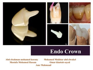
- 1. A + Endo Crown Abd elrahman mohamed korany Mohamed Mokhtar abd elwakel Mostafa Mohamed Hassan Omar khattab sayed Amr Mahmoud
- 2. OVERVIEW • Introduction • Definition • • • • Advantages & Dis-advamtages Indications & Contra-indications Preparation Discussion
- 3. Introduction Traditionally, the functional and esthetic recovery of endo-dontically treated teeth with extensive coronal loss has been achieved by fabricating total crowns supported on cast metal cores But with the development of intraradicular posts made of glass fiber, and of the technique of bonding to dentin, resulting from the development of resin materials The restoration of endodontically treated teeth became : simpler more economical and biocompatible.
- 4. Introduction with the advent. of adhesive dentistry and the new resources That may be used to perform restorations of total crowns in endodontically treated teeth, as is the case of ENDOCROWNS .
- 5. What Is endo-crown ! • Definition The endo-crown is an onlay restoration on endo-donticaly treated teeth , which is more conservative than traditional post and core system and uses the adhesive resin with mono-block technique
- 6. What Is endo-crown ! • Concept To engage the large pulpal chamber of root of treated molar teeth with ceramic etched restoration which provides cuspal coverage.
- 7. What Is endo-crown ! • History The first study published on endocrown restoration (oradhesive endodontic restoration) was conducted by Pissis in 1995 In it,he described the ceramic Monoblock effect for teeth with extensive loss of coronal structure. it was Bindl and Mörmann who named this restorative procedure “endocrown” in 1999 as adhesive endodontic crowns and characterized as total porcelain crowns fixed to endodontically treated posterior teeth .
- 8. Endo-Crown Indications • Succesfully root treated molar. Endo Crown : Indications & Contra-indications • Excessive loss of coronal dental tissue and limited inter-occlusal space
- 9. Endo-Crown Indications • Molar with short Calcified Canals Endo Crown : Indications & Contra-indications
- 10. Endo-Crown Contra-indications • Para Functional Habbits Endo Crown : Indications & Contra-indications • Can’t obtain adequate isolation From salive • Depth of pulp chamber less than 3mm • Cervical Margin less than 2 mm wide • If adhesion can’t be assured
- 11. Endo-Crown Advantages • Less Complex , More practical and easier to perform Endo Crown : Advantages & Dis-advantages • Allow minimal tooth wear thus strengthens the tooth • Preparation design is conservative and biological width is minimal • Allows re-entery to canals if required without post removal • Reduce patient cost and chairside time
- 12. Endo-Crown Dis-advantages • Risk of debonding and root fracture In endo-crown Endo Crown : Advantages & Dis-advantages • Limitation maybe restricted to the ceramic material which must be acid etchable ceramics . Can’t be used in centrals and premolars
- 13. Endo-Crown Preparation Method • Occlusal Preparation The goal in preparation is to achieve an overall reduction in the height of the occlusal surface of at least 2 mm in the axial direction. This reduction can be achieved by drilling 2-mm-deep grooves as guides , then using a green diamond wheel bur to reduce the occlusal surface Making the guide grooves in an isolated tooth and in situ Endo Crown : Preparation
- 14. Endo-Crown Preparation Method • Occlusal Preparation The goal in preparation is to achieve an overall reduction in the height of the occlusal surface of at least 2 mm in the axial direction. This reduction can be achieved by drilling 2-mm-deep grooves as guides , then using a green diamond wheel bur to reduce the occlusal surface Preparation of the cervical margin or “cervical sidewalk” using a wheel bur held parallel to the occlusal plane. Endo Crown : Preparation
- 15. Endo-Crown Preparation Method • Axial Preparation Involves eliminating undercuts in the access cavity. A cylindrical-conical green diamond bur with a total occlusal convergence of 7° is used to make the coronal pulp chamber and endodontic access cavity continuous The depth of the cavity should be at least 3 mm. Axial preparation using a cylindro-conical drill to make the coronal pulp chamber continuous with the access cavity Endo Crown : Preparation
- 16. Endo-Crown Preparation Method • Polishing The cervical band The bur used in this step has the same taper as the one used in axial preparation, but a larger diameter and a finer particle size. It should be guided around the entire surface of the cervical band to remove micro-irregularities and produce a flat, polished surface. The margin line should appear as a regular line with a sharp edge . Cervical margin before (a) and after (b) polishing. Endo Crown : Preparation
- 17. Endo-Crown Preparation Method • Preparation of the cavity floor The entrance to the pulpal canal is opened. Gutta percha is removed to a depth not exceeding 2 mm to take advantage of the saddle-like anatomy of the cavity floor. This should be done with a nonabrasive instrument to maintain the integrity of the canals entrance. No drilling of dentin is carried out. • Cleaning of pulp chamber Ultrasound is recommended to clean the pulp chamber and its floor thoroughly. Abrasion is not indicated. Endo Crown : Preparation
- 18. Endo-Crown Preparation Method Endo Crown : Preparation • Bonding Adhesives such as self-adhesive RelyX Unicem (3M, St. Paul, Minn.) or composites such as Multilink (Ivoclar, Schaan, Liechtenstein) are used for bonding the endocrown to the prepared tooth Prepared tooth (a), endocrown (b) and final result after bonding (c).
- 19. Endo-Crown Discussion Endo Crown : Discussion • Longevity and Effectiveness Several studies concluded that endocrowns were more resistant to compressive forces than conventional crowns More recently, finite element analysis highlighted the role of endocrowns in stress distribution • Choice of Materials Glass-ceramic has the advantages of biocompatibility and biomimicry, and its wear coefficient is close to that of the natural tooth. In addition, the single interface of a 1- piece restoration enhances cohesion