SlideShare uma empresa Scribd logo
1 de 115
Ultrasound examination of the ankle joint.
Dr/ ABD ALLAH NAZEER. MD.
ULTRASOUND OF THE ANKLE – Normal.
Lateral Ankle
Peroneus longus and brevis tendons.
Calcaneo-fibula ligament.
Anterior Talo-Fibula ligament
Peroneus tendons scan plane.
Peroneus longus and brevis tendons.
Transverse at the medial malleolus.
Scan plane for the Peroneus Brevis
insertion to the base of the 5th metatarsal.
Peroneus brevis insertion onto
the base of the 5th metatarsal.
Calcaneo fibula ligament scan plane. Calcaneo-fibular ligament.
ATFL scan plane. Anterior Talo-fibula ligament (ATFL).
Ultrasound of the Peroneus Longus tendon insertion. The insertion is to the lateral edge
of the 1st cuneiform, with some fibers extending to the base of the 1st metatarsal.
Anterior Ankle
Tibio-fibula ligament
Extensor Digitorum tendon(s)
Extensor Hallucis Longus
Tibialis anterior
Extensor retinaculum
Tibio fibula ligament scan plane. Normal Tibio fibula ligament.
Scan plane for the extensor
digitorum tendon of the foot.
Extensor digitorum tendon. There is a common
tendon until the level of the extensor retinaculum
at the anterior ankle crease. After this, the tendon
divides to the 4 lateral toes. (The big toe has it's
own tendon - the extensor hallucis).
Extensor hallucis scan plane Longitudinal extensor hallucis longus tendon.
Tibialis Anterior scan plane. Longitudinal Tibialis Anterior tendon.
Medial Ankle
Tibialis Posterior tendon
Flexor Digitorum tendon
Flexor Hallucis Longus tendon
Deltoid ligament
Posterior tibial nerve
Scan plane to see Tibialis Posterior, Flexor
Digitorum and Flexor Hallucis Longus (you
may need to adjust the probe posteriorly
to view the deeper FHL).
Tibialis posterior, flexor Digitorum and flexor
Hallucis longus tendons (known as "Tom, Dick
& Harry"). If including the neurovascular
bundle - Tom Dick And Very Nervous Harry.
Deltoid Ligament scan plane: Use
plenty of gel and have the probe
tip overlie the malleolar tip.
Because of it's obliquity, you cannot
readily see all aspects of the ligament.
Posterior Ankle
Achilles tendon
Retrocalcaneal (Kager's) fat-pad
Scan plane for the Achilles tendon. Normal Achilles tendon longitudinal panorama.
ULTRASOUND OF THE ANKLE PROTOCOL
ROLE OF ULTRASOUND
Ultrasound is essentially used for the external structures of the ankle. Ultrasound
is a valuable diagnostic tool in assessing the following indications; Muscular,
tendinous and ligamentous damage (chronic and acute) Bursitis Joint effusion
Vascular pathology Haematomas Soft tissue masses such as ganglia, lipomas
Classification of a mass e.g. solid, cystic, mixed Post surgical complications e.g
abscess, edema Guidance of injection, aspiration or biopsy Relationship of normal
anatomy and pathology to each other Some bony pathology.
LIMITATIONS
It is recognised that ultrasound cannot adequately assess the deltoid ligament,
the ankle mortice and some inter-tarsal ligaments.
EQUIPMENT SELECTION AND TECHNIQUE
Use of a high resolution, (8-15MHZ)small footprint probe , is essential when
assessing the superficial structures of the ankle. Careful scanning technique to
avoid anisotropy (and possible misdiagnosis) Beam steering or compounding can
help to overcome anisotropy in linear structures such as tendons. Good colour /
power / Doppler capabilities when assessing vessels or vascularity of a structure.
Be prepared to change frequency output of probe (or probes) to adequately assess
both superficial and deeper structures.
SCANNING TECHNIQUE
LATERAL ANKLE
Patient sits on the side of a raised bed with foot resting on the Sonographers knee for
support. Alternatively, the patient may sit or lie supine on bed with their foot flat.
Peroneus longus and brevis tendons: Begin in transverse, posterior to the lateral malleolus.
The two tendons are easily seen with the peroneus brevis closest to the bone. Follow them
proximally to the muscle and distally with particular attention to the insertion of the
peroneus brevis tendon onto the base of the 5th metatarsal.
Calcaneo-fibular ligament: With the probe diagonally under the malleolus, angled from the
fibula to the heel, you will see the peroneus tendons in transverse and the calcaneo-fibula
ligament underlying them.
ANTERIOR ANKLE
Patient positioned as above.
Anterior talo-fibula ligament (ATFL): Place the heel of the probe on the anterior aspect of
the distal fibula and rotate the toe of the probe towards the 2nd toe. The ligament should
be readily visible as a 2cm long, 3mm thick tight fibrillar structure.
Tibio-Fibula ligament (TFL): From the ATAF, further rotate the toe of the probe until just
above the horizontal. The TFL has a similar appearance to the ATAF.
Extensor tendons: In transverse across the anterior ankle crease, you will see, from lateral
to medial, the extensor digitorum, extensor hallucis and tibialis anterior tendons. By
independently mobilizing each of the toes and watching the tendons glide, you can identify
which is which and exclude tendon rupture The overlying extensor retinaculum should also
be observed.
MEDIAL ANKLE
Patient positioned as above.
Tibialis posterior, flexor digitorum and flexor hallucis longus tendons: Assess the
tendons along their length in longitudinal and transverse. Pay particular
attention to the insertion of the Tibialis Posterior tendon with caution not to
mistake the often present accessory ossicle, for an avulsed fragment. Begin
diagonally under the medial malleolus with the toe of the probe on the malleolus.
You will see the tibialis posterior and flexor digitorum tendons in transverse. Slide
the probe postero-inferiorly and you will see the flexor hallucis longus tendon
deeper against the calcaneum.
The deltoid ligament: Is poorly assessed with ultrasound.
Posterior tibial nerve: The neurovascular bundle is easily seen posterior to the
medial malleolus
POSTERIOR ANKLE
Patient positioned prone with the feet off the end of the bed.
Achilles tendon: Perform a survey scan ,in transverse, from the calcaneum up to
the musculo-tendinous junction (of both medial and lateral gastrocnemii). Rotate
into longitudinal and examine for thickening and integrity. At the same time
assess the retrocalcaneal (Kager's) fat pad.
The calf muscles and plantaris tendon should also be assessed as clinically
indicated.
An ankle series should include the following minimum images;
Peroneus tendons - long, trans + peroneus brevis insertion
Calcaneo-fibular ligament
Anterior talo-fibula ligament
Tibio-Fibula ligament
Extensor digitorum tendon
Extensor hallucis longus tendon
Tibialis anterior tendon
Tibialis posterior tendon
Flexor digitorum tendon
Flexor hallucis longus
Achilles tendon and retro calcaneal bursa
Document the normal anatomy. Any pathology found in 2
planes(ligament tears or thickening, synovial bulging/cysts, joint
effusion, gross bony changes), including measurements and any
vascularity.
MAIN INDICATIONS OFTHE ANKLE ULTRASONOGRAPHIC STUDY
Tendon pathology of the different compartments
of the ankle.
• Evaluation of ligament injuries.
• Bone and joint disorders (synovitis, chondral
and osteochondral lesions, occult fractures)
• Retroachilles and preachilles bursitis.
• Characterization of tumors (accessory muscles, ganglia,
neurogenic tumours, soft tissue abscesses, etc.)
• Localization of foreign bodies.
ABNORMALITIES OF TENDONS
Tendon injuries include tenosynovitis, tendinosis,
rupture and instability.
POSTERIOR COMPARTMENT
ACHILLES TENDINOPATHY
Can be classified as tendinosis and paratendinitis. The isolated
paratendinitis shows intratendinous normal structure, exist paratendinitis
spill, shown irregularities in the edges of the tendon, adhesions and
scarring associated paratendon and a heterogeneous aspect preachilles
fat pad. In the tendinosis there is in swelling of the tendon, usually
bilateral, and textural heterogeneity intratendinous focal hypoechoic areas.
TEAR OF ACHILLES
The rupture site is located generally between the 2 and 6 cm from the insertion
into the calcaneous, in the called critical zone of relative hypovascularity.
In ultrasound, complete rupture of the Achilles tendon is seen as a focal defect
between the broken ends of the tendon. In the acute phase ends are
contiguous, but the defect may be filled by the anechoic or hypoechoic
hematoma. In most cases paratendon remains intact as a envelope straight
echogenic contouring breakage. As a sign associated can exist distortion of the
fibrillar configuration and loss of parallelism of the tendon fibers. Other signs
are indicative fat herniation into the defect, better visualization of the plantar
tendon and the existence of a posterior acoustic shadowing at the site of the
tear (useful sign to differentiate partial thickness tears).
Complete rupture of the Achilles tendon with focal defect between
the ends of the tendon and posterior acoustic shadowing at the
site of the tear, useful sign to differentiate partial thickness tears.
Patient with psoriasis which identifies tendinosis and Achilles tendon swelling
bilateral and textural heterogeneity with intratendinous focal hypoechoic areas.
Ultrasound and radiological correlation calcified Achilles
enthesitis. Calcaneal spur as associated finding.
Discreetly thickened right Achilles tendon at its insertion, combining linear hyperechoic
and hypoechoic areas on small breaks intrasubstance and calcifications of the enthesis.
Fat herniation into the defect by complete tear of the Achilles tendon.
Complete tear of the Achilles tendon with retraction
of ends and integrity thin plantaris tendon.
Tendinosis versus partial tear of Achilles tendon.
•PREACHILLES AND RETROACHILLES BURSITIS
Although bursitis can occur in isolation, often are
related and systemic inflammatory diseases.
In the ultrasound examination the distended
retrocalcaneal and preachilles appears are a
hypoechoic structure shaped coma, interposed
between the Achilles tendon and the posterosuperior
aspect of the calcaneous. Care must be taken not to
confuse it with the fatty space Kager containing oval
lobules of hyperechoic fat. When bursitis is a
manifestation of synovitis is appreciated
hypervascular area with Doppler ultrasound.
In the retroachilles bursitis exists thickening and
collection of fluid in the subcutaneous tissue
superficial to the tendon retrocalcaneal portion.
Bursitis preachilles with typical morphology, appreciating accumulation of fluid
in a "coma" between the anterior portion of the Achilles tendon and the calcaneous.
Bursitis preachílea that associated synovitis, showing hyperemia with increased Doppler flow.
Rheumatoid arthritis with retrocalcaneal bursitis (*1) and calcaneal erosions (*2)
ACCESSORY NAVICULAR BONE
In the distal insertion site may exist TP 2 types of bones
accessories:
Accessory navicular bone type I (os tibiale externum): size
between the 2 and 6 mm, can be contained within the TP and
being positioned immediately proximal posteromedial to the
navicular bone. It generally produces symptoms and should
not be confused with a tendon calcification or avulsion
fracture.
Type II accessory navicular bone: is an accessory
ossification centre of the navicular bone with size between
9 and 12 mm triangular shaped and articulated through a
synchondrosis of the posterior and medial navicular bone.
It insertion site of some fibers of TP and is associated with a
syndrome of pain and increased incidence of tendon rupture
caused by abnormal overloads. Osteoarthritic changes may
underlie this synchondrosis which mimic tendon pathology.
Accessory navicular bone within the posterior tibial tendon, as anatomical variant.
ANTERIOR COMPARTMENT
The tendons of the anterior ankle, compared
with the rest of the ankle tendons are rarely
affected by disease. The anterior tibial tendon
is the most prone to abnormalities, like
tendinopathy, tenosynovitis and its place
between the most frequent rupture is extensor
retinaculum and insertion into the first
cuneiform and the base of the first metatarsal.
Sometimes the retracted tendon stump causes
a nodule on the anteromedial aspect of the
distal portion of the leg, making clinically
confused with a tumour or cyst.
Distal insertion tendinopathy of the anterior tibial tendon with increased vascularity
Tenosynovitis of the anterior tibial tendon with effusion.
Anterior tibial tendon tenosynovitis with intratendinous ruptures.
Anterior tibial tendon rupture with retraction of the tendon.
Tenosynovitis of the extensor digitorum tendons with a synovial effusion.
Tenosynovitis of the extensor digitorum tendon with
hypervascularized thickened tendon and synovium and synovial fluid.
Rheumatoid arthritis with tenosynovitis of the extensor digitorum and peroneal tendons.
Thickening of the extensor hallucis longus tendon caused by osteosynthesis material.
Multicystic non vascularized intramuscular mass in the extensor digitorum brevis muscle.
LATERAL COMPARTMENT
TENOSYNOVITIS OF THE PERONEAL TENDONS
The main sign of ultrasound is the presence of liquid inside the
common synovial sheath, whereas usually tendinous
morphology is preserved We must differentiate tenosynovitis of
a spill within the common peroneal sheath secondary to a tear of
calcaneofibular ligament (PCL). The ultrasound diagnosis is the
detection of the tendons in a lateral plane relative to the distal portion of
the lateral malleolus, instead of behind it. The dynamic exploration foot
dorsiflexion as both eversion may help identify cases of intermittent
subluxation. In long-standing disease can be observed fusiform thickening
of the peroneal tendons at the tip of the malleolus. The complete tears
of the peroneus brevis tendon (PC) and peroneus longus (PL) are
rare, occurring in the area of the lateral malleolus or midfoot. Seen
in patients with ankle sprains or history of chronic instability,
especially if they suffer widespread diseases (RA, DM,) or treated
with corticosteroids. The peroneal tendon ruptures causing the
inability to eversion of the foot and cavus foot varus.
Important thickening of both peroneal tendons (longus and brevis) associated
with moderate amount of fluid and thickening of the synovial sheath.
Discreet amount of fluid in the common sheath of the peroneal tendons
associated with disorganization and a heterogeneous appearance
peroneus brevis tendon related to longitudinal rupture.
Peroneal tenosynovitis and osteosynthesis
Tenosynovitis of the peroneal tendons and an intratendinous rupture of the short peroneal tendon.
Intratendinous rupture and cyst in the peroneus brevis tendon.
Peroneus quartus with an accessory muscle next to the peroneus longus and brevis tendons.
Tumor next to the peroneal tendons that proved to be a benign fibrous mass.
MEDIAL COMPARTMENT
ANOMALIES POSTERIOR TIBIAL TENDON (TP)
It is the most frequently injured in this compartment, with ruptures
in asymptomatic middle-aged obese women as a result of
widespread disease (RA, seronegative Spondyloarthropathy) or
associated with bone fractures. It causes a gradual collapse of the
medial longitudinal arch with hindfoot valgus deformity and
excessive forefoot pronation.
The presence of small vessels in inflammatory diseases
intratendinosis can simulate a fissuration tendon, confusion can be
ignored using colour Doppler. Another possible diagnostic difficulty is
when the undamaged tendon flexor digitorum longus (FLD) moves back and
simulates the TP unbroken, but it is smaller and in this case we only see in the
groove retromalleolar 1 only tendon.
Ultrasound has also proven to be an effective resource for identifying
alterations associated tendon tenosynovitis serosa and hypertrophic.
Subluxation and dislocation anteriorly and medial TP relative to internal
malleolus is rare and valued sonographically placing the foot in dorsiflexion
with forced supination.
Thickening, moderate amount of fluid surrounding, calcifications and
hyperemia affecting the sheath and the posterior tibial tendon related
to chronic tenosynovitis in patient affects rheumatoid arthritis.
Thickening, moderate amount of surrounding fluid, and calcifications (arrow) affecting the
posterior tibial tendon related to chronic tenosynovitis in patient affects rheumatoid arthritis.
Tenosynovitis of the posterior tibial tendon caused by a bony ridge at the insertion
of the retinaculum. The retinaculum is thickened and the tendon subluxates.
Psoriatic arthritis. Ankle. Grayscale examination.
Posterior tibial tendon on longitudinal (a) and
transverse (b) scan. Marked tendon sheath
widening with homogeneous anechoic aspect
(asterisks) of the content indicating an exudative
tenosynovitis. Note as the normal fibrillar
echotexture is conserved. Hand. Flexor tendons of
second finger. The volar longitudinal (c) and
transverse scan (d) shows a tendon sheath
widening with signs of synovial proliferation,
presence of intense power Doppler signal
surrounding the tendon, and micro interruption of
the margin (arrow). e Wrist. Extensor carpi ulnaris
tendon (sixth compartment of the extensor
tendons; ecu) on lateral longitudinal (e) and
transverse (f) scan. Chronic tenosynovitis with clear
areas of low of echogenicity and loss of the
continuity of tendon fibrils indicative of partial
tendon tear (arrows). Moreover, note the presence
of power Doppler signal within the interruption
indicating still activity of the inflammatory
process. g Dactylitis. Volar longitudinal scan using
the “extended view” technique, showing
proliferative tenosynovitis of the finger flexor
tendon (circle), exudative synovitis of both
proximal and distal interphalangeal joint (white
asterisks) and edema of the peritendinous tissue
(black asterisks). TP posterior tibial tendon, mm
medial malleolus, DP distal phalanx, mp middle
phalanx, cu cubital bone, ft flexor tendons.
Tenosynovitis of the posterior tibial (*1) and flexor digitorum longus tendon (*2)
Tibialis Posterior Tendon rupture.
Tenosynovitis of the posterior tibial tendon, the flexor digitorum tendon and flexor hallucis longus tendon
Tenosynovitis of the tendons on the medial side in the left ankle.
•LIGAMENT INJURIES
The ligament partial tear ligament shows a hypoechoic
areas swollen with internal focal or diffuse. In the complete
ruptures within the substance of the divided ligament rift is
observed corresponding to the hematoma hypoechoic, and
the free ends of the divided ligament can be and retracted
appreciated corrugated, in contrast with normal
appearance straight.
Grade I: Mild stretching of the ligament, without breakage
or instability.
Grade II: Partial tearing of the ligament
Grade III: Complete tearing.
Degrees depending on the severity of the injury and the
place of employment.
LATERAL COMPARTMENT
These injuries occur secondary to inversion sprains, with internal rotation of
the foot combined with ankle plantar flexion.
The anterior talar fibular ligament tears (ATFL) usually occur as isolated
involvement (70%) or associated with calcaneal fibular ligament (CFL) (20-40%),
but the posterior talar fibular ligament (PTFL) affects only major trauma
involving ankle dislocation.
ATFL breaks is associated with breakage of the joint capsule and synovial fluid
extravasation into the anterolateral soft ankle, whereas the complete tearing of
CFL can communicate the ankle joint and synovial sheath peroneal tendons.
Rupture of CFL is rarely associated with superior peroneal retinaculum tear.
The CFL is tensioned during dorsiflexion pulling on peroneal tendons laterally,
so that absence of its displacement is sign of a complete tear.
Within a damaged ligament can also observe calcifications that often
correspond to fragments of avulsion bone.
During the ultrasound can be performed forced maneuvers to detect to joint
laxity and ligament injuries. The anterior drawer test is performed with the feet
hanging over the edge of the examination table while the forefoot is pulled
anteriorly when the foot is in plantar flexion and inversion. This maneuver
helps differentiate partial tears (grade II) of the ATFL to complete (grade III),
where the anterior displacement of the talus on the tibia open a crack in the
substance becoming more visible the ligament injury.
The sindesmosys sprains are up to 10% of ankle
injuries, happening in eversion and pronation
movements (like the deltoid ligament injury) and
primarily affect the anterior tibiofibular ligament (ATFL),
the failure is frequently associated with fracture of the
fibula.
According to the place of employment are 4 degrees,
useful for prognostic evaluation and therapeutic strategy
choice.
Grade I: stretch or partial tear of the ATFL
Grade II: complete tearing but only the ATFL
Grade III: complete tear of the ATFL and partial CFL
Grade IV: complete tear of the ATFL and CFL.
The grade I and II injuries usually scarred without any
significant instability, whereas grade III and IV injuries
can cause chronic pain and require surgical treatment.
Thickened but continued ATFL right in relation to the partial rupture.
Grade 111 sprain of right ATFL.
Thickened anterior talofibular ligament with calcifications and a partial rupture.
Thickened tibiofibular ligament after trauma.
Anterior Talofibular Ligament Partial Tear and Elastography.
Tibio fibular ligament rupture with bony avulsion.
Complete rupture of right ATFL.
Bony avulsion with radiographic evidence as visible calcification in a grade III sprain ATFL.
Moderately thickened and hypoechoic ATFL.
Discontinuity of the ATFL with fluid surrounding the torn ends (arrow).
Thickening and hypoechogenicity of the fibular end of the ligament seen
here passing over the subtalar joint which contains a small of fluid.
Complete disruption to the tibiofibular ligament with bony
irregularity of particularly the tibial margin and fluid in the gap.
MEDIAL COMPARTMENT
Because of the low incidence of eversion ankle
sprains and the thickness of the deltoid ligament
is rarely injured in isolation and when the injury
does not usually full thickness. Usually
accompanied by lesions of the medial malleolus
and lateral displacement of the talus, with
consequent widening of the ankle mortise.
Ultrasound is useful for differentiating ligament
injury of the posterior tibial tendon injury
(TP) adjacent, they have similar symptoms.
The inability to visualize the deltoid ligament may
indicate tear , but this is not considered a reliable
sign as its full ultrasound is not always possible.
Partial medial ligament rupture.
Thickened heterogeneous abnormal deltoid demonstrating hyperemia.
Ultrasonogram showing a disrupted deltoid ligament with the probe in the coronal plane. The
white arrow indicates the medial malleolus, the blue arrow indicates the talus, and the red
arrows indicate the disrupted deltoid ligament (A). Radiograph showing the same injury (B).
•JOINT AND BONE DISORDERS
Ultrasound reliably detect spills mild (> 2 ml) inside
of the ankle joint, by exploring the front and rear
recess as well as areas of synovial proliferation, and
may even display using the colour Doppler
hyperemic areas, in arthritis patients.
The intraarticular joints are displayed surrounded
fluid in one of the recesses of the ankle or subtalar
joint. Sonographically diagnosed when changing
position to perform flexion and extension. Fracture of
the lateral process of the talus is overlooked up to 50% of
cases with plain radiography. Ultrasound may suspect the
presence of focal cortical disruption, helping further to
exclude concomitant ligament injury
Rheumatoid arthritis with synovial thickening (*1) and erosion of the tibia (*2)
Infectious arthritis with a pus filled anterior recess of the ankle.
Arthritis and tenosynovitis of the ankle in a patient with rheumatoid arthritis.
•TUMOURS
Ultrasound can differentiate between a mass complex
and solid and a cystic, in addition to verifying the
existence of accessory muscles (peroneus fourth,
accessory flexor digitorum longus and accessory
soleus). As in any other location can be found
neoformative soft tissue tumors , inflammatory,
infectious, etc.
Ganglions of this location are more often symptomatic
and larger with multiple partitions
branched and lobed edges. Differential diagnosis must
be made with tenosynovitis, abscesses, seromas and
varicosities.
Neurogenic tumours are described as pathognomonic
homogeneous hypoechoic oval mass in continuity with
a nerve of origin
Ganglion cysts are non-malignant cystic masses that occur
in association with musculoskeletal structures . They are
sometimes also simply referred to as ganglia or a
ganglion, but should not be confused with the anatomical
term ganglion.
Ultrasound
The vast majority are anechoic to hypoechoic on
ultrasound and have well defined margins. Many
demonstrate internal septations as well as acoustic
enhancement.
A synovial cyst is a small, fluid-filled sac or pouch that can
develop over a tendon or joint, creating a mass under the
skin. Synovial cyst is rare in ankle joint. A synovial cyst
may or may not be painful, depending on their size and
location.
Ganglion cyst in the foot with bony erosion between navicular and cuneiform bones
Tarsal synovial cyst.
Anechoic ganglion situated in the sinus tarsi fat between the talus and calcaneous.
Plantar fasciitis (PF) refers to inflammation of the
plantar fascia of the foot. It is considered the most
common cause of heel pain.
Clinical presentation
Pain on the undersurface of the heel on weight
bearing is the principal complaint. It can be worse
when weight is borne after a period of rest (e.g. in the
morning) and eases with walking. Passive dorsiflexion
of the toes may exacerbates discomfort.
Ultrasound
Often the initial imaging modality of choice.
Ultrasound typically shows increased thickness of the
fascia and a hypoechoic fascia.
Thickened hypoechoic origin of the plantar fascia which has a convex superior margin.
Complete avulsion of the plantar fascia from the calcaneal tubercle.
Plantar fasciitis
• LOCATING FOREIGN BODIES AND FRACTURES.
As in any other location, the ankle
area is also subsidiary host foreign
whose classification, location and
existence can be defined perfectly by
ultrasound. Being a focused study to
the area of interest and with great
resolution for surface structures, can
be considered more resolute
examination to screen these cases.
Osseous diseases and fractures | Sonography discovered Medial Malleolus Fracture.
Ultrasound
examination of left
ankle. A: Longitudinal
sonogram, left ankle,
demonstrates a
wooden foreign body;
B: Transverse view,
left ankle. Note the
hypervascularity in
the inflamed area; C:
Corresponding X-ray
of left ankle. Note the
swelling on the lateral
aspect. No foreign
body is visible.
Thank You.

Mais conteúdo relacionado

Mais procurados

Mri anatomy of ankle radiology ppt pk
Mri anatomy of ankle radiology ppt pkMri anatomy of ankle radiology ppt pk
Mri anatomy of ankle radiology ppt pkDr pradeep Kumar
 
Post-graduate Certifcate Musculoskeletal Ultrasound - The Shoulder
Post-graduate Certifcate Musculoskeletal Ultrasound - The ShoulderPost-graduate Certifcate Musculoskeletal Ultrasound - The Shoulder
Post-graduate Certifcate Musculoskeletal Ultrasound - The ShoulderDr. Peter Resteghini
 
MRI KNEE JOINT ANATOMY
MRI KNEE JOINT ANATOMYMRI KNEE JOINT ANATOMY
MRI KNEE JOINT ANATOMYNikhil Bansal
 
Transitional vertebrae radiology
Transitional vertebrae radiologyTransitional vertebrae radiology
Transitional vertebrae radiologyDr. Mohit Goel
 
Presentation1.pptx, ultrasound of the hand and fingers.
Presentation1.pptx, ultrasound of the hand and fingers.Presentation1.pptx, ultrasound of the hand and fingers.
Presentation1.pptx, ultrasound of the hand and fingers.Abdellah Nazeer
 
Presentation1.pptx. ultrasound examination of the foot
Presentation1.pptx. ultrasound examination of the footPresentation1.pptx. ultrasound examination of the foot
Presentation1.pptx. ultrasound examination of the footAbdellah Nazeer
 
Ultrasound of the abdominal wall hernias
Ultrasound of the abdominal wall herniasUltrasound of the abdominal wall hernias
Ultrasound of the abdominal wall herniasSamir Haffar
 
Presentation1.pptx, radiological imaging of sacroiliac joint diseases.
Presentation1.pptx, radiological imaging of sacroiliac joint diseases.Presentation1.pptx, radiological imaging of sacroiliac joint diseases.
Presentation1.pptx, radiological imaging of sacroiliac joint diseases.Abdellah Nazeer
 
The elbow joint in concern of diagnostic imaging .pptx 1
The elbow joint in concern of diagnostic imaging .pptx 1The elbow joint in concern of diagnostic imaging .pptx 1
The elbow joint in concern of diagnostic imaging .pptx 1DR Laith
 
Doppler ultrasound of acute scrotum
Doppler ultrasound of acute scrotumDoppler ultrasound of acute scrotum
Doppler ultrasound of acute scrotumSamir Haffar
 
Basics of msk ultrasound By Dr. Raham Bacha
Basics of msk ultrasound  By Dr. Raham BachaBasics of msk ultrasound  By Dr. Raham Bacha
Basics of msk ultrasound By Dr. Raham BachaMedical Ultrasound
 
Muscloskeletal Ultrasound of the shoulder (basic level)
Muscloskeletal Ultrasound of the shoulder (basic level)Muscloskeletal Ultrasound of the shoulder (basic level)
Muscloskeletal Ultrasound of the shoulder (basic level)Samar Tharwat
 
Presentation1, radiological imaging of anterior knee pain.
Presentation1, radiological imaging of anterior knee pain.Presentation1, radiological imaging of anterior knee pain.
Presentation1, radiological imaging of anterior knee pain.Abdellah Nazeer
 
Presentation1.pptx, radiological imaging of intusussception.
Presentation1.pptx, radiological imaging of intusussception.Presentation1.pptx, radiological imaging of intusussception.
Presentation1.pptx, radiological imaging of intusussception.Abdellah Nazeer
 
Presentation1.pptx, radiological anatomy of the knee joint.
Presentation1.pptx, radiological anatomy of the knee joint.Presentation1.pptx, radiological anatomy of the knee joint.
Presentation1.pptx, radiological anatomy of the knee joint.Abdellah Nazeer
 

Mais procurados (20)

Mri anatomy of ankle radiology ppt pk
Mri anatomy of ankle radiology ppt pkMri anatomy of ankle radiology ppt pk
Mri anatomy of ankle radiology ppt pk
 
Post-graduate Certifcate Musculoskeletal Ultrasound - The Shoulder
Post-graduate Certifcate Musculoskeletal Ultrasound - The ShoulderPost-graduate Certifcate Musculoskeletal Ultrasound - The Shoulder
Post-graduate Certifcate Musculoskeletal Ultrasound - The Shoulder
 
MRI KNEE JOINT ANATOMY
MRI KNEE JOINT ANATOMYMRI KNEE JOINT ANATOMY
MRI KNEE JOINT ANATOMY
 
Transitional vertebrae radiology
Transitional vertebrae radiologyTransitional vertebrae radiology
Transitional vertebrae radiology
 
Presentation1.pptx, ultrasound of the hand and fingers.
Presentation1.pptx, ultrasound of the hand and fingers.Presentation1.pptx, ultrasound of the hand and fingers.
Presentation1.pptx, ultrasound of the hand and fingers.
 
Presentation1.pptx. ultrasound examination of the foot
Presentation1.pptx. ultrasound examination of the footPresentation1.pptx. ultrasound examination of the foot
Presentation1.pptx. ultrasound examination of the foot
 
Radiology of Bone Tumours
Radiology of Bone TumoursRadiology of Bone Tumours
Radiology of Bone Tumours
 
Ultrasound of the abdominal wall hernias
Ultrasound of the abdominal wall herniasUltrasound of the abdominal wall hernias
Ultrasound of the abdominal wall hernias
 
Presentation1.pptx, radiological imaging of sacroiliac joint diseases.
Presentation1.pptx, radiological imaging of sacroiliac joint diseases.Presentation1.pptx, radiological imaging of sacroiliac joint diseases.
Presentation1.pptx, radiological imaging of sacroiliac joint diseases.
 
Renal doppler
Renal dopplerRenal doppler
Renal doppler
 
Mri of knee
Mri of kneeMri of knee
Mri of knee
 
The elbow joint in concern of diagnostic imaging .pptx 1
The elbow joint in concern of diagnostic imaging .pptx 1The elbow joint in concern of diagnostic imaging .pptx 1
The elbow joint in concern of diagnostic imaging .pptx 1
 
Doppler ultrasound of acute scrotum
Doppler ultrasound of acute scrotumDoppler ultrasound of acute scrotum
Doppler ultrasound of acute scrotum
 
Mri anatomy of ankle
Mri anatomy of ankleMri anatomy of ankle
Mri anatomy of ankle
 
Basics of msk ultrasound By Dr. Raham Bacha
Basics of msk ultrasound  By Dr. Raham BachaBasics of msk ultrasound  By Dr. Raham Bacha
Basics of msk ultrasound By Dr. Raham Bacha
 
Ultrasound - Bone, muscle, soft tissue
Ultrasound - Bone, muscle, soft tissueUltrasound - Bone, muscle, soft tissue
Ultrasound - Bone, muscle, soft tissue
 
Muscloskeletal Ultrasound of the shoulder (basic level)
Muscloskeletal Ultrasound of the shoulder (basic level)Muscloskeletal Ultrasound of the shoulder (basic level)
Muscloskeletal Ultrasound of the shoulder (basic level)
 
Presentation1, radiological imaging of anterior knee pain.
Presentation1, radiological imaging of anterior knee pain.Presentation1, radiological imaging of anterior knee pain.
Presentation1, radiological imaging of anterior knee pain.
 
Presentation1.pptx, radiological imaging of intusussception.
Presentation1.pptx, radiological imaging of intusussception.Presentation1.pptx, radiological imaging of intusussception.
Presentation1.pptx, radiological imaging of intusussception.
 
Presentation1.pptx, radiological anatomy of the knee joint.
Presentation1.pptx, radiological anatomy of the knee joint.Presentation1.pptx, radiological anatomy of the knee joint.
Presentation1.pptx, radiological anatomy of the knee joint.
 

Semelhante a Presentation1.pptx. ultrasound examination of the ankle joint.

Imaging of foot in non trauma and non neoplastic diseases
Imaging of foot in non trauma and non neoplastic diseasesImaging of foot in non trauma and non neoplastic diseases
Imaging of foot in non trauma and non neoplastic diseasesShail Padmani
 
Role of sonography in knee joint diseases
Role of sonography in knee joint diseasesRole of sonography in knee joint diseases
Role of sonography in knee joint diseasesREKHAKHARE
 
Median Nerve Injury and Carpal Tunnel Syndrome
Median Nerve Injury and Carpal Tunnel Syndrome Median Nerve Injury and Carpal Tunnel Syndrome
Median Nerve Injury and Carpal Tunnel Syndrome Veer Abhishek Goud
 
Radiology of the lower extremity dr asif ali khan
Radiology of the lower extremity   dr asif ali khanRadiology of the lower extremity   dr asif ali khan
Radiology of the lower extremity dr asif ali khanYangtze university
 
Lecture_10_new-_Radiography_of_Lower_Limb_I.ppt
Lecture_10_new-_Radiography_of_Lower_Limb_I.pptLecture_10_new-_Radiography_of_Lower_Limb_I.ppt
Lecture_10_new-_Radiography_of_Lower_Limb_I.pptARNKadunaState
 
sajithankle-160130132535.pdf
sajithankle-160130132535.pdfsajithankle-160130132535.pdf
sajithankle-160130132535.pdfa7med7amdy2
 
Elbow Anatomy And Examination
Elbow Anatomy And ExaminationElbow Anatomy And Examination
Elbow Anatomy And Examinationmed027972
 
X Ray and MRI of Knee Joint
X Ray and MRI of Knee JointX Ray and MRI of Knee Joint
X Ray and MRI of Knee JointGaurav Purohit
 
Talus fractures and mx
Talus fractures and mxTalus fractures and mx
Talus fractures and mxAnup Maurya
 
Presentation1 radiological imaging of tarsal tunnel syndrome.
Presentation1 radiological imaging of tarsal tunnel syndrome.Presentation1 radiological imaging of tarsal tunnel syndrome.
Presentation1 radiological imaging of tarsal tunnel syndrome.Abdellah Nazeer
 
Approach to foot x ray.pdf
Approach to foot x ray.pdfApproach to foot x ray.pdf
Approach to foot x ray.pdfmkakmal2012
 
usg shoulder by dr. kanishka.pptx
usg shoulder by dr. kanishka.pptxusg shoulder by dr. kanishka.pptx
usg shoulder by dr. kanishka.pptxRajVaghasia
 

Semelhante a Presentation1.pptx. ultrasound examination of the ankle joint. (20)

Imaging of foot in non trauma and non neoplastic diseases
Imaging of foot in non trauma and non neoplastic diseasesImaging of foot in non trauma and non neoplastic diseases
Imaging of foot in non trauma and non neoplastic diseases
 
Role of sonography in knee joint diseases
Role of sonography in knee joint diseasesRole of sonography in knee joint diseases
Role of sonography in knee joint diseases
 
Ultrasonido de tobillo anatomía
Ultrasonido de tobillo anatomíaUltrasonido de tobillo anatomía
Ultrasonido de tobillo anatomía
 
Ctev
CtevCtev
Ctev
 
Median Nerve Injury and Carpal Tunnel Syndrome
Median Nerve Injury and Carpal Tunnel Syndrome Median Nerve Injury and Carpal Tunnel Syndrome
Median Nerve Injury and Carpal Tunnel Syndrome
 
Ankle arthrodesis
Ankle arthrodesisAnkle arthrodesis
Ankle arthrodesis
 
Radiology of the lower extremity dr asif ali khan
Radiology of the lower extremity   dr asif ali khanRadiology of the lower extremity   dr asif ali khan
Radiology of the lower extremity dr asif ali khan
 
Wrist and hand examination
Wrist and hand examinationWrist and hand examination
Wrist and hand examination
 
Lecture_10_new-_Radiography_of_Lower_Limb_I.ppt
Lecture_10_new-_Radiography_of_Lower_Limb_I.pptLecture_10_new-_Radiography_of_Lower_Limb_I.ppt
Lecture_10_new-_Radiography_of_Lower_Limb_I.ppt
 
sajithankle-160130132535.pdf
sajithankle-160130132535.pdfsajithankle-160130132535.pdf
sajithankle-160130132535.pdf
 
Sajith ankle
Sajith ankleSajith ankle
Sajith ankle
 
Club foot
Club footClub foot
Club foot
 
Ctev
CtevCtev
Ctev
 
Elbow Anatomy And Examination
Elbow Anatomy And ExaminationElbow Anatomy And Examination
Elbow Anatomy And Examination
 
Surgery 6th year, Tutorial (Dr. Ali A. Nabi)
Surgery 6th year, Tutorial (Dr. Ali A. Nabi)Surgery 6th year, Tutorial (Dr. Ali A. Nabi)
Surgery 6th year, Tutorial (Dr. Ali A. Nabi)
 
X Ray and MRI of Knee Joint
X Ray and MRI of Knee JointX Ray and MRI of Knee Joint
X Ray and MRI of Knee Joint
 
Talus fractures and mx
Talus fractures and mxTalus fractures and mx
Talus fractures and mx
 
Presentation1 radiological imaging of tarsal tunnel syndrome.
Presentation1 radiological imaging of tarsal tunnel syndrome.Presentation1 radiological imaging of tarsal tunnel syndrome.
Presentation1 radiological imaging of tarsal tunnel syndrome.
 
Approach to foot x ray.pdf
Approach to foot x ray.pdfApproach to foot x ray.pdf
Approach to foot x ray.pdf
 
usg shoulder by dr. kanishka.pptx
usg shoulder by dr. kanishka.pptxusg shoulder by dr. kanishka.pptx
usg shoulder by dr. kanishka.pptx
 

Mais de Abdellah Nazeer

Muculoskeletal Pediatic Imaging..pptx
Muculoskeletal Pediatic Imaging..pptxMuculoskeletal Pediatic Imaging..pptx
Muculoskeletal Pediatic Imaging..pptxAbdellah Nazeer
 
Presentation1, Ultrasound of the bowel loops and the lymph nodes..pptx
Presentation1, Ultrasound of the bowel loops and the lymph nodes..pptxPresentation1, Ultrasound of the bowel loops and the lymph nodes..pptx
Presentation1, Ultrasound of the bowel loops and the lymph nodes..pptxAbdellah Nazeer
 
Presentation1 Short cases MD..pptx
Presentation1 Short cases MD..pptxPresentation1 Short cases MD..pptx
Presentation1 Short cases MD..pptxAbdellah Nazeer
 
Presentation1, MD MCQ Cases..pptx
Presentation1, MD MCQ Cases..pptxPresentation1, MD MCQ Cases..pptx
Presentation1, MD MCQ Cases..pptxAbdellah Nazeer
 
Presentation1, Short Cases Quiz..pptx
Presentation1, Short Cases Quiz..pptxPresentation1, Short Cases Quiz..pptx
Presentation1, Short Cases Quiz..pptxAbdellah Nazeer
 
Presentation1, radiological imaging of lateral hindfoot impingement.
Presentation1, radiological imaging of lateral hindfoot impingement.Presentation1, radiological imaging of lateral hindfoot impingement.
Presentation1, radiological imaging of lateral hindfoot impingement.Abdellah Nazeer
 
Presentation2, radiological anatomy of the liver and spleen.
Presentation2, radiological anatomy of the liver and spleen.Presentation2, radiological anatomy of the liver and spleen.
Presentation2, radiological anatomy of the liver and spleen.Abdellah Nazeer
 
Presentation1, artifacts and pitfalls of the wrist and elbow joints.
Presentation1, artifacts and pitfalls of the wrist and elbow joints.Presentation1, artifacts and pitfalls of the wrist and elbow joints.
Presentation1, artifacts and pitfalls of the wrist and elbow joints.Abdellah Nazeer
 
Presentation1, artifact and pitfalls of the knee, hip and ankle joints.
Presentation1, artifact and pitfalls of the knee, hip and ankle joints.Presentation1, artifact and pitfalls of the knee, hip and ankle joints.
Presentation1, artifact and pitfalls of the knee, hip and ankle joints.Abdellah Nazeer
 
Presentation1, radiological imaging of artifact and pitfalls in shoulder join...
Presentation1, radiological imaging of artifact and pitfalls in shoulder join...Presentation1, radiological imaging of artifact and pitfalls in shoulder join...
Presentation1, radiological imaging of artifact and pitfalls in shoulder join...Abdellah Nazeer
 
Presentation1, radiological imaging of internal abdominal hernia.
Presentation1, radiological imaging of internal abdominal hernia.Presentation1, radiological imaging of internal abdominal hernia.
Presentation1, radiological imaging of internal abdominal hernia.Abdellah Nazeer
 
Presentation11, radiological imaging of ovarian torsion.
Presentation11, radiological imaging of ovarian torsion.Presentation11, radiological imaging of ovarian torsion.
Presentation11, radiological imaging of ovarian torsion.Abdellah Nazeer
 
Presentation1, musculoskeletal anatomy.
Presentation1, musculoskeletal anatomy.Presentation1, musculoskeletal anatomy.
Presentation1, musculoskeletal anatomy.Abdellah Nazeer
 
Presentation1, new mri techniques in the diagnosis and monitoring of multiple...
Presentation1, new mri techniques in the diagnosis and monitoring of multiple...Presentation1, new mri techniques in the diagnosis and monitoring of multiple...
Presentation1, new mri techniques in the diagnosis and monitoring of multiple...Abdellah Nazeer
 
Presentation1, radiological application of diffusion weighted mri in neck mas...
Presentation1, radiological application of diffusion weighted mri in neck mas...Presentation1, radiological application of diffusion weighted mri in neck mas...
Presentation1, radiological application of diffusion weighted mri in neck mas...Abdellah Nazeer
 
Presentation1, radiological application of diffusion weighted images in breas...
Presentation1, radiological application of diffusion weighted images in breas...Presentation1, radiological application of diffusion weighted images in breas...
Presentation1, radiological application of diffusion weighted images in breas...Abdellah Nazeer
 
Presentation1, radiological application of diffusion weighted images in abdom...
Presentation1, radiological application of diffusion weighted images in abdom...Presentation1, radiological application of diffusion weighted images in abdom...
Presentation1, radiological application of diffusion weighted images in abdom...Abdellah Nazeer
 
Presentation1, radiological application of diffusion weighted imges in neuror...
Presentation1, radiological application of diffusion weighted imges in neuror...Presentation1, radiological application of diffusion weighted imges in neuror...
Presentation1, radiological application of diffusion weighted imges in neuror...Abdellah Nazeer
 
Presentation1, mr physics.
Presentation1, mr physics.Presentation1, mr physics.
Presentation1, mr physics.Abdellah Nazeer
 
Presentation1. ct physics.
Presentation1. ct physics.Presentation1. ct physics.
Presentation1. ct physics.Abdellah Nazeer
 

Mais de Abdellah Nazeer (20)

Muculoskeletal Pediatic Imaging..pptx
Muculoskeletal Pediatic Imaging..pptxMuculoskeletal Pediatic Imaging..pptx
Muculoskeletal Pediatic Imaging..pptx
 
Presentation1, Ultrasound of the bowel loops and the lymph nodes..pptx
Presentation1, Ultrasound of the bowel loops and the lymph nodes..pptxPresentation1, Ultrasound of the bowel loops and the lymph nodes..pptx
Presentation1, Ultrasound of the bowel loops and the lymph nodes..pptx
 
Presentation1 Short cases MD..pptx
Presentation1 Short cases MD..pptxPresentation1 Short cases MD..pptx
Presentation1 Short cases MD..pptx
 
Presentation1, MD MCQ Cases..pptx
Presentation1, MD MCQ Cases..pptxPresentation1, MD MCQ Cases..pptx
Presentation1, MD MCQ Cases..pptx
 
Presentation1, Short Cases Quiz..pptx
Presentation1, Short Cases Quiz..pptxPresentation1, Short Cases Quiz..pptx
Presentation1, Short Cases Quiz..pptx
 
Presentation1, radiological imaging of lateral hindfoot impingement.
Presentation1, radiological imaging of lateral hindfoot impingement.Presentation1, radiological imaging of lateral hindfoot impingement.
Presentation1, radiological imaging of lateral hindfoot impingement.
 
Presentation2, radiological anatomy of the liver and spleen.
Presentation2, radiological anatomy of the liver and spleen.Presentation2, radiological anatomy of the liver and spleen.
Presentation2, radiological anatomy of the liver and spleen.
 
Presentation1, artifacts and pitfalls of the wrist and elbow joints.
Presentation1, artifacts and pitfalls of the wrist and elbow joints.Presentation1, artifacts and pitfalls of the wrist and elbow joints.
Presentation1, artifacts and pitfalls of the wrist and elbow joints.
 
Presentation1, artifact and pitfalls of the knee, hip and ankle joints.
Presentation1, artifact and pitfalls of the knee, hip and ankle joints.Presentation1, artifact and pitfalls of the knee, hip and ankle joints.
Presentation1, artifact and pitfalls of the knee, hip and ankle joints.
 
Presentation1, radiological imaging of artifact and pitfalls in shoulder join...
Presentation1, radiological imaging of artifact and pitfalls in shoulder join...Presentation1, radiological imaging of artifact and pitfalls in shoulder join...
Presentation1, radiological imaging of artifact and pitfalls in shoulder join...
 
Presentation1, radiological imaging of internal abdominal hernia.
Presentation1, radiological imaging of internal abdominal hernia.Presentation1, radiological imaging of internal abdominal hernia.
Presentation1, radiological imaging of internal abdominal hernia.
 
Presentation11, radiological imaging of ovarian torsion.
Presentation11, radiological imaging of ovarian torsion.Presentation11, radiological imaging of ovarian torsion.
Presentation11, radiological imaging of ovarian torsion.
 
Presentation1, musculoskeletal anatomy.
Presentation1, musculoskeletal anatomy.Presentation1, musculoskeletal anatomy.
Presentation1, musculoskeletal anatomy.
 
Presentation1, new mri techniques in the diagnosis and monitoring of multiple...
Presentation1, new mri techniques in the diagnosis and monitoring of multiple...Presentation1, new mri techniques in the diagnosis and monitoring of multiple...
Presentation1, new mri techniques in the diagnosis and monitoring of multiple...
 
Presentation1, radiological application of diffusion weighted mri in neck mas...
Presentation1, radiological application of diffusion weighted mri in neck mas...Presentation1, radiological application of diffusion weighted mri in neck mas...
Presentation1, radiological application of diffusion weighted mri in neck mas...
 
Presentation1, radiological application of diffusion weighted images in breas...
Presentation1, radiological application of diffusion weighted images in breas...Presentation1, radiological application of diffusion weighted images in breas...
Presentation1, radiological application of diffusion weighted images in breas...
 
Presentation1, radiological application of diffusion weighted images in abdom...
Presentation1, radiological application of diffusion weighted images in abdom...Presentation1, radiological application of diffusion weighted images in abdom...
Presentation1, radiological application of diffusion weighted images in abdom...
 
Presentation1, radiological application of diffusion weighted imges in neuror...
Presentation1, radiological application of diffusion weighted imges in neuror...Presentation1, radiological application of diffusion weighted imges in neuror...
Presentation1, radiological application of diffusion weighted imges in neuror...
 
Presentation1, mr physics.
Presentation1, mr physics.Presentation1, mr physics.
Presentation1, mr physics.
 
Presentation1. ct physics.
Presentation1. ct physics.Presentation1. ct physics.
Presentation1. ct physics.
 

Presentation1.pptx. ultrasound examination of the ankle joint.

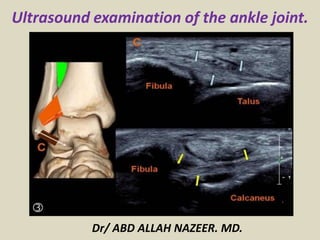
  • 1. Ultrasound examination of the ankle joint. Dr/ ABD ALLAH NAZEER. MD.
  • 2. ULTRASOUND OF THE ANKLE – Normal. Lateral Ankle Peroneus longus and brevis tendons. Calcaneo-fibula ligament. Anterior Talo-Fibula ligament Peroneus tendons scan plane. Peroneus longus and brevis tendons. Transverse at the medial malleolus.
  • 3. Scan plane for the Peroneus Brevis insertion to the base of the 5th metatarsal. Peroneus brevis insertion onto the base of the 5th metatarsal.
  • 4. Calcaneo fibula ligament scan plane. Calcaneo-fibular ligament.
  • 5. ATFL scan plane. Anterior Talo-fibula ligament (ATFL).
  • 6. Ultrasound of the Peroneus Longus tendon insertion. The insertion is to the lateral edge of the 1st cuneiform, with some fibers extending to the base of the 1st metatarsal.
  • 7. Anterior Ankle Tibio-fibula ligament Extensor Digitorum tendon(s) Extensor Hallucis Longus Tibialis anterior Extensor retinaculum Tibio fibula ligament scan plane. Normal Tibio fibula ligament.
  • 8. Scan plane for the extensor digitorum tendon of the foot. Extensor digitorum tendon. There is a common tendon until the level of the extensor retinaculum at the anterior ankle crease. After this, the tendon divides to the 4 lateral toes. (The big toe has it's own tendon - the extensor hallucis).
  • 9. Extensor hallucis scan plane Longitudinal extensor hallucis longus tendon.
  • 10. Tibialis Anterior scan plane. Longitudinal Tibialis Anterior tendon.
  • 11. Medial Ankle Tibialis Posterior tendon Flexor Digitorum tendon Flexor Hallucis Longus tendon Deltoid ligament Posterior tibial nerve Scan plane to see Tibialis Posterior, Flexor Digitorum and Flexor Hallucis Longus (you may need to adjust the probe posteriorly to view the deeper FHL). Tibialis posterior, flexor Digitorum and flexor Hallucis longus tendons (known as "Tom, Dick & Harry"). If including the neurovascular bundle - Tom Dick And Very Nervous Harry.
  • 12. Deltoid Ligament scan plane: Use plenty of gel and have the probe tip overlie the malleolar tip. Because of it's obliquity, you cannot readily see all aspects of the ligament.
  • 13. Posterior Ankle Achilles tendon Retrocalcaneal (Kager's) fat-pad Scan plane for the Achilles tendon. Normal Achilles tendon longitudinal panorama.
  • 14.
  • 15.
  • 16.
  • 17.
  • 18.
  • 19.
  • 20.
  • 21.
  • 22.
  • 23.
  • 24.
  • 25.
  • 26. ULTRASOUND OF THE ANKLE PROTOCOL ROLE OF ULTRASOUND Ultrasound is essentially used for the external structures of the ankle. Ultrasound is a valuable diagnostic tool in assessing the following indications; Muscular, tendinous and ligamentous damage (chronic and acute) Bursitis Joint effusion Vascular pathology Haematomas Soft tissue masses such as ganglia, lipomas Classification of a mass e.g. solid, cystic, mixed Post surgical complications e.g abscess, edema Guidance of injection, aspiration or biopsy Relationship of normal anatomy and pathology to each other Some bony pathology. LIMITATIONS It is recognised that ultrasound cannot adequately assess the deltoid ligament, the ankle mortice and some inter-tarsal ligaments. EQUIPMENT SELECTION AND TECHNIQUE Use of a high resolution, (8-15MHZ)small footprint probe , is essential when assessing the superficial structures of the ankle. Careful scanning technique to avoid anisotropy (and possible misdiagnosis) Beam steering or compounding can help to overcome anisotropy in linear structures such as tendons. Good colour / power / Doppler capabilities when assessing vessels or vascularity of a structure. Be prepared to change frequency output of probe (or probes) to adequately assess both superficial and deeper structures.
  • 27. SCANNING TECHNIQUE LATERAL ANKLE Patient sits on the side of a raised bed with foot resting on the Sonographers knee for support. Alternatively, the patient may sit or lie supine on bed with their foot flat. Peroneus longus and brevis tendons: Begin in transverse, posterior to the lateral malleolus. The two tendons are easily seen with the peroneus brevis closest to the bone. Follow them proximally to the muscle and distally with particular attention to the insertion of the peroneus brevis tendon onto the base of the 5th metatarsal. Calcaneo-fibular ligament: With the probe diagonally under the malleolus, angled from the fibula to the heel, you will see the peroneus tendons in transverse and the calcaneo-fibula ligament underlying them. ANTERIOR ANKLE Patient positioned as above. Anterior talo-fibula ligament (ATFL): Place the heel of the probe on the anterior aspect of the distal fibula and rotate the toe of the probe towards the 2nd toe. The ligament should be readily visible as a 2cm long, 3mm thick tight fibrillar structure. Tibio-Fibula ligament (TFL): From the ATAF, further rotate the toe of the probe until just above the horizontal. The TFL has a similar appearance to the ATAF. Extensor tendons: In transverse across the anterior ankle crease, you will see, from lateral to medial, the extensor digitorum, extensor hallucis and tibialis anterior tendons. By independently mobilizing each of the toes and watching the tendons glide, you can identify which is which and exclude tendon rupture The overlying extensor retinaculum should also be observed.
  • 28. MEDIAL ANKLE Patient positioned as above. Tibialis posterior, flexor digitorum and flexor hallucis longus tendons: Assess the tendons along their length in longitudinal and transverse. Pay particular attention to the insertion of the Tibialis Posterior tendon with caution not to mistake the often present accessory ossicle, for an avulsed fragment. Begin diagonally under the medial malleolus with the toe of the probe on the malleolus. You will see the tibialis posterior and flexor digitorum tendons in transverse. Slide the probe postero-inferiorly and you will see the flexor hallucis longus tendon deeper against the calcaneum. The deltoid ligament: Is poorly assessed with ultrasound. Posterior tibial nerve: The neurovascular bundle is easily seen posterior to the medial malleolus POSTERIOR ANKLE Patient positioned prone with the feet off the end of the bed. Achilles tendon: Perform a survey scan ,in transverse, from the calcaneum up to the musculo-tendinous junction (of both medial and lateral gastrocnemii). Rotate into longitudinal and examine for thickening and integrity. At the same time assess the retrocalcaneal (Kager's) fat pad. The calf muscles and plantaris tendon should also be assessed as clinically indicated.
  • 29. An ankle series should include the following minimum images; Peroneus tendons - long, trans + peroneus brevis insertion Calcaneo-fibular ligament Anterior talo-fibula ligament Tibio-Fibula ligament Extensor digitorum tendon Extensor hallucis longus tendon Tibialis anterior tendon Tibialis posterior tendon Flexor digitorum tendon Flexor hallucis longus Achilles tendon and retro calcaneal bursa Document the normal anatomy. Any pathology found in 2 planes(ligament tears or thickening, synovial bulging/cysts, joint effusion, gross bony changes), including measurements and any vascularity.
  • 30. MAIN INDICATIONS OFTHE ANKLE ULTRASONOGRAPHIC STUDY Tendon pathology of the different compartments of the ankle. • Evaluation of ligament injuries. • Bone and joint disorders (synovitis, chondral and osteochondral lesions, occult fractures) • Retroachilles and preachilles bursitis. • Characterization of tumors (accessory muscles, ganglia, neurogenic tumours, soft tissue abscesses, etc.) • Localization of foreign bodies. ABNORMALITIES OF TENDONS Tendon injuries include tenosynovitis, tendinosis, rupture and instability.
  • 31. POSTERIOR COMPARTMENT ACHILLES TENDINOPATHY Can be classified as tendinosis and paratendinitis. The isolated paratendinitis shows intratendinous normal structure, exist paratendinitis spill, shown irregularities in the edges of the tendon, adhesions and scarring associated paratendon and a heterogeneous aspect preachilles fat pad. In the tendinosis there is in swelling of the tendon, usually bilateral, and textural heterogeneity intratendinous focal hypoechoic areas. TEAR OF ACHILLES The rupture site is located generally between the 2 and 6 cm from the insertion into the calcaneous, in the called critical zone of relative hypovascularity. In ultrasound, complete rupture of the Achilles tendon is seen as a focal defect between the broken ends of the tendon. In the acute phase ends are contiguous, but the defect may be filled by the anechoic or hypoechoic hematoma. In most cases paratendon remains intact as a envelope straight echogenic contouring breakage. As a sign associated can exist distortion of the fibrillar configuration and loss of parallelism of the tendon fibers. Other signs are indicative fat herniation into the defect, better visualization of the plantar tendon and the existence of a posterior acoustic shadowing at the site of the tear (useful sign to differentiate partial thickness tears).
  • 32. Complete rupture of the Achilles tendon with focal defect between the ends of the tendon and posterior acoustic shadowing at the site of the tear, useful sign to differentiate partial thickness tears.
  • 33. Patient with psoriasis which identifies tendinosis and Achilles tendon swelling bilateral and textural heterogeneity with intratendinous focal hypoechoic areas.
  • 34. Ultrasound and radiological correlation calcified Achilles enthesitis. Calcaneal spur as associated finding.
  • 35. Discreetly thickened right Achilles tendon at its insertion, combining linear hyperechoic and hypoechoic areas on small breaks intrasubstance and calcifications of the enthesis.
  • 36. Fat herniation into the defect by complete tear of the Achilles tendon.
  • 37. Complete tear of the Achilles tendon with retraction of ends and integrity thin plantaris tendon.
  • 38. Tendinosis versus partial tear of Achilles tendon.
  • 39. •PREACHILLES AND RETROACHILLES BURSITIS Although bursitis can occur in isolation, often are related and systemic inflammatory diseases. In the ultrasound examination the distended retrocalcaneal and preachilles appears are a hypoechoic structure shaped coma, interposed between the Achilles tendon and the posterosuperior aspect of the calcaneous. Care must be taken not to confuse it with the fatty space Kager containing oval lobules of hyperechoic fat. When bursitis is a manifestation of synovitis is appreciated hypervascular area with Doppler ultrasound. In the retroachilles bursitis exists thickening and collection of fluid in the subcutaneous tissue superficial to the tendon retrocalcaneal portion.
  • 40. Bursitis preachilles with typical morphology, appreciating accumulation of fluid in a "coma" between the anterior portion of the Achilles tendon and the calcaneous.
  • 41. Bursitis preachílea that associated synovitis, showing hyperemia with increased Doppler flow.
  • 42. Rheumatoid arthritis with retrocalcaneal bursitis (*1) and calcaneal erosions (*2)
  • 43. ACCESSORY NAVICULAR BONE In the distal insertion site may exist TP 2 types of bones accessories: Accessory navicular bone type I (os tibiale externum): size between the 2 and 6 mm, can be contained within the TP and being positioned immediately proximal posteromedial to the navicular bone. It generally produces symptoms and should not be confused with a tendon calcification or avulsion fracture. Type II accessory navicular bone: is an accessory ossification centre of the navicular bone with size between 9 and 12 mm triangular shaped and articulated through a synchondrosis of the posterior and medial navicular bone. It insertion site of some fibers of TP and is associated with a syndrome of pain and increased incidence of tendon rupture caused by abnormal overloads. Osteoarthritic changes may underlie this synchondrosis which mimic tendon pathology.
  • 44. Accessory navicular bone within the posterior tibial tendon, as anatomical variant.
  • 45. ANTERIOR COMPARTMENT The tendons of the anterior ankle, compared with the rest of the ankle tendons are rarely affected by disease. The anterior tibial tendon is the most prone to abnormalities, like tendinopathy, tenosynovitis and its place between the most frequent rupture is extensor retinaculum and insertion into the first cuneiform and the base of the first metatarsal. Sometimes the retracted tendon stump causes a nodule on the anteromedial aspect of the distal portion of the leg, making clinically confused with a tumour or cyst.
  • 46. Distal insertion tendinopathy of the anterior tibial tendon with increased vascularity
  • 47. Tenosynovitis of the anterior tibial tendon with effusion.
  • 48. Anterior tibial tendon tenosynovitis with intratendinous ruptures.
  • 49. Anterior tibial tendon rupture with retraction of the tendon.
  • 50. Tenosynovitis of the extensor digitorum tendons with a synovial effusion.
  • 51. Tenosynovitis of the extensor digitorum tendon with hypervascularized thickened tendon and synovium and synovial fluid.
  • 52. Rheumatoid arthritis with tenosynovitis of the extensor digitorum and peroneal tendons.
  • 53. Thickening of the extensor hallucis longus tendon caused by osteosynthesis material.
  • 54. Multicystic non vascularized intramuscular mass in the extensor digitorum brevis muscle.
  • 55. LATERAL COMPARTMENT TENOSYNOVITIS OF THE PERONEAL TENDONS The main sign of ultrasound is the presence of liquid inside the common synovial sheath, whereas usually tendinous morphology is preserved We must differentiate tenosynovitis of a spill within the common peroneal sheath secondary to a tear of calcaneofibular ligament (PCL). The ultrasound diagnosis is the detection of the tendons in a lateral plane relative to the distal portion of the lateral malleolus, instead of behind it. The dynamic exploration foot dorsiflexion as both eversion may help identify cases of intermittent subluxation. In long-standing disease can be observed fusiform thickening of the peroneal tendons at the tip of the malleolus. The complete tears of the peroneus brevis tendon (PC) and peroneus longus (PL) are rare, occurring in the area of the lateral malleolus or midfoot. Seen in patients with ankle sprains or history of chronic instability, especially if they suffer widespread diseases (RA, DM,) or treated with corticosteroids. The peroneal tendon ruptures causing the inability to eversion of the foot and cavus foot varus.
  • 56. Important thickening of both peroneal tendons (longus and brevis) associated with moderate amount of fluid and thickening of the synovial sheath.
  • 57. Discreet amount of fluid in the common sheath of the peroneal tendons associated with disorganization and a heterogeneous appearance peroneus brevis tendon related to longitudinal rupture.
  • 58. Peroneal tenosynovitis and osteosynthesis
  • 59. Tenosynovitis of the peroneal tendons and an intratendinous rupture of the short peroneal tendon.
  • 60. Intratendinous rupture and cyst in the peroneus brevis tendon.
  • 61. Peroneus quartus with an accessory muscle next to the peroneus longus and brevis tendons.
  • 62. Tumor next to the peroneal tendons that proved to be a benign fibrous mass.
  • 63. MEDIAL COMPARTMENT ANOMALIES POSTERIOR TIBIAL TENDON (TP) It is the most frequently injured in this compartment, with ruptures in asymptomatic middle-aged obese women as a result of widespread disease (RA, seronegative Spondyloarthropathy) or associated with bone fractures. It causes a gradual collapse of the medial longitudinal arch with hindfoot valgus deformity and excessive forefoot pronation. The presence of small vessels in inflammatory diseases intratendinosis can simulate a fissuration tendon, confusion can be ignored using colour Doppler. Another possible diagnostic difficulty is when the undamaged tendon flexor digitorum longus (FLD) moves back and simulates the TP unbroken, but it is smaller and in this case we only see in the groove retromalleolar 1 only tendon. Ultrasound has also proven to be an effective resource for identifying alterations associated tendon tenosynovitis serosa and hypertrophic. Subluxation and dislocation anteriorly and medial TP relative to internal malleolus is rare and valued sonographically placing the foot in dorsiflexion with forced supination.
  • 64. Thickening, moderate amount of fluid surrounding, calcifications and hyperemia affecting the sheath and the posterior tibial tendon related to chronic tenosynovitis in patient affects rheumatoid arthritis.
  • 65. Thickening, moderate amount of surrounding fluid, and calcifications (arrow) affecting the posterior tibial tendon related to chronic tenosynovitis in patient affects rheumatoid arthritis.
  • 66. Tenosynovitis of the posterior tibial tendon caused by a bony ridge at the insertion of the retinaculum. The retinaculum is thickened and the tendon subluxates.
  • 67. Psoriatic arthritis. Ankle. Grayscale examination. Posterior tibial tendon on longitudinal (a) and transverse (b) scan. Marked tendon sheath widening with homogeneous anechoic aspect (asterisks) of the content indicating an exudative tenosynovitis. Note as the normal fibrillar echotexture is conserved. Hand. Flexor tendons of second finger. The volar longitudinal (c) and transverse scan (d) shows a tendon sheath widening with signs of synovial proliferation, presence of intense power Doppler signal surrounding the tendon, and micro interruption of the margin (arrow). e Wrist. Extensor carpi ulnaris tendon (sixth compartment of the extensor tendons; ecu) on lateral longitudinal (e) and transverse (f) scan. Chronic tenosynovitis with clear areas of low of echogenicity and loss of the continuity of tendon fibrils indicative of partial tendon tear (arrows). Moreover, note the presence of power Doppler signal within the interruption indicating still activity of the inflammatory process. g Dactylitis. Volar longitudinal scan using the “extended view” technique, showing proliferative tenosynovitis of the finger flexor tendon (circle), exudative synovitis of both proximal and distal interphalangeal joint (white asterisks) and edema of the peritendinous tissue (black asterisks). TP posterior tibial tendon, mm medial malleolus, DP distal phalanx, mp middle phalanx, cu cubital bone, ft flexor tendons.
  • 68. Tenosynovitis of the posterior tibial (*1) and flexor digitorum longus tendon (*2)
  • 70. Tenosynovitis of the posterior tibial tendon, the flexor digitorum tendon and flexor hallucis longus tendon
  • 71. Tenosynovitis of the tendons on the medial side in the left ankle.
  • 72. •LIGAMENT INJURIES The ligament partial tear ligament shows a hypoechoic areas swollen with internal focal or diffuse. In the complete ruptures within the substance of the divided ligament rift is observed corresponding to the hematoma hypoechoic, and the free ends of the divided ligament can be and retracted appreciated corrugated, in contrast with normal appearance straight. Grade I: Mild stretching of the ligament, without breakage or instability. Grade II: Partial tearing of the ligament Grade III: Complete tearing. Degrees depending on the severity of the injury and the place of employment.
  • 73. LATERAL COMPARTMENT These injuries occur secondary to inversion sprains, with internal rotation of the foot combined with ankle plantar flexion. The anterior talar fibular ligament tears (ATFL) usually occur as isolated involvement (70%) or associated with calcaneal fibular ligament (CFL) (20-40%), but the posterior talar fibular ligament (PTFL) affects only major trauma involving ankle dislocation. ATFL breaks is associated with breakage of the joint capsule and synovial fluid extravasation into the anterolateral soft ankle, whereas the complete tearing of CFL can communicate the ankle joint and synovial sheath peroneal tendons. Rupture of CFL is rarely associated with superior peroneal retinaculum tear. The CFL is tensioned during dorsiflexion pulling on peroneal tendons laterally, so that absence of its displacement is sign of a complete tear. Within a damaged ligament can also observe calcifications that often correspond to fragments of avulsion bone. During the ultrasound can be performed forced maneuvers to detect to joint laxity and ligament injuries. The anterior drawer test is performed with the feet hanging over the edge of the examination table while the forefoot is pulled anteriorly when the foot is in plantar flexion and inversion. This maneuver helps differentiate partial tears (grade II) of the ATFL to complete (grade III), where the anterior displacement of the talus on the tibia open a crack in the substance becoming more visible the ligament injury.
  • 74. The sindesmosys sprains are up to 10% of ankle injuries, happening in eversion and pronation movements (like the deltoid ligament injury) and primarily affect the anterior tibiofibular ligament (ATFL), the failure is frequently associated with fracture of the fibula. According to the place of employment are 4 degrees, useful for prognostic evaluation and therapeutic strategy choice. Grade I: stretch or partial tear of the ATFL Grade II: complete tearing but only the ATFL Grade III: complete tear of the ATFL and partial CFL Grade IV: complete tear of the ATFL and CFL. The grade I and II injuries usually scarred without any significant instability, whereas grade III and IV injuries can cause chronic pain and require surgical treatment.
  • 75. Thickened but continued ATFL right in relation to the partial rupture.
  • 76. Grade 111 sprain of right ATFL.
  • 77. Thickened anterior talofibular ligament with calcifications and a partial rupture.
  • 79. Anterior Talofibular Ligament Partial Tear and Elastography.
  • 80. Tibio fibular ligament rupture with bony avulsion.
  • 81. Complete rupture of right ATFL.
  • 82.
  • 83. Bony avulsion with radiographic evidence as visible calcification in a grade III sprain ATFL.
  • 84.
  • 85. Moderately thickened and hypoechoic ATFL.
  • 86. Discontinuity of the ATFL with fluid surrounding the torn ends (arrow).
  • 87. Thickening and hypoechogenicity of the fibular end of the ligament seen here passing over the subtalar joint which contains a small of fluid.
  • 88. Complete disruption to the tibiofibular ligament with bony irregularity of particularly the tibial margin and fluid in the gap.
  • 89. MEDIAL COMPARTMENT Because of the low incidence of eversion ankle sprains and the thickness of the deltoid ligament is rarely injured in isolation and when the injury does not usually full thickness. Usually accompanied by lesions of the medial malleolus and lateral displacement of the talus, with consequent widening of the ankle mortise. Ultrasound is useful for differentiating ligament injury of the posterior tibial tendon injury (TP) adjacent, they have similar symptoms. The inability to visualize the deltoid ligament may indicate tear , but this is not considered a reliable sign as its full ultrasound is not always possible.
  • 91. Thickened heterogeneous abnormal deltoid demonstrating hyperemia.
  • 92. Ultrasonogram showing a disrupted deltoid ligament with the probe in the coronal plane. The white arrow indicates the medial malleolus, the blue arrow indicates the talus, and the red arrows indicate the disrupted deltoid ligament (A). Radiograph showing the same injury (B).
  • 93. •JOINT AND BONE DISORDERS Ultrasound reliably detect spills mild (> 2 ml) inside of the ankle joint, by exploring the front and rear recess as well as areas of synovial proliferation, and may even display using the colour Doppler hyperemic areas, in arthritis patients. The intraarticular joints are displayed surrounded fluid in one of the recesses of the ankle or subtalar joint. Sonographically diagnosed when changing position to perform flexion and extension. Fracture of the lateral process of the talus is overlooked up to 50% of cases with plain radiography. Ultrasound may suspect the presence of focal cortical disruption, helping further to exclude concomitant ligament injury
  • 94.
  • 95.
  • 96. Rheumatoid arthritis with synovial thickening (*1) and erosion of the tibia (*2)
  • 97. Infectious arthritis with a pus filled anterior recess of the ankle.
  • 98. Arthritis and tenosynovitis of the ankle in a patient with rheumatoid arthritis.
  • 99. •TUMOURS Ultrasound can differentiate between a mass complex and solid and a cystic, in addition to verifying the existence of accessory muscles (peroneus fourth, accessory flexor digitorum longus and accessory soleus). As in any other location can be found neoformative soft tissue tumors , inflammatory, infectious, etc. Ganglions of this location are more often symptomatic and larger with multiple partitions branched and lobed edges. Differential diagnosis must be made with tenosynovitis, abscesses, seromas and varicosities. Neurogenic tumours are described as pathognomonic homogeneous hypoechoic oval mass in continuity with a nerve of origin
  • 100.
  • 101.
  • 102.
  • 103. Ganglion cysts are non-malignant cystic masses that occur in association with musculoskeletal structures . They are sometimes also simply referred to as ganglia or a ganglion, but should not be confused with the anatomical term ganglion. Ultrasound The vast majority are anechoic to hypoechoic on ultrasound and have well defined margins. Many demonstrate internal septations as well as acoustic enhancement. A synovial cyst is a small, fluid-filled sac or pouch that can develop over a tendon or joint, creating a mass under the skin. Synovial cyst is rare in ankle joint. A synovial cyst may or may not be painful, depending on their size and location.
  • 104. Ganglion cyst in the foot with bony erosion between navicular and cuneiform bones
  • 106. Anechoic ganglion situated in the sinus tarsi fat between the talus and calcaneous.
  • 107. Plantar fasciitis (PF) refers to inflammation of the plantar fascia of the foot. It is considered the most common cause of heel pain. Clinical presentation Pain on the undersurface of the heel on weight bearing is the principal complaint. It can be worse when weight is borne after a period of rest (e.g. in the morning) and eases with walking. Passive dorsiflexion of the toes may exacerbates discomfort. Ultrasound Often the initial imaging modality of choice. Ultrasound typically shows increased thickness of the fascia and a hypoechoic fascia.
  • 108. Thickened hypoechoic origin of the plantar fascia which has a convex superior margin.
  • 109. Complete avulsion of the plantar fascia from the calcaneal tubercle.
  • 111. • LOCATING FOREIGN BODIES AND FRACTURES. As in any other location, the ankle area is also subsidiary host foreign whose classification, location and existence can be defined perfectly by ultrasound. Being a focused study to the area of interest and with great resolution for surface structures, can be considered more resolute examination to screen these cases.
  • 112.
  • 113. Osseous diseases and fractures | Sonography discovered Medial Malleolus Fracture.
  • 114. Ultrasound examination of left ankle. A: Longitudinal sonogram, left ankle, demonstrates a wooden foreign body; B: Transverse view, left ankle. Note the hypervascularity in the inflamed area; C: Corresponding X-ray of left ankle. Note the swelling on the lateral aspect. No foreign body is visible.