SlideShare uma empresa Scribd logo
1 de 70
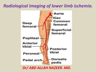
Radiological imaging of lower limb ischemia.
Dr/ ABD ALLAH NAZEER. MD.
Acute limb ischemia is defined as “Any sudden decrease in limb perfusion
causing a potential threat to limb viability”. By convention this usually
refers to patients presenting with symptoms for less than 2 weeks. The
spectrum of acute limb ischemia therefore ranges from the patient with a
few hours history of a painful cold white leg, to the patient with a few days
history of short distance claudication or the patient with a sudden increase
in ischemic symptoms on a background of peripheral arterial disease.
Acute limb ischemia.
Pathophysiology
An embolus is defined as a material (gas, solid or liquid) that is carried
within the circulation and lodges in a blood vessel in another part of the
circulation, causing occlusion of the blood vessel. Radiologically the upper
border of an embolus is classically concave, known as the “meniscus” sign.
Acute : varies but up to 2 weeks
Chronic : > 2 weeks
Acute on Chronic : Sudden and rapid deterioration in symptoms
Common etiologies
1. Embolic – Causes 15% of ALI cases
• 80% from cardiac source - valve, mural thrombus
• 20% non-cardiac source - thrombosed aneurysm, ulcerated atherosclerotic plaque
• Decreasing incidence with fewer cases of rheumatic heart disease
• Most commonly lodged at: Femoral artery bifurcation, aortoiliac arterial system,
brachial artery
• Unique condition = Blue Toe Syndrome
-Sudden appearance of cool, painful foot with cyanosis/petechaie of toes and
plantar foot
- Paradoxically: normal pedal pulses
- Etiology: emboli to the digital arteries of the foot
- Management: Requires imaging for more proximal atherosclerotic lesion
- Should consider popliteal artery aneurysm
- Treatment: Antiocoagulation alone +/- surgical bypass of atherosclerotic lesion
2. Thrombotic – Causes 85% of ALI cases
• Of native or aneurysmal artery
• Most commonly located at: Bypass grant, common femoral artery, popliteal artery
• Of indwelling bypass graft
Less common etiologies
1. Extrinsic compression of arterial lumen
• Aortic or vascular dissection, creating pseudo lumen which compromises true lumen
• Compartment syndrome
• Thoracic outlet syndrome
2. Vasospasm
• Raynaud’s disease
- Vasospastic condition causing well-demarcated ischemia to fingers and toes
- Three stages: Pallor (hypoperfusion), then cyanotic (hypoxemia), then hyperemia
(reperfusion)
- Usually bilateral limb involvement
3. Vasculitis
• Buerger’s disease (Thromboangiitis obliterans)
- Segmental, inflammatory vaso-occlusive disease of small and medium arteries
-Heavy tobacco use is a significant risk factor
-• Temporal arteritis (Giant cell arteritis)
- Inflammatory condition of medium and large arteries
• Takayasu’s arteritis
- Chronic, inflammatory, large-vessel disease of aorta and its large branches
-Predominantly found in young women (age 20-30)
-4. Low intravascular volume +/- mild peripheral vascular disease (PVD)
• Congestive heart failure, dehydration, sepsis
5. Trauma
Imaging options
1. Digital subtraction angiography (DSA)
• Gold standard for limb ischemia imaging
• Advantages: During angiography, may be able to immediately start therapeutic
intervention (intra-arterial lysis) without losing time
• Disadvantages: 8% incidence of contrast nephrotoxicity .
- Relatively invasive
2. Duplex ultrasonography:
• Ultrasound with color flow and Doppler waveform information regarding vascular
flow and patency.
• Advantages: - Noninvasive and non-nephrotoxic
-Fairly accurate for infrainguinal arterial occlusive disease, especially of bypass grafts
in this location
• Disadvantages: - Time intensive (30-40 min)
- Suprainguinal arterial occlusions and distal run-off vessels not well-visualized
-Calcifications of arterial walls (in diabetic patients, especially) can create artifacts and
obscure visualization.
• Comparisons between duplex ultrasound versus angiography pre-operatively for
chronic limb ischemia showed a high correlation rate between the two.
• May be considered as a second-line approach to ALI imaging if angiography is not
possible for infrainguinal arterial occlusion.
3. CT angiography (CTA)
• Advantages: - Noninvasive
-Can concurrently evaluate for aortic dissection while looking at runoff vessels
in the extremities.
-Promising literature regarding usefulness in staging severity of chronic
peripheral arterial disease (PAD):
Sensitivity of CTA = 96% and specificity = 97%
• Disadvantages: - Time intensive (30-40 min)
-Exposure to IV contrast –> If going to angiography after CTA for intra-arterial
lysis of clot, patient will now receive a 2nd contrast bolus, increasing the risk
of renal failure.
• Not well-studied in acute limb ischemia
4. MR angiography (MRA)
• Advantages: - Less contrast load than angiography
-Gadolinium IV contrast is safe for the kidneys
-No ionizing irradiation
• Disadvantages: - Very time intensive and often unavailable during weekend and
night hours.
-Promising literature regarding usefulness in staging severity of chronic PAD in
the femoral/popliteal system:
Thrombosis or embolism at femoral artery.
FA lumen is filled with hypoechoic thrombus or embolus . Normal flow in adjacent FV.
Occlusion of the CIA with reserved flow
in IIA (blue) to supply flow to EIA(red).
Short occlusion of the mid-SFA(large arrow) with
large collateral at both end of occlusion(small arrow).
Tardus et parvus = small amplitude + slow rising pulse
Color Doppler imaging: thrombosis
of the left common iliac artery.
Arteriography: severe stenosis
of the left common iliac artery.
Complete
occlusion of
the aorta and
proximal iliac
arteries
Angio-imaging of the popliteal trifurcation before
and after selective balloon embolectomy.
Thrombus of the left atrium with complete popliteal artery occlusion.
Angiogram showing bilateral occlusions of superficial femoral
arteries in thighs. Collaterals arising from the profunda
femoris artery can functionally bypass this occlusion.
Obstruction of the CFA with engorgement of the collateral vessels.
Acute thrombosis of the
femoropopliteal and trifurcation
vessels in an 84-year-old woman
with an acutely cool right leg. The
patient refused angiography. (a)
Transverse CT angiographic image
at the level of the adductor canal
shows rounded filling defect in the
right popliteal artery (arrow). The
contralateral left popliteal artery
(P) is patent at this level. (b) Large
field of view multipath CPR and (c)
enlarged image of popliteal region
show the extent of right-sided
thrombus (arrowheads). In
addition to the popliteal artery
(POP), the anterior tibial artery
(AT), tibioperoneal trunk (TPT),
and posterior tibial artery (PT) are
occluded. F, femoral condyle.
High-grade left popliteal
stenosis (asterisk) is also noted.
No flow at
the PT artery
Images of adventitial cystic
disease in a 55-year-old
patient. (a) Axial source
image from peripheral CT
angiography shows marked
compression of the popliteal
artery (P) by an ovoid fluid-
density lesion (arrowheads).
Note the predominant
transverse compression of the
flow lumen. F, fabella. (b)
Corresponding sagittal thin-
slab MIP image shows
craniocaudal extent of fluid-
density lesion in the popliteal
arterial wall (arrowheads). (c)
VR image viewed obliquely
from the posterior direction
demonstrates severe
narrowing of the right
popliteal artery (arrows).
CT Angiography. Digital Subtraction Angiography
CT angiography showing the presence of an intraluminal aortic
(short arrow) and iliac (long arrow) saddle embolus.
CT angiography of the lower limbs showing distal posterior and anterior tibial
artery occlusion of the left side and three-vessel occlusion of the right side.
Digital subtraction angiogram (DSA) illustrates a high-grade short-
segment stenosis of the lumen of the right superficial femoral artery (a).
(MRA) of the lower extremities was obtained by using the bolus-chase
technique. A short-segment high-grade stenosis is present in the middle
of the left superficial femoral artery. Note the collateral arterial supply.
Occlusion of the
left common
femoral artery.
Acute limb ischemia.
Acute right lower extremity pain and absent
popliteal and pedal pulses. There is an occlusion
of the popliteal artery caused by an embolus.
Acute limb ischemia with occlusion
of the SFA by embolus.
Occlusion of the popliteal artery
caused by an embolus.
Anterior tibial artery occluded
proximally with limited contrast filing of
ATA distally. The TPT and PTA are patent
Peripheral vascular disease, (chronic limb ischemia).
ABI Interpretation
1.00–1.29 Normal
0.91–0.99 Borderline
0.41–0.90 Mild-to-moderate
disease
≤0.40 Severe disease
≥1.30 Non compressible
Interpreting the Ankle-Brachial Index.
Segmental Pressures (mm Hg).
Pulse Volume Recordings
ABI in diabetics patient with calcification of
vessel wall and beaded appearance of color flow.
Severe stenosis of the SFA with area of color
flow disturbance and aliasing(Arrows).
Calcified atheroma of the SFA with drop-out
of color flow signal in parts of the lumen.
This elderly male diabetic patient had severe pain in the right leg. Colour Doppler ultrasound and
spectral Doppler tracing reveal severe diffuse stenotic disease of the arterial system starting from the
right popliteal artery to the dorsalis pedis artery. This kind of a picture is seen in long-standing
diabetes mellitus resulting in severe arterial disease or arteriopathy. The waveform of the arteries
affected including the popliteal artery, the anterior and posterior tibial arteries show loss of tri-phasic
spectral waveform which is replaced with spectral broadening and an almost venous like flow pattern.
This is known as severe dampening of the waveform and this also known as tardus parvus flow.
Peripheral CT angiogram of
a 62-year-old man with
abdominal and bilateral
common iliac artery
aneurysms and subacute
onset of right foot pain. (a)
Oblique (45° left anterior
oblique) MIP image of
entire data set. Box
indicates magnified views.
(b–d) axial CT images
through the right proximal
calf show embolic filling
defects in the anterior
tibial artery (arrowheads)
and the tibioperoneal trunk
(arrows). (e,f)
Corresponding CPR images
from the popliteal artery
through the anterior tibial
artery (e) and posterior
tibial artery (f) display
intraluminal filling defects.
DSA confirms CT
angiography findings (g).
Peripheral CT angiogram in an
83-year-old man with right
side–dominant calf and foot
claudication. Acceptable image
noise level in the abdomen and
unprecedented spatial
resolution down to the plantar
arch and metatarsal branches.
MIP of the entire data set (a);
VR views of the abdomen (b)
and right leg runoff (c–f) show
distal aortic calcific plaque and
patent renal arteries (b)
occlusion of the right popliteal
trifurcation with reconstitution
of the peroneal artery (c),
which reconstitutes the right
posterior tibial artery above
the ankle via a communicating
branch (d), and supplies the
foot through the common and
lateral plantar arteries (e)
which fill the plantar arch (f).
Peripheral CT angiography in a 72-
year-old woman with non healing left
forefoot ulcer. (a) VR image of the left
superficial femoral artery shows
excessive vessel wall calcifications,
precluding the assessment of the flow
channel. Cross-sectional views were
required to visualize the vessel lumen.
Axial CT images (b,c) through the
mid-superficial femoral artery (dotted
line in a and d) with viewing window
settings (level/width) of 200 HU/600
HU (b) does not allow us to
distinguish between opacified vessel
lumen and vessel calcification, which
can be distinguished only when an
adequately wide window width (300
HU/1,200 HU) is used (c). Similar wide
window settings are also used for a
CPR through the same vessel (d),
displaying several areas of wall
calcification with and without
stenosis.
Peripheral CT angiography (16
0.75 mm, 2.0 mm/1.0 mm) in a 73-
year-old woman with intermittent
claudication bilaterally. MIP (a)
shows long right femoropopliteal
occlusion (curved arrow) and
diffuse disease of the left
superficial femoral artery
with a short distal near-occlusion.
CPR (b) through left iliofemoral
arteries demonstrates multiple
mild stenosis of the external iliac
artery (arrowheads), a diffusely
diseased left superficial femoral
artery, and short (3 cm) distal left
superficial femoral artery
occlusion.
Corresponding selective DSA
images of the left external iliac
artery (c) and the distal left
superficial femoral artery (d) were
obtained immediately before
angioplasty/stent implantation.
Peripheral CT angiography (16 0.75
mm, 2.0 mm/1.0 mm) of a diabetic
male patient with bilateral
claudication. MIP (a) shows arterial
calcifications near the aortic bifurcation
(arrow), as well as in the right
(arrowheads) and left common femoral
arteries, in the right femoropopliteal
region, and in the crural vessels. A long
stent is seen in the left femoropopliteal
segment (curved arrow).Frontal view
(b) and magnified 45° left anterior
oblique (c) multipath CPR images
provide simultaneous CPRs through the
aorta and bilateral iliac through crural
arteries. Note that prominent
calcifications cause luminal narrowing
in the proximal left common iliac artery
(arrow) and in the right common
femoral artery (arrowheads). The left
common femoral artery is normal; the
long femoropopliteal stent is patent
(curved arrow). Mixed calcified and non
calcified occlusion of the right distal
femoral artery is also seen (open
arrow).
Intermittent left leg claudication in a 62-year-old
woman with a history of tobacco use and
aortobifemoral bypass grafting. The ankle-brachial
index was 0.65. (a– d) MIP images with bone
segmentation; (e– h) DSA images obtained before
treatment. (a) Oblique MIP image shows high-
grade stenosis (arrows) at the origin of the left
profunda femoris artery (P); a previously placed
aortobifemoral graft (G) is noted, as is a patent
superficial femoral artery (SFA). (b) Coronal MIP of
the left thigh demonstrates multifocal moderate to
severe stenosis in the SFA (arrowheads). The SFA is
small in caliber with soft and calcified plaque
present. (c) Coronal MIP of the calf shows a one-
vessel runoff (peroneal; PER) to the left foot. Mild
venous contamination (V) is present. (d) Sagittal
MIP image of the left foot shows collateral vessel
reconstitution (arrowheads) of the dorsalis pedis
(DP) above the ankle from the peroneal artery. (e)
DSA image from selective catheterization of the left
profunda femoris artery corroborates the CT
angiographic finding of high-grade profunda
femoris artery (P) stenosis (arrows). (f) DSA image
of left superficial femoral artery shows multiple
focal stenoses (arrowheads) in the same segment
of the superficial femoral artery as demonstrated
by CT angiography. Note the lack of calcium
visualization on the subtracted image from DSA. (g)
DSA image of the calf confirms single peroneal
vessel runoff. (h) DSA image of left foot confirms
reconstitution of the dorsalis pedis artery (DP) from
the peroneal artery (arrowheads).
Magnetic
Resonance
Angiography
(MRA) show
occlusion of
the left CIA
with filling
of the SFA
from
collateral.
(MRA) of the lower extremities was obtained by using the bolus-chase technique.
Atherosclerotic disease involves the bilateral superficial femoral arteries. Note the multiple
lesions, which are primarily in the middle portions, and the large collateral arterial supply.
67-year-old man
with more than
10-year history of
diabetes mellitus
presented with
symptoms of limb
ischemia.
Thank You.

Mais conteúdo relacionado

Mais procurados

Imaging of Aortic Dissection
Imaging of Aortic DissectionImaging of Aortic Dissection
Imaging of Aortic DissectionSakher Alkhaderi
 
Doppler ultrasound of A-V access for hemodialysis
Doppler ultrasound of A-V access for hemodialysisDoppler ultrasound of A-V access for hemodialysis
Doppler ultrasound of A-V access for hemodialysisSamir Haffar
 
Imaging in stroke
Imaging in stroke Imaging in stroke
Imaging in stroke Deepak Garg
 
Magnetic Resonance Angiography and Venography
Magnetic Resonance Angiography and VenographyMagnetic Resonance Angiography and Venography
Magnetic Resonance Angiography and VenographyAnjan Dangal
 
Presentation1.pptx, radiological vascular anatomy of the upper and lower limbs.
Presentation1.pptx, radiological vascular anatomy of the upper and lower limbs.Presentation1.pptx, radiological vascular anatomy of the upper and lower limbs.
Presentation1.pptx, radiological vascular anatomy of the upper and lower limbs.Abdellah Nazeer
 
Peripheral arterial doppler
Peripheral  arterial dopplerPeripheral  arterial doppler
Peripheral arterial dopplerAnish Choudhary
 
Radiology Spots PPT- 2 by Dr Chandni Wadhwani
Radiology Spots PPT- 2 by Dr Chandni WadhwaniRadiology Spots PPT- 2 by Dr Chandni Wadhwani
Radiology Spots PPT- 2 by Dr Chandni WadhwaniChandni Wadhwani
 
HEPATOCELLULAR CARCINOMA RADIOLOGY
HEPATOCELLULAR CARCINOMA RADIOLOGYHEPATOCELLULAR CARCINOMA RADIOLOGY
HEPATOCELLULAR CARCINOMA RADIOLOGYRMLIMS
 
Presentation1, radiological imaging of thoracic aortic aneurysm.
Presentation1, radiological imaging of thoracic aortic aneurysm.Presentation1, radiological imaging of thoracic aortic aneurysm.
Presentation1, radiological imaging of thoracic aortic aneurysm.Abdellah Nazeer
 
Presentation1.pptx, radiological imaging of skeletal dysplasia
Presentation1.pptx, radiological imaging of skeletal dysplasiaPresentation1.pptx, radiological imaging of skeletal dysplasia
Presentation1.pptx, radiological imaging of skeletal dysplasiaAbdellah Nazeer
 
Radiology Spotters mixed collection ppt
Radiology Spotters mixed collection pptRadiology Spotters mixed collection ppt
Radiology Spotters mixed collection pptDr pradeep Kumar
 
Venous Doppler Lower limb Dr Mukesh Tilgam
Venous Doppler Lower limb Dr Mukesh TilgamVenous Doppler Lower limb Dr Mukesh Tilgam
Venous Doppler Lower limb Dr Mukesh TilgamDrmukesh Tilgam
 
Doppler ultrasound of the kidneys
Doppler ultrasound of the kidneysDoppler ultrasound of the kidneys
Doppler ultrasound of the kidneysSamir Haffar
 
portal doppler ppt .pptx
portal doppler ppt .pptxportal doppler ppt .pptx
portal doppler ppt .pptxdypradio
 

Mais procurados (20)

Imaging of Aortic Dissection
Imaging of Aortic DissectionImaging of Aortic Dissection
Imaging of Aortic Dissection
 
Radiology spotters
Radiology spottersRadiology spotters
Radiology spotters
 
Doppler ultrasound of A-V access for hemodialysis
Doppler ultrasound of A-V access for hemodialysisDoppler ultrasound of A-V access for hemodialysis
Doppler ultrasound of A-V access for hemodialysis
 
Imaging in stroke
Imaging in stroke Imaging in stroke
Imaging in stroke
 
Magnetic Resonance Angiography and Venography
Magnetic Resonance Angiography and VenographyMagnetic Resonance Angiography and Venography
Magnetic Resonance Angiography and Venography
 
Spots with keys (2)
Spots with keys (2)Spots with keys (2)
Spots with keys (2)
 
Presentation1.pptx, radiological vascular anatomy of the upper and lower limbs.
Presentation1.pptx, radiological vascular anatomy of the upper and lower limbs.Presentation1.pptx, radiological vascular anatomy of the upper and lower limbs.
Presentation1.pptx, radiological vascular anatomy of the upper and lower limbs.
 
Hip joint
Hip jointHip joint
Hip joint
 
Aortic aneurysm imaging
Aortic aneurysm imagingAortic aneurysm imaging
Aortic aneurysm imaging
 
Mediastinal mass
Mediastinal massMediastinal mass
Mediastinal mass
 
Peripheral arterial doppler
Peripheral  arterial dopplerPeripheral  arterial doppler
Peripheral arterial doppler
 
Radiology Spots PPT- 2 by Dr Chandni Wadhwani
Radiology Spots PPT- 2 by Dr Chandni WadhwaniRadiology Spots PPT- 2 by Dr Chandni Wadhwani
Radiology Spots PPT- 2 by Dr Chandni Wadhwani
 
HEPATOCELLULAR CARCINOMA RADIOLOGY
HEPATOCELLULAR CARCINOMA RADIOLOGYHEPATOCELLULAR CARCINOMA RADIOLOGY
HEPATOCELLULAR CARCINOMA RADIOLOGY
 
Presentation1, radiological imaging of thoracic aortic aneurysm.
Presentation1, radiological imaging of thoracic aortic aneurysm.Presentation1, radiological imaging of thoracic aortic aneurysm.
Presentation1, radiological imaging of thoracic aortic aneurysm.
 
Presentation1.pptx, radiological imaging of skeletal dysplasia
Presentation1.pptx, radiological imaging of skeletal dysplasiaPresentation1.pptx, radiological imaging of skeletal dysplasia
Presentation1.pptx, radiological imaging of skeletal dysplasia
 
Radiology Spotters mixed collection ppt
Radiology Spotters mixed collection pptRadiology Spotters mixed collection ppt
Radiology Spotters mixed collection ppt
 
Venous Doppler Lower limb Dr Mukesh Tilgam
Venous Doppler Lower limb Dr Mukesh TilgamVenous Doppler Lower limb Dr Mukesh Tilgam
Venous Doppler Lower limb Dr Mukesh Tilgam
 
Cardiac CT
Cardiac CT Cardiac CT
Cardiac CT
 
Doppler ultrasound of the kidneys
Doppler ultrasound of the kidneysDoppler ultrasound of the kidneys
Doppler ultrasound of the kidneys
 
portal doppler ppt .pptx
portal doppler ppt .pptxportal doppler ppt .pptx
portal doppler ppt .pptx
 

Semelhante a Presentation1.pptx, radiological imaging of lower limb ischemia.

Carotid artery disease
Carotid artery diseaseCarotid artery disease
Carotid artery diseaseBlerim Ademi
 
Presentation1.pptx, radiological imaging of upper limb ischemia.
Presentation1.pptx, radiological imaging of upper limb ischemia.Presentation1.pptx, radiological imaging of upper limb ischemia.
Presentation1.pptx, radiological imaging of upper limb ischemia.Abdellah Nazeer
 
Aortic dissection 01
Aortic dissection 01Aortic dissection 01
Aortic dissection 01Rakesh Sharma
 
Aortic dissection Nikhil
Aortic dissection NikhilAortic dissection Nikhil
Aortic dissection NikhilNikhil Vaishnav
 
Aortic dissection nikku ppt
Aortic dissection nikku pptAortic dissection nikku ppt
Aortic dissection nikku pptNikhil Vaishnav
 
Peripheral artery diseases by Dr. Garvit.pptx
Peripheral artery diseases by Dr. Garvit.pptxPeripheral artery diseases by Dr. Garvit.pptx
Peripheral artery diseases by Dr. Garvit.pptxgarvitnanecha
 
Amol gulhane -peripheral vascular disease
Amol gulhane -peripheral vascular diseaseAmol gulhane -peripheral vascular disease
Amol gulhane -peripheral vascular diseaseAmol Gulhane
 
03 msu disease of the vessels hajhamad m
03 msu disease of the vessels hajhamad m03 msu disease of the vessels hajhamad m
03 msu disease of the vessels hajhamad mMohammed M. H. Hajhamad
 
Management of aorto arteritis
Management of aorto arteritisManagement of aorto arteritis
Management of aorto arteritisIndia CTVS
 
Diagnosis and management of aortic dissection
Diagnosis and management of aortic dissectionDiagnosis and management of aortic dissection
Diagnosis and management of aortic dissectionIndia CTVS
 
Role of doppler in acute vasclar emergencies dr.rupa
Role of doppler in acute vasclar emergencies dr.rupaRole of doppler in acute vasclar emergencies dr.rupa
Role of doppler in acute vasclar emergencies dr.rupaTeleradiology Solutions
 
Mcq in cardiology 2015 magdi sasi
Mcq  in cardiology  2015  magdi  sasiMcq  in cardiology  2015  magdi  sasi
Mcq in cardiology 2015 magdi sasicardilogy
 
Role of ct angiography in diagnosis of coronary anomalies
Role of ct angiography in diagnosis of coronary anomalies Role of ct angiography in diagnosis of coronary anomalies
Role of ct angiography in diagnosis of coronary anomalies GhadaSheta
 
Echo and CAD-2.pptx
Echo and CAD-2.pptxEcho and CAD-2.pptx
Echo and CAD-2.pptxAnayaAnaya14
 
Anaesthetic management of a patient with mitral stenosis put for non-cardiac ...
Anaesthetic management of a patient with mitral stenosis put for non-cardiac ...Anaesthetic management of a patient with mitral stenosis put for non-cardiac ...
Anaesthetic management of a patient with mitral stenosis put for non-cardiac ...Ankur Khandelwal
 
Congenital heart diseases in adults
Congenital heart diseases in adults Congenital heart diseases in adults
Congenital heart diseases in adults anoop k r
 

Semelhante a Presentation1.pptx, radiological imaging of lower limb ischemia. (20)

Carotid artery disease
Carotid artery diseaseCarotid artery disease
Carotid artery disease
 
Presentation1.pptx, radiological imaging of upper limb ischemia.
Presentation1.pptx, radiological imaging of upper limb ischemia.Presentation1.pptx, radiological imaging of upper limb ischemia.
Presentation1.pptx, radiological imaging of upper limb ischemia.
 
Aortic dissection 01
Aortic dissection 01Aortic dissection 01
Aortic dissection 01
 
Aortic dissection Nikhil
Aortic dissection NikhilAortic dissection Nikhil
Aortic dissection Nikhil
 
Aortic dissection nikku ppt
Aortic dissection nikku pptAortic dissection nikku ppt
Aortic dissection nikku ppt
 
Peripheral artery diseases by Dr. Garvit.pptx
Peripheral artery diseases by Dr. Garvit.pptxPeripheral artery diseases by Dr. Garvit.pptx
Peripheral artery diseases by Dr. Garvit.pptx
 
Amol gulhane -peripheral vascular disease
Amol gulhane -peripheral vascular diseaseAmol gulhane -peripheral vascular disease
Amol gulhane -peripheral vascular disease
 
03 msu disease of the vessels hajhamad m
03 msu disease of the vessels hajhamad m03 msu disease of the vessels hajhamad m
03 msu disease of the vessels hajhamad m
 
Coarctation of aorta
Coarctation of aortaCoarctation of aorta
Coarctation of aorta
 
Hocm
HocmHocm
Hocm
 
Management of aorto arteritis
Management of aorto arteritisManagement of aorto arteritis
Management of aorto arteritis
 
Diagnosis and management of aortic dissection
Diagnosis and management of aortic dissectionDiagnosis and management of aortic dissection
Diagnosis and management of aortic dissection
 
Coarctation of aorta
Coarctation of aortaCoarctation of aorta
Coarctation of aorta
 
Role of doppler in acute vasclar emergencies dr.rupa
Role of doppler in acute vasclar emergencies dr.rupaRole of doppler in acute vasclar emergencies dr.rupa
Role of doppler in acute vasclar emergencies dr.rupa
 
Mcq in cardiology 2015 magdi sasi
Mcq  in cardiology  2015  magdi  sasiMcq  in cardiology  2015  magdi  sasi
Mcq in cardiology 2015 magdi sasi
 
Role of ct angiography in diagnosis of coronary anomalies
Role of ct angiography in diagnosis of coronary anomalies Role of ct angiography in diagnosis of coronary anomalies
Role of ct angiography in diagnosis of coronary anomalies
 
Echo and CAD-2.pptx
Echo and CAD-2.pptxEcho and CAD-2.pptx
Echo and CAD-2.pptx
 
Anaesthetic management of a patient with mitral stenosis put for non-cardiac ...
Anaesthetic management of a patient with mitral stenosis put for non-cardiac ...Anaesthetic management of a patient with mitral stenosis put for non-cardiac ...
Anaesthetic management of a patient with mitral stenosis put for non-cardiac ...
 
Cardiac tamponade Toufiqur Rahman
Cardiac tamponade Toufiqur RahmanCardiac tamponade Toufiqur Rahman
Cardiac tamponade Toufiqur Rahman
 
Congenital heart diseases in adults
Congenital heart diseases in adults Congenital heart diseases in adults
Congenital heart diseases in adults
 

Mais de Abdellah Nazeer

Muculoskeletal Pediatic Imaging..pptx
Muculoskeletal Pediatic Imaging..pptxMuculoskeletal Pediatic Imaging..pptx
Muculoskeletal Pediatic Imaging..pptxAbdellah Nazeer
 
Presentation1, Ultrasound of the bowel loops and the lymph nodes..pptx
Presentation1, Ultrasound of the bowel loops and the lymph nodes..pptxPresentation1, Ultrasound of the bowel loops and the lymph nodes..pptx
Presentation1, Ultrasound of the bowel loops and the lymph nodes..pptxAbdellah Nazeer
 
Presentation1 Short cases MD..pptx
Presentation1 Short cases MD..pptxPresentation1 Short cases MD..pptx
Presentation1 Short cases MD..pptxAbdellah Nazeer
 
Presentation1, MD MCQ Cases..pptx
Presentation1, MD MCQ Cases..pptxPresentation1, MD MCQ Cases..pptx
Presentation1, MD MCQ Cases..pptxAbdellah Nazeer
 
Presentation1, Short Cases Quiz..pptx
Presentation1, Short Cases Quiz..pptxPresentation1, Short Cases Quiz..pptx
Presentation1, Short Cases Quiz..pptxAbdellah Nazeer
 
Presentation1, radiological imaging of lateral hindfoot impingement.
Presentation1, radiological imaging of lateral hindfoot impingement.Presentation1, radiological imaging of lateral hindfoot impingement.
Presentation1, radiological imaging of lateral hindfoot impingement.Abdellah Nazeer
 
Presentation2, radiological anatomy of the liver and spleen.
Presentation2, radiological anatomy of the liver and spleen.Presentation2, radiological anatomy of the liver and spleen.
Presentation2, radiological anatomy of the liver and spleen.Abdellah Nazeer
 
Presentation1, artifacts and pitfalls of the wrist and elbow joints.
Presentation1, artifacts and pitfalls of the wrist and elbow joints.Presentation1, artifacts and pitfalls of the wrist and elbow joints.
Presentation1, artifacts and pitfalls of the wrist and elbow joints.Abdellah Nazeer
 
Presentation1, artifact and pitfalls of the knee, hip and ankle joints.
Presentation1, artifact and pitfalls of the knee, hip and ankle joints.Presentation1, artifact and pitfalls of the knee, hip and ankle joints.
Presentation1, artifact and pitfalls of the knee, hip and ankle joints.Abdellah Nazeer
 
Presentation1, radiological imaging of artifact and pitfalls in shoulder join...
Presentation1, radiological imaging of artifact and pitfalls in shoulder join...Presentation1, radiological imaging of artifact and pitfalls in shoulder join...
Presentation1, radiological imaging of artifact and pitfalls in shoulder join...Abdellah Nazeer
 
Presentation1, radiological imaging of internal abdominal hernia.
Presentation1, radiological imaging of internal abdominal hernia.Presentation1, radiological imaging of internal abdominal hernia.
Presentation1, radiological imaging of internal abdominal hernia.Abdellah Nazeer
 
Presentation11, radiological imaging of ovarian torsion.
Presentation11, radiological imaging of ovarian torsion.Presentation11, radiological imaging of ovarian torsion.
Presentation11, radiological imaging of ovarian torsion.Abdellah Nazeer
 
Presentation1, musculoskeletal anatomy.
Presentation1, musculoskeletal anatomy.Presentation1, musculoskeletal anatomy.
Presentation1, musculoskeletal anatomy.Abdellah Nazeer
 
Presentation1, new mri techniques in the diagnosis and monitoring of multiple...
Presentation1, new mri techniques in the diagnosis and monitoring of multiple...Presentation1, new mri techniques in the diagnosis and monitoring of multiple...
Presentation1, new mri techniques in the diagnosis and monitoring of multiple...Abdellah Nazeer
 
Presentation1, radiological application of diffusion weighted mri in neck mas...
Presentation1, radiological application of diffusion weighted mri in neck mas...Presentation1, radiological application of diffusion weighted mri in neck mas...
Presentation1, radiological application of diffusion weighted mri in neck mas...Abdellah Nazeer
 
Presentation1, radiological application of diffusion weighted images in breas...
Presentation1, radiological application of diffusion weighted images in breas...Presentation1, radiological application of diffusion weighted images in breas...
Presentation1, radiological application of diffusion weighted images in breas...Abdellah Nazeer
 
Presentation1, radiological application of diffusion weighted images in abdom...
Presentation1, radiological application of diffusion weighted images in abdom...Presentation1, radiological application of diffusion weighted images in abdom...
Presentation1, radiological application of diffusion weighted images in abdom...Abdellah Nazeer
 
Presentation1, radiological application of diffusion weighted imges in neuror...
Presentation1, radiological application of diffusion weighted imges in neuror...Presentation1, radiological application of diffusion weighted imges in neuror...
Presentation1, radiological application of diffusion weighted imges in neuror...Abdellah Nazeer
 
Presentation1, mr physics.
Presentation1, mr physics.Presentation1, mr physics.
Presentation1, mr physics.Abdellah Nazeer
 
Presentation1. ct physics.
Presentation1. ct physics.Presentation1. ct physics.
Presentation1. ct physics.Abdellah Nazeer
 

Mais de Abdellah Nazeer (20)

Muculoskeletal Pediatic Imaging..pptx
Muculoskeletal Pediatic Imaging..pptxMuculoskeletal Pediatic Imaging..pptx
Muculoskeletal Pediatic Imaging..pptx
 
Presentation1, Ultrasound of the bowel loops and the lymph nodes..pptx
Presentation1, Ultrasound of the bowel loops and the lymph nodes..pptxPresentation1, Ultrasound of the bowel loops and the lymph nodes..pptx
Presentation1, Ultrasound of the bowel loops and the lymph nodes..pptx
 
Presentation1 Short cases MD..pptx
Presentation1 Short cases MD..pptxPresentation1 Short cases MD..pptx
Presentation1 Short cases MD..pptx
 
Presentation1, MD MCQ Cases..pptx
Presentation1, MD MCQ Cases..pptxPresentation1, MD MCQ Cases..pptx
Presentation1, MD MCQ Cases..pptx
 
Presentation1, Short Cases Quiz..pptx
Presentation1, Short Cases Quiz..pptxPresentation1, Short Cases Quiz..pptx
Presentation1, Short Cases Quiz..pptx
 
Presentation1, radiological imaging of lateral hindfoot impingement.
Presentation1, radiological imaging of lateral hindfoot impingement.Presentation1, radiological imaging of lateral hindfoot impingement.
Presentation1, radiological imaging of lateral hindfoot impingement.
 
Presentation2, radiological anatomy of the liver and spleen.
Presentation2, radiological anatomy of the liver and spleen.Presentation2, radiological anatomy of the liver and spleen.
Presentation2, radiological anatomy of the liver and spleen.
 
Presentation1, artifacts and pitfalls of the wrist and elbow joints.
Presentation1, artifacts and pitfalls of the wrist and elbow joints.Presentation1, artifacts and pitfalls of the wrist and elbow joints.
Presentation1, artifacts and pitfalls of the wrist and elbow joints.
 
Presentation1, artifact and pitfalls of the knee, hip and ankle joints.
Presentation1, artifact and pitfalls of the knee, hip and ankle joints.Presentation1, artifact and pitfalls of the knee, hip and ankle joints.
Presentation1, artifact and pitfalls of the knee, hip and ankle joints.
 
Presentation1, radiological imaging of artifact and pitfalls in shoulder join...
Presentation1, radiological imaging of artifact and pitfalls in shoulder join...Presentation1, radiological imaging of artifact and pitfalls in shoulder join...
Presentation1, radiological imaging of artifact and pitfalls in shoulder join...
 
Presentation1, radiological imaging of internal abdominal hernia.
Presentation1, radiological imaging of internal abdominal hernia.Presentation1, radiological imaging of internal abdominal hernia.
Presentation1, radiological imaging of internal abdominal hernia.
 
Presentation11, radiological imaging of ovarian torsion.
Presentation11, radiological imaging of ovarian torsion.Presentation11, radiological imaging of ovarian torsion.
Presentation11, radiological imaging of ovarian torsion.
 
Presentation1, musculoskeletal anatomy.
Presentation1, musculoskeletal anatomy.Presentation1, musculoskeletal anatomy.
Presentation1, musculoskeletal anatomy.
 
Presentation1, new mri techniques in the diagnosis and monitoring of multiple...
Presentation1, new mri techniques in the diagnosis and monitoring of multiple...Presentation1, new mri techniques in the diagnosis and monitoring of multiple...
Presentation1, new mri techniques in the diagnosis and monitoring of multiple...
 
Presentation1, radiological application of diffusion weighted mri in neck mas...
Presentation1, radiological application of diffusion weighted mri in neck mas...Presentation1, radiological application of diffusion weighted mri in neck mas...
Presentation1, radiological application of diffusion weighted mri in neck mas...
 
Presentation1, radiological application of diffusion weighted images in breas...
Presentation1, radiological application of diffusion weighted images in breas...Presentation1, radiological application of diffusion weighted images in breas...
Presentation1, radiological application of diffusion weighted images in breas...
 
Presentation1, radiological application of diffusion weighted images in abdom...
Presentation1, radiological application of diffusion weighted images in abdom...Presentation1, radiological application of diffusion weighted images in abdom...
Presentation1, radiological application of diffusion weighted images in abdom...
 
Presentation1, radiological application of diffusion weighted imges in neuror...
Presentation1, radiological application of diffusion weighted imges in neuror...Presentation1, radiological application of diffusion weighted imges in neuror...
Presentation1, radiological application of diffusion weighted imges in neuror...
 
Presentation1, mr physics.
Presentation1, mr physics.Presentation1, mr physics.
Presentation1, mr physics.
 
Presentation1. ct physics.
Presentation1. ct physics.Presentation1. ct physics.
Presentation1. ct physics.
 

Presentation1.pptx, radiological imaging of lower limb ischemia.

  • 1. Radiological imaging of lower limb ischemia. Dr/ ABD ALLAH NAZEER. MD.
  • 2. Acute limb ischemia is defined as “Any sudden decrease in limb perfusion causing a potential threat to limb viability”. By convention this usually refers to patients presenting with symptoms for less than 2 weeks. The spectrum of acute limb ischemia therefore ranges from the patient with a few hours history of a painful cold white leg, to the patient with a few days history of short distance claudication or the patient with a sudden increase in ischemic symptoms on a background of peripheral arterial disease. Acute limb ischemia. Pathophysiology An embolus is defined as a material (gas, solid or liquid) that is carried within the circulation and lodges in a blood vessel in another part of the circulation, causing occlusion of the blood vessel. Radiologically the upper border of an embolus is classically concave, known as the “meniscus” sign. Acute : varies but up to 2 weeks Chronic : > 2 weeks Acute on Chronic : Sudden and rapid deterioration in symptoms
  • 3. Common etiologies 1. Embolic – Causes 15% of ALI cases • 80% from cardiac source - valve, mural thrombus • 20% non-cardiac source - thrombosed aneurysm, ulcerated atherosclerotic plaque • Decreasing incidence with fewer cases of rheumatic heart disease • Most commonly lodged at: Femoral artery bifurcation, aortoiliac arterial system, brachial artery • Unique condition = Blue Toe Syndrome -Sudden appearance of cool, painful foot with cyanosis/petechaie of toes and plantar foot - Paradoxically: normal pedal pulses - Etiology: emboli to the digital arteries of the foot - Management: Requires imaging for more proximal atherosclerotic lesion - Should consider popliteal artery aneurysm - Treatment: Antiocoagulation alone +/- surgical bypass of atherosclerotic lesion 2. Thrombotic – Causes 85% of ALI cases • Of native or aneurysmal artery • Most commonly located at: Bypass grant, common femoral artery, popliteal artery • Of indwelling bypass graft
  • 4. Less common etiologies 1. Extrinsic compression of arterial lumen • Aortic or vascular dissection, creating pseudo lumen which compromises true lumen • Compartment syndrome • Thoracic outlet syndrome 2. Vasospasm • Raynaud’s disease - Vasospastic condition causing well-demarcated ischemia to fingers and toes - Three stages: Pallor (hypoperfusion), then cyanotic (hypoxemia), then hyperemia (reperfusion) - Usually bilateral limb involvement 3. Vasculitis • Buerger’s disease (Thromboangiitis obliterans) - Segmental, inflammatory vaso-occlusive disease of small and medium arteries -Heavy tobacco use is a significant risk factor -• Temporal arteritis (Giant cell arteritis) - Inflammatory condition of medium and large arteries • Takayasu’s arteritis - Chronic, inflammatory, large-vessel disease of aorta and its large branches -Predominantly found in young women (age 20-30) -4. Low intravascular volume +/- mild peripheral vascular disease (PVD) • Congestive heart failure, dehydration, sepsis 5. Trauma
  • 5. Imaging options 1. Digital subtraction angiography (DSA) • Gold standard for limb ischemia imaging • Advantages: During angiography, may be able to immediately start therapeutic intervention (intra-arterial lysis) without losing time • Disadvantages: 8% incidence of contrast nephrotoxicity . - Relatively invasive 2. Duplex ultrasonography: • Ultrasound with color flow and Doppler waveform information regarding vascular flow and patency. • Advantages: - Noninvasive and non-nephrotoxic -Fairly accurate for infrainguinal arterial occlusive disease, especially of bypass grafts in this location • Disadvantages: - Time intensive (30-40 min) - Suprainguinal arterial occlusions and distal run-off vessels not well-visualized -Calcifications of arterial walls (in diabetic patients, especially) can create artifacts and obscure visualization. • Comparisons between duplex ultrasound versus angiography pre-operatively for chronic limb ischemia showed a high correlation rate between the two. • May be considered as a second-line approach to ALI imaging if angiography is not possible for infrainguinal arterial occlusion.
  • 6. 3. CT angiography (CTA) • Advantages: - Noninvasive -Can concurrently evaluate for aortic dissection while looking at runoff vessels in the extremities. -Promising literature regarding usefulness in staging severity of chronic peripheral arterial disease (PAD): Sensitivity of CTA = 96% and specificity = 97% • Disadvantages: - Time intensive (30-40 min) -Exposure to IV contrast –> If going to angiography after CTA for intra-arterial lysis of clot, patient will now receive a 2nd contrast bolus, increasing the risk of renal failure. • Not well-studied in acute limb ischemia 4. MR angiography (MRA) • Advantages: - Less contrast load than angiography -Gadolinium IV contrast is safe for the kidneys -No ionizing irradiation • Disadvantages: - Very time intensive and often unavailable during weekend and night hours. -Promising literature regarding usefulness in staging severity of chronic PAD in the femoral/popliteal system:
  • 7.
  • 8.
  • 9. Thrombosis or embolism at femoral artery. FA lumen is filled with hypoechoic thrombus or embolus . Normal flow in adjacent FV.
  • 10. Occlusion of the CIA with reserved flow in IIA (blue) to supply flow to EIA(red).
  • 11. Short occlusion of the mid-SFA(large arrow) with large collateral at both end of occlusion(small arrow).
  • 12.
  • 13. Tardus et parvus = small amplitude + slow rising pulse
  • 14.
  • 15.
  • 16. Color Doppler imaging: thrombosis of the left common iliac artery. Arteriography: severe stenosis of the left common iliac artery.
  • 17. Complete occlusion of the aorta and proximal iliac arteries
  • 18.
  • 19.
  • 20. Angio-imaging of the popliteal trifurcation before and after selective balloon embolectomy.
  • 21. Thrombus of the left atrium with complete popliteal artery occlusion.
  • 22. Angiogram showing bilateral occlusions of superficial femoral arteries in thighs. Collaterals arising from the profunda femoris artery can functionally bypass this occlusion.
  • 23. Obstruction of the CFA with engorgement of the collateral vessels.
  • 24. Acute thrombosis of the femoropopliteal and trifurcation vessels in an 84-year-old woman with an acutely cool right leg. The patient refused angiography. (a) Transverse CT angiographic image at the level of the adductor canal shows rounded filling defect in the right popliteal artery (arrow). The contralateral left popliteal artery (P) is patent at this level. (b) Large field of view multipath CPR and (c) enlarged image of popliteal region show the extent of right-sided thrombus (arrowheads). In addition to the popliteal artery (POP), the anterior tibial artery (AT), tibioperoneal trunk (TPT), and posterior tibial artery (PT) are occluded. F, femoral condyle. High-grade left popliteal stenosis (asterisk) is also noted.
  • 25. No flow at the PT artery
  • 26. Images of adventitial cystic disease in a 55-year-old patient. (a) Axial source image from peripheral CT angiography shows marked compression of the popliteal artery (P) by an ovoid fluid- density lesion (arrowheads). Note the predominant transverse compression of the flow lumen. F, fabella. (b) Corresponding sagittal thin- slab MIP image shows craniocaudal extent of fluid- density lesion in the popliteal arterial wall (arrowheads). (c) VR image viewed obliquely from the posterior direction demonstrates severe narrowing of the right popliteal artery (arrows).
  • 27. CT Angiography. Digital Subtraction Angiography
  • 28. CT angiography showing the presence of an intraluminal aortic (short arrow) and iliac (long arrow) saddle embolus.
  • 29. CT angiography of the lower limbs showing distal posterior and anterior tibial artery occlusion of the left side and three-vessel occlusion of the right side.
  • 30. Digital subtraction angiogram (DSA) illustrates a high-grade short- segment stenosis of the lumen of the right superficial femoral artery (a).
  • 31. (MRA) of the lower extremities was obtained by using the bolus-chase technique. A short-segment high-grade stenosis is present in the middle of the left superficial femoral artery. Note the collateral arterial supply.
  • 32. Occlusion of the left common femoral artery.
  • 34.
  • 35.
  • 36. Acute right lower extremity pain and absent popliteal and pedal pulses. There is an occlusion of the popliteal artery caused by an embolus. Acute limb ischemia with occlusion of the SFA by embolus.
  • 37. Occlusion of the popliteal artery caused by an embolus. Anterior tibial artery occluded proximally with limited contrast filing of ATA distally. The TPT and PTA are patent
  • 38.
  • 39. Peripheral vascular disease, (chronic limb ischemia).
  • 40.
  • 41.
  • 42.
  • 43.
  • 44.
  • 45.
  • 46.
  • 47.
  • 48.
  • 49. ABI Interpretation 1.00–1.29 Normal 0.91–0.99 Borderline 0.41–0.90 Mild-to-moderate disease ≤0.40 Severe disease ≥1.30 Non compressible Interpreting the Ankle-Brachial Index.
  • 52. ABI in diabetics patient with calcification of vessel wall and beaded appearance of color flow.
  • 53. Severe stenosis of the SFA with area of color flow disturbance and aliasing(Arrows).
  • 54. Calcified atheroma of the SFA with drop-out of color flow signal in parts of the lumen.
  • 55. This elderly male diabetic patient had severe pain in the right leg. Colour Doppler ultrasound and spectral Doppler tracing reveal severe diffuse stenotic disease of the arterial system starting from the right popliteal artery to the dorsalis pedis artery. This kind of a picture is seen in long-standing diabetes mellitus resulting in severe arterial disease or arteriopathy. The waveform of the arteries affected including the popliteal artery, the anterior and posterior tibial arteries show loss of tri-phasic spectral waveform which is replaced with spectral broadening and an almost venous like flow pattern. This is known as severe dampening of the waveform and this also known as tardus parvus flow.
  • 56.
  • 57.
  • 58.
  • 59.
  • 60.
  • 61. Peripheral CT angiogram of a 62-year-old man with abdominal and bilateral common iliac artery aneurysms and subacute onset of right foot pain. (a) Oblique (45° left anterior oblique) MIP image of entire data set. Box indicates magnified views. (b–d) axial CT images through the right proximal calf show embolic filling defects in the anterior tibial artery (arrowheads) and the tibioperoneal trunk (arrows). (e,f) Corresponding CPR images from the popliteal artery through the anterior tibial artery (e) and posterior tibial artery (f) display intraluminal filling defects. DSA confirms CT angiography findings (g).
  • 62. Peripheral CT angiogram in an 83-year-old man with right side–dominant calf and foot claudication. Acceptable image noise level in the abdomen and unprecedented spatial resolution down to the plantar arch and metatarsal branches. MIP of the entire data set (a); VR views of the abdomen (b) and right leg runoff (c–f) show distal aortic calcific plaque and patent renal arteries (b) occlusion of the right popliteal trifurcation with reconstitution of the peroneal artery (c), which reconstitutes the right posterior tibial artery above the ankle via a communicating branch (d), and supplies the foot through the common and lateral plantar arteries (e) which fill the plantar arch (f).
  • 63. Peripheral CT angiography in a 72- year-old woman with non healing left forefoot ulcer. (a) VR image of the left superficial femoral artery shows excessive vessel wall calcifications, precluding the assessment of the flow channel. Cross-sectional views were required to visualize the vessel lumen. Axial CT images (b,c) through the mid-superficial femoral artery (dotted line in a and d) with viewing window settings (level/width) of 200 HU/600 HU (b) does not allow us to distinguish between opacified vessel lumen and vessel calcification, which can be distinguished only when an adequately wide window width (300 HU/1,200 HU) is used (c). Similar wide window settings are also used for a CPR through the same vessel (d), displaying several areas of wall calcification with and without stenosis.
  • 64. Peripheral CT angiography (16 0.75 mm, 2.0 mm/1.0 mm) in a 73- year-old woman with intermittent claudication bilaterally. MIP (a) shows long right femoropopliteal occlusion (curved arrow) and diffuse disease of the left superficial femoral artery with a short distal near-occlusion. CPR (b) through left iliofemoral arteries demonstrates multiple mild stenosis of the external iliac artery (arrowheads), a diffusely diseased left superficial femoral artery, and short (3 cm) distal left superficial femoral artery occlusion. Corresponding selective DSA images of the left external iliac artery (c) and the distal left superficial femoral artery (d) were obtained immediately before angioplasty/stent implantation.
  • 65. Peripheral CT angiography (16 0.75 mm, 2.0 mm/1.0 mm) of a diabetic male patient with bilateral claudication. MIP (a) shows arterial calcifications near the aortic bifurcation (arrow), as well as in the right (arrowheads) and left common femoral arteries, in the right femoropopliteal region, and in the crural vessels. A long stent is seen in the left femoropopliteal segment (curved arrow).Frontal view (b) and magnified 45° left anterior oblique (c) multipath CPR images provide simultaneous CPRs through the aorta and bilateral iliac through crural arteries. Note that prominent calcifications cause luminal narrowing in the proximal left common iliac artery (arrow) and in the right common femoral artery (arrowheads). The left common femoral artery is normal; the long femoropopliteal stent is patent (curved arrow). Mixed calcified and non calcified occlusion of the right distal femoral artery is also seen (open arrow).
  • 66. Intermittent left leg claudication in a 62-year-old woman with a history of tobacco use and aortobifemoral bypass grafting. The ankle-brachial index was 0.65. (a– d) MIP images with bone segmentation; (e– h) DSA images obtained before treatment. (a) Oblique MIP image shows high- grade stenosis (arrows) at the origin of the left profunda femoris artery (P); a previously placed aortobifemoral graft (G) is noted, as is a patent superficial femoral artery (SFA). (b) Coronal MIP of the left thigh demonstrates multifocal moderate to severe stenosis in the SFA (arrowheads). The SFA is small in caliber with soft and calcified plaque present. (c) Coronal MIP of the calf shows a one- vessel runoff (peroneal; PER) to the left foot. Mild venous contamination (V) is present. (d) Sagittal MIP image of the left foot shows collateral vessel reconstitution (arrowheads) of the dorsalis pedis (DP) above the ankle from the peroneal artery. (e) DSA image from selective catheterization of the left profunda femoris artery corroborates the CT angiographic finding of high-grade profunda femoris artery (P) stenosis (arrows). (f) DSA image of left superficial femoral artery shows multiple focal stenoses (arrowheads) in the same segment of the superficial femoral artery as demonstrated by CT angiography. Note the lack of calcium visualization on the subtracted image from DSA. (g) DSA image of the calf confirms single peroneal vessel runoff. (h) DSA image of left foot confirms reconstitution of the dorsalis pedis artery (DP) from the peroneal artery (arrowheads).
  • 67. Magnetic Resonance Angiography (MRA) show occlusion of the left CIA with filling of the SFA from collateral.
  • 68. (MRA) of the lower extremities was obtained by using the bolus-chase technique. Atherosclerotic disease involves the bilateral superficial femoral arteries. Note the multiple lesions, which are primarily in the middle portions, and the large collateral arterial supply.
  • 69. 67-year-old man with more than 10-year history of diabetes mellitus presented with symptoms of limb ischemia.