Presentation1.pptx, radiological imaging of hydrocephalus.
•Transferir como PPTX, PDF•
94 gostaram•13,786 visualizações


Recomendados
Recomendados
Mais conteúdo relacionado
Mais procurados
Mais procurados (20)
Destaque
Destaque (20)
Semelhante a Presentation1.pptx, radiological imaging of hydrocephalus.
Semelhante a Presentation1.pptx, radiological imaging of hydrocephalus. (20)
Mais de Abdellah Nazeer
Mais de Abdellah Nazeer (20)
Presentation1.pptx, radiological imaging of hydrocephalus.
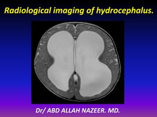
- 1. Radiological imaging of hydrocephalus. Dr/ ABD ALLAH NAZEER. MD.
- 21. The contrast among a normal brain in a normal adult (left), the brain of a normal man with impressive hydrocephalus and an equally impressive hydrocephalus in a 54-year-old man with deep cognitive and motor impairment since childhood (right;
- 22. A 25-week premature male with an intraventricular hemorrhage and subsequent development of hydrocephalus: ( a ) CUS shows the right-sided intraventricular hemorrhage; ( b ) CT also shows parenchymal hemorrhage
- 26. A 4-month-old male with vein of Galen malformation: ( a ) MRI shows the dilated vein of Galen; ( b ) cerebral angiogram shows the dilated vein of Galen and the surrounding vasculature
- 27. ( a – b ) A 8-year-old female with a posterior fossa brain tumor and hydrocephalus.
- 28. A 13-year-old male with hydrocephalus secondary to aqueductal stenosis, status post endoscopic third ventriculostomy (ETV). CSF cine flow study demonstrates CSF flow across the fenestration in the anterior third ventricle: ( a ) phase-contrast magnitude cine MRI; ( b ) phase contrast directional cine MRI.
- 29. CT/MRI axial scans demonstrating ventriculomegaly with relatively well- identified sulci. Periventricular edema is observed in case 4 which suggest acute onset.
- 30. Pineal body tumour with hydrocephalus.
- 38. Tectal Glioma.
- 40. Giant cell astrocytoma with hydrocephalus.
- 42. Choroid plexus tumors with hydrocephalus.
- 43. Choroid plexus carcinoma with hydrocephalus.
- 44. Fourth ventricular medulloblastoma with hydrocephalus.
- 45. Fourth ventricle tumour with hydrocephalus.
- 46. Posterior fossa immature teratoma with hydrocephalus.
- 47. Posterior fossa cystic mass with hydrocephalus.
- 48. Tuberculous meningitis with hydrocephalus.
- 49. Normal Pressure Hydrocephalus • Described by Hakim, Adams, et al (1965). • 50% known cause (SAH, meningitis). • 50% idiopathic (older). • Diagnosis primarily clinical: Gait apraxia, dementia, incontinence. • Radiology: communicating hydrocephalus.
- 51. Physiologic Tests for NPH • Nuclear cisternography – Communicating hydrocephalus • Pressure monitoring – Water hammer pulse – Plateau waves • Saline Infusion (outpatient) – If resistance > 4mmHg/ml/min, 82% respond to ELD • 50 cc Tap test: PPV 73-100%; sensitivity 26-62% • External lumbar drainage (inpatient)
- 53. External Lumbar Drainage • 16 gauge lumbar puncture; catheter drainage • 10 cc/hour for 3 days; gait assessed. • Of 151 patients* with iNPH,100 improved with ELD. – Gait only (88%), gait/dementia (84%), triad (59%). • Of responders, 90% improve with VP shunt. • Of nonresponders, 22% improve with shunt.
- 55. What Causes Idiopathic NPH? • Consider normal bulk flow of water in brain • Consider association of deep white matter ischemia (DWMI) and NPH. Normal Bulk Flow of Extracellular Brain Water • Water leaves upstream arterioles under pressure-osmotic gradients (e.g, mannitol). • Normal and excess water resorbed by downstream capillaries and venules. • Vasogenic edema flows centripetally to be absorbed by ventricles • Interstitial edema flows centrifugally to subarachnoid space via extracellular space.
- 56. Possible Etiology of NPH • Hypothesis: NPH patients have always had large ventricles (“slightly enlarged”) – Decreased CSF resorption (saline infusion test) – Unrecognized benign external hydrocephalus? • No evidence for previous SAH or meningitis • Significant CSF resorption pathway is via extracellular space of brain (like tectal gliomas) • Everything fine until “second hit”: DWMI DWMI is “Second Hit” in NPH • No symptoms until DWMI occurs later in life • Resistance to peripheral CSF flow through extracellular space increases slightly due to DWMI. – loss of myelin lipid: more hydrophilic environment – Greater attraction of out flowing CSF to myelin protein • CSF production continues unabated – Accumulates in ventricles -hydrocephalus worsens – Increased tangential shearing forces. – NPH symptoms begin.
- 57. NPH.
- 59. NPH.
- 63. CSF Flow Void.
- 64. Proposed Causes of CSF Motion • Production by choroid plexus (500 ml/day). • Cardiac pulsations. – Choroid plexus (Bering, 1959). – Large arteries. – Cerebral hemispheres (phase-contrast MRI).
- 65. Normal flow.
- 69. Enlarged Sylvian cisterns in NPH
- 70. Association of DWMI and NPH: Clinical • Some cases of L’etat Lacunaire (Marie, 1901) may have been NPH (Fischer, 1981) • Coexistence of NPH and Hypertensive Cerebrovascular Disease Noted Previously (Earnest 1974, Goto 1977, Graff-Radford 1987) • CSF flow void is useful indicator of favorable response to CSF diversion • Presence of DWMI is not contraindication to shunting • NPH and DWMI may be related
- 71. CSF Flow Measurement • CSF flow void on conventional MR images represents average motion during 256 spin echo acquisitions. • Since CSF motion is due to cardiac pulsations, it is better evaluated using cardiac gated techniques. Phase Contrast CSF Velocity Imaging • “Velocity” is speed plus direction • Flow sensitization along craniocaudal axis – Flow up: shades of black – Flow down: shades of white – No flow: gray – Set aliasing velocity – Quantification of velocity or flow
- 72. Qualitative CSF Velocity Imaging.
- 73. Quantitative CSF Flow Study • 512x512; 16 cm FOV • .32 mm pixels • 4mm slice angled perpendicular to aqueduct • Velocity-encode in slice direction • Retrospective cardiac-gating (not EKG triggering) Through-plane flow-encoding • Venc= 10, 20, 30 cm/sec (NPH) • Venc= 5 cm/sec (shunt malfunction)
- 77. Quantitative CSF Velocity Imaging • Calculates “Aqueductal CSF stroke volume” • Stroke volume: microliters of CSF flowing back or forth over cardiac cycle • Verified by pulsatile flow phantom using ultrasound flow meter (Mullin, 1993) Quantitative CSF Velocity Imaging
- 80. CSF Flow Study: No Flow.
- 84. Conclusions • NPH diagnosed by symptoms, not MRI • MRI used to confirm diagnosis of shunt-responsive NPH • Asymptomatic patients may have dilated ventricles and elevated CSF flow: Pre NPH? • Not everyone with benign external hydrocephalus gets NPH • Keep your extracellular space open
- 85. Thank You.