Presentation1.pptx ankle joint..
•Transferir como PPTX, PDF•
74 gostaram•11,788 visualizações


Recomendados
Mais conteúdo relacionado
Mais procurados
Mais procurados (20)
Destaque
Destaque (13)
Semelhante a Presentation1.pptx ankle joint..
Semelhante a Presentation1.pptx ankle joint.. (20)
Mais de Abdellah Nazeer
Mais de Abdellah Nazeer (20)
Presentation1.pptx ankle joint..
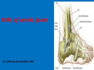
- 1. Dr/ ABD ALLAH NAZEER. MD. MRI of ankle joint
- 7. X-Ray Ankle anatomy. Hind foot
- 12. MRI ANATOMY.
- 13. TA= tibialis anterior, EHL= extensor hallucis longus, EDL= extensor digitorum longus; PL= peroneus longus, PB= peroneus brevis; TP= tibialis posterior, FDL= flexor digitorum longus, FHL= flexor hallucis longus; TC(A)= tendo calcanei (Achilles), pla= plantaris tendon.
- 15. Anterior tendons. (a, b) Axial and coronal T1. From medial to lateral – TA (blue arrows), EHL (yellow arrows), EDL (red arrows), and PT (orange arrows). The asterisk (a) represents the extensor retinaculum.
- 16. Achilles tendon. (a) Axial T1. The Achilles tendon (red arrow) is concave anteriorly and convex along the posterior margin. (b) Sagittal PD FS. The normal Achilles tendon inserts onto the posterior surface of the calcaneous (blue arrow).
- 17. Medial tendons. FHL (blue arrow), FDH (red arrow), TP (red arrow). The neurovascular bundle (purple ellipse) is located between the FHL and FDL. (b). The PT (yellow arrow) and FHL (red arrow) pass posterior to the medial malleolus (MM), with the FHL coursing posterolateral to the PT in the region of the tarsal tunnel. (c) . PT (yellow arrow), FDL (red arrow), FHL (blue arrow). The FHL (blue arrow). The PT (yellow arrow) is identified inserting onto the navicular (N). (f) The FHL (blue arrow) FDL (red arrow) within the plantar midfoot, an intersection known as Henry’s knot
- 18. Lateral tendons. The PB tendon (blue arrow) and PL tendon (red arrow) course posterior to the lateral malleolus (LM).
- 19. (a-d) Ankle ligaments. Coronal and axial T1 weighted MR images. The ankle is bound by the deltoid ligament medially (red), the lateral collateral ligamentous complex laterally (blue), and the syndesmotic ligaments (yellow). Normal ligaments are predominately low in signal intensity, but may contain longitudinal streaks of intermediate signal intensity
- 21. Lateral ligaments. (a) Axial T1. The ATFL (blue arrow) extends from the distal lateral malleolus to the lateral talus. (b) Axial T2. The PTFL (orange arrow) is a broad band between the posterior tip of the LM and the lateral talus. (c) Coronal T2. The PTFL (orange arrow) is again seen extending from the distal LM to the talus. The posterior tibiofibular ligament (yellow arrow) lies superior to the PTFL, and attaches the LM to the tibial plafond . The normal inhomogeneous appearance of these ligaments is secondary to intervening fat. (d) Coronal T1. The CN ligament (red arrow) arises from the tip of the LM, and courses medial to the peroneal tendons (purple ellipse) to attach to the lateral calcaneous
- 22. Syndesmotic ligaments. The anterior tibiofibular ligament extends from the distal fibular to the distal tibia, just proximal to the tibiotalar joint. The posterior tibiofibular ligament (yellow arrows) is an inhomogeneous band that joins the posterior distal tibia and fibula. The interosseous ligament (red ellipse) is the distal continuation of the interosseous membrane, uniting the distal tibia and fibula.
- 23. Sinus tarsi. (a) Coronal T1. The normal fat is visualized within the sinus tarsi (yellow ellipse). (b) Sagittal PD FS. The fat within the tarsal sinus suppresses normally (blue arrow). (c) Axial T1. The talocalcaneal interosseous ligament (red arrow) courses within the sinus tarsi, and functions to maintain apposition of the talus and calcaneous.
- 24. Posterior aspect. Tendons: Achilles Tendon Ligaments: No
- 30. (A) The clinical photo shows swelling, ecchymosis and dimpling in the posterior aspect of the ankle. (B) Simple lateral ankle image shows loss of Kager's triangle and bony fragments (arrow). (C) Sagittal T2 magnetic resonance image shows rupture of the Achilles tendon at calcaneal insertion site and enthesopathic spur.
- 34. Achilles tendon pathology. (a) Sagittal PD FS. A near complete tear of the Achilles tendon is visualized (blue ellipse). The tendon proximal and distal to the rupture demonstrates intermediate signal intensity (yellow arrows), consistent with superimposed severe tendinopathy. (b) Sagittal PD FS. Thickening of the Achilles tendon (orange arrow) is noted, with minimal intrasubstance intermediate signal intensity, compatible with tendinitis. (c) Sagittal PD FS. Minimal thickening of the Achilles tendon is evident, with increased peritendinous signal intensity present, consistent with peritendinitis. Minimal retrocalcaneal bursitis is also identified (purple ellipse).
- 43. Bursitis in rheumatoid arthritis.
- 46. Haglund's syndrome Refers to the triad of insertional Achilles tendinopathy with Haglund's calcaneal deformity. Retrocalcaneal bursitis Superficial retro-tendo-Achilles bursitis. Plain film Loss of Kager triangle due to retrocalcaneal bursitis. prominent bursal projection of the calcaneum. ("pump-bump") or Haglund deformity. MRI Focal enlargement and abnormal signal at achilis tendon insertion segment. Retrocalcaneal and retro achilis bursal fluid collection. Calcaneal bony spur better appreciated at T1 sagittal images. Marrow edema of the posterior calcaneal tuberosity.
- 51. Lateral aspect Tendons: Peroneal tendons Ligaments : Tibiofibular Syndesmotic complex Lateral collateral ligament
- 53. Tendinitis. (a) Axial T1. The PL tendon is mildly thickened (blue ellipse). (b) Sagittal PD FS. Again noted is focal, mild thickening of the PL tendon (red arrow).
- 54. Axial T1 image shows a C-shaped brevis tendon around the longus, a patient with rheumatoid arthritis with Tenosynovitis.
- 55. Splitting of the PB tendon with lateral displacement of PL.
- 56. Partial peroneus brevis tendon tear.
- 57. Complete rupture of PB tendon.
- 58. Complete rupture of PL tendon.
- 59. High grade partial rupture of the PT.
- 65. Complete (Grade III) tear of the anterior talofibular ligament with an accompanying proximal partial tear (Grade II) of the deep posterior tibiotalar component of the deltoid ligament complex.
- 66. Anterior talofibular ligament tear with bony avulsion of the talar attachment
- 67. Chronic anterior talo-fibular ligament injury.
- 68. Tibiofibular syndesmotic complex, (Anterior , posterior tibio-fibular and inferior transverse ligament).
- 69. The AITFL (arrow) and PITFL (arrowheads) are both torn with discontinuity, irregular morphology and increased intrasubstance signal.
- 70. Complete tear of the AT &AITFL (arrow).
- 72. Anterior aspect. Tendons: Tibialis anterior Extensor H. longus extensor digitorum longus Ligaments: No
- 73. Anterior Tibialis Tendon Rupture. Result of either laceration of the tendon or blunt trauma most often occurs in middle-aged patients following an eccentric loading of a degenerated tibialis anterior tendon against a plantar flexed foot Delay in diagnosis is common because of intact ankle dorsiflexion that occurs as a result of secondary function of the extensor hallucis longus and extensor digitorum longus muscles.
- 74. Acute rupture of the distal anterior tibial tendon at the insertion on the medial cuneiform and base of first metatarsal
- 75. Tibialis tendon anterior acute injury.
- 76. Tibialis tendon anterior acute injury
- 77. Tendinosis of the anterior tibial tendon.
- 78. Tendinosis of the extensor hallucis longus.
- 80. Extensor digitorum brevis chronic tear.
- 81. Medial aspect Tendon: Tibialis posterior. Flexor digitorum longus Flexor H. longus Ligaments: Deltoid ligament Deltoid ligament: Superficial and deep portion.
- 82. Posterior tibial tendon rupture. The posterior tibial tendon maintains the arch of the foot and posterior tibial tendon rupture is one of the most common causes of acquired flat foot in adults. The foot may become so deformed that severe ankle arthritis develops. Types I. partial tear with hypertrophy of the (4-5 times FDL, FHL) II.Partial tear with attenuated tendon or splitted tendon III.Complete tear with gap and non visualization of tendon
- 83. Type 1 tear of the posterior tibialis tendon.
- 84. Rupture of the tibialis posterior tendon. Partial tibialis posterior tendon tear.
- 85. Tenosynovitis is the inflammation of the fluid-filled sheath (called the synovuim) that surrounds a tendon. Symptoms of tenosynovitis include pain, swelling and difficulty moving the particular joint where the inflammation occurs. Acute: Synovial fluid around the tendon Chronic: Fluid + thickened tendon & synovitis FHL: Ballet dancers TP: Rheumatoid arthritis, old patients PL,PB: Spastic flat foot, young patients Tenosynovitis is also linked to infections arthritis caused by bacteria such as Neisseria gonorrhea
- 86. Tenosynovitis. (a) Axial T2 FS. Increased T2 weighted signal intensity is visualized about the FHL (blue arrow) greater than the FDL (red arrow) and PT (yellow arrow) tendons. (b) Sagittal T2 FS. Increased fluid is again noted within the FHL tendon sheath (blue arrow). (c) Sagittal T2 FS. A trace amount of increased fluid is noted within the PT tendon sheath.
- 87. Tenosynovitis of the FHL.
- 88. Tenosynovitis of the anterior tibial tendon.
- 89. Tenosynovitis of the extensor hallucis longus.
- 90. Tenosynovitis of the extensor digitorum longus and peroneus tertius
- 91. Stenosing tenosynovitis; sagittal PD-fat-saturation-weighted sequence (A and B). Stenosing tenosynovitis of the FHL tendon (arrows) secondary to os trigonum syndrome (arrowhead B).
- 92. Tenosynovitis of the peroneous tendons.
- 94. Tenosynovitis of the tibialis posterior and peroneous tendons.
- 95. Deltoid ligament. Normal T1& T2. 1.Tibio navicular, Tibio spring, Tibio calcaneal 2.Post. Tibio-talar Springs ant. Tibiotalar
- 96. Deltoid Sprain.
- 100. Plantar fasciitis, a self-limiting condition, is a common cause of heel pain in adults. It affects more than 1 million persons per year, and two-thirds of patients with plantar fasciitis will seek care from their family physician. Plantar fasciitis affects sedentary and athletic populations. Obesity, excessive foot pronation, excessive running, and prolonged standing are risk factors for developing plantar fasciitis. Diagnosis is primarily based on history and physical examination. Patients may present with heel pain with their first steps in the morning or after prolonged sitting, and sharp pain with palpation of the medial plantar calcaneal region.
- 101. Plantar fasciitis.
- 103. Sinus tarsi syndrome (STS) is a clinical finding that mainly consists of pain and tenderness of the lateral side of the hindfoot, between the ankle and the heel. Radiographic features Plain film Osteoarthritis of the subtalar joint and intraosseous cysts may be present in advanced cases. CT Shows secondary bony changes earlier than plain films. Bone scan - scintigraphy Inflammatory changes may be attributed to the sinus tarsi/subtalar region. MRI Probably the best test to show changes in the tissues of the sinus tarsi including inflammation, scar tissue formation or ligamentous injuries. The T1-hyperintense fat in the sinus tarsi space is replaced by either fluid or scar tissue, and the ligaments may be disrupted. Ganglion cysts in the region of the sinus tarsi may compress the posterior tibial nerve.
- 104. Sinus tarsi syndrome. Sagittal T1-weighted (A) and T2-weighted (B) images showing abnormal signal intensity in the tarsal sinus and bone erosions.
- 105. Sinus tarsi syndrome (STS).
- 107. Sinus tarsi syndrome, with absence of fat signal at the sinus tarsi
- 108. Tarsal tunnel fibro-osseous canal. Fibro-osseous tunnel found in the medial aspect of the ankle extending . Bony floor [talus, calcaneous, sustentaculum tali] Fibrous roof [Flexor retinaculum] Multiple septations divide the tunnel into small compartments Contents TPT,FHL,FDL,AVN.
- 109. Tarsal tunnel syndrome is a compression, or squeezing, on the posterior tibial nerve that produces symptoms anywhere along the path of the nerve. The posterior tibial nerve runs along the inside of the ankle into the foot. Etiology: Idiopathic (50% of cases). Osteoarthritis, post- traumatic ankle deformities (scar tissue may also restrict movement in the tarsal tunnel and cause nerve entrapment) or tenosynovitis. It may also be associated with rheumatoid arthritis and diabetes. Compression may also result from a cyst, lipoma, ganglion, exostosis or neoplasms within the tarsal tunnel. People with severely flat feet are at increased risk of developing tarsal tunnel syndrome. Trauma to the ankle.
- 110. Ganglion cyst resulting in tarsal tunnel syndrome.
- 111. Tarsal tunnel syndrome secondary to schwanoma (arrows)
- 112. Tarsal tunnel syndrome secondary to varicose veins.
- 113. Tarsal tunnel syndrome. (a) Axial T1. There is marked enlargement of the tibial nerve (blue ellipse). (b) Axial T1. Post-surgical changes of prior flexor retinaculum release are visualized (red arrow), as evidenced by thickening and discontinuity of the retinaculum and overlying scarring of the subcutaneous tissues. (c) Axial T2 FS. Enlargement of and intermediated T2 weighted signal intensity within the abductor hallucis muscle (yellow arrow) are evident. (d) Sagittal PD FS. Enlargement of and edema within the flexor digitorum braves muscle (purple arrow) are also identified. This constellation of findings is consistent with tarsal tunnel syndrome and compression of the plantar branch of the tibial nerve.
- 114. Baxter Neuropathy. (a,b) Sagittal and axial T1. Isolated fatty atrophy of the abductor digiti minimi muscle, consistent with chronic compression of the inferior calcaneal nerve.
- 115. Tarsal tunnel syndrome secondary to hemangioma.
- 116. Talocalcaneal coalition. (a) Sagittal PD FS. The middle facet of the subtalar joint is broadened, with osseous (blue arrow) and fibrocartilagenous (yellow arrows) bridging identified. Associated mild edema is also noted in the talus and calcaneous (green arrows). Axial T2 FS. Talocalcaneal coalition of the middle facet is again evident (orange arrow). Increased T2 weighted signal intensity within the adjacent talus and calcaneous (orange arrows) is compatible with edema.
- 117. Os Trigonum syndrome. Definition: An os trigonum is a small, round bone that sits just behind the ankle joint. The os trigonum is present in about 5-15% of people. An os trigonum occurs when one area of bone does not fuse with the rest of the talus (ankle bone) during growth.
- 120. Osteochondral lesions Causes: Spontaneous. Trauma. Corticosteroids. Sites: Talus most common Navicular Kohler’s disease (Child) Mueller-Weiss syndrome (adult) 2nd metatarsal head Freiberg’s disease Calcaneous Steroid treatment
- 121. Osteochondral Lesion of the talar dome(stage1 ).
- 122. Osteochondral Lesion of the talar dome stage 11.
- 123. Talus Osteochondritis Dissecans(stage 111).
- 124. Stage IV Detached displaced Fragment.
- 125. Koehler's Bone Disease. Koehler's bone disease is rare, referring to childhood onset avascular necrosis of the navicular bone in the foot. It commonly affects children aged 3 to 5 years old, but is seen any time between age 2 and 10 years. It is more common in boys; however, girls with this condition are often younger than boys with the disease. This is probably due to the onset of ossification in girls, which occurs at age 18-24 months. In boys ossification occurs at age 24-30 months.
- 128. Mueller Weiss syndrome refers to spontaneous osteonecrosis of the tarsal navicular in adults. This syndrome is distinct from Koehler's disease, the osteochondrosis of the tarsal navicular bone that occurs in children.
- 130. Lateral hindfoot impingement. Lateral hindfoot impingement is an extra-articular osseous impingement affecting the talus, calcaneus and distal fibula. Its development relates to hindfoot valgus malalignment, and a lateral shift of the calcaneus which may lead to abnormal bony contact between the talus and calcaneus specifically at the posterior peripheral margin of the sinus tarsi, and sometimes also the development of “neofacets” at the sinus tarsi, as well as at the fibula and adjacent calcaneus. The predominant MR features of this diagnosis are bone marrow edema and bony cystic changes, located at the apex of the lateral talar process and the calcaneus at the apex of the angle of Gissane. Diagnosis is usually clinical.
- 131. A 3D representation of the normal appearance of the structures involved with lateral hindfoot impingement. 3D representation in the coronal plane just anterior to the posterior subtalar joint demonstrates changes of lateral hindfoot impingement.
- 132. Lateral hindfoot impingement, with extra-articular talocalcaneal impingement and subfibular (calcaneofibular) impingement.