SlideShare uma empresa Scribd logo
1 de 50
Artifacts and pitfalls in shoulder magnetic resonance imaging.
Dr/ ABD ALLAH NAZEER. MD.
TENDONS:
Long head of the biceps brachii
The vincula of the biceps tendon (synovial
bands attaching to the biceps within the
tendon sheath) may be seen within the
bicipital groove.
Blood vessels can also be depicted in the
bicipital groove.
Bifid or duplicated biceps brachii tendon
constitutes a normal variation, it may be
mistaken for longitudinal split tendon tears.
Biceps vincula (mesotendon). The biceps tendon obtains its blood
supply from a mesotendon (arrow), also called vinculatendinum
Vessels adjacent to the long head of the biceps tendon (arrows).
Coronal fat-suppressed T2-weighted image shows thin, high-signal-
intensity linear structures (arrows) in supraspinatus tendon and muscle.
These are blood vessels that should not be misinterpreted for tears.
Sagittal fat-suppressed T2-weighted MR image shows dilated veins (arrow)
in spinoglenoid notch. These should not be mistaken for paralabral cysts.
Bifid biceps tendon (arrow).
Rotator cuff
The supraspinatus tendon usually inserts into the
greater tubercle of the humerus. However, rarely
it may be ectopically inserted into the bicipital
groove. Mochizuki et al. have demonstrated that
the supraspinatus tendon may also insert into the
lesser tuberosity of the humerus.
Also, Clark et al. have demonstrated the difficulty
in separating the fibers of the supraspinatus and
infraspinatus tendons. In some cases, one just
cannot say exactly which tendon has a tear, but
this should not be a source of distress.
Frequently, in the area of the insertion, one can hardly determine where
the supraspinatus tendon ends and where the infraspinatus tendon starts.
Coronal (A) and abduction external rotation (B) fat-suppressed T1-weighted MR
arthrograms show rotator cable (arrow) underlying supraspinatus tendon.
A magic angle artifact may also occur in the
supraspinatus tendon at short TE spin echo
sequences; likewise seen in the intracapsular
long head of the biceps brachii. In such cases,
T2-weighted sequences are very useful to
distinguish between magic angle artifact
and rotator cuff tendinosis or partial tendon
tear). The area of bright signal within the
tendon does not persist or is decreased on
T2-weighted images with longer TE.
Magic angle and rotator cuff. Increased signal in the supraspinatus tendon at short TE sequence.
Magic angle and rotator cuff. Coronal PD and T2-weighted images of the
supraspinatus insertion. At left, increased signal on the short TE sequence
(arrow), which is not seen on the long TE sequence at right (arrow).
Another pitfall involving the supraspinatus tendon is the small sulcus of uncovered bone located between the
supraspinatus insertion and the joint cartilage. The presence of this sulcus is important because this small
area of exposed bone should not be mistaken for a chondral articular surface injury or even a partial tear.
A small sulcus between the osseous insertion of the supraspinatus and the articular cartilage (arrow) is a normal finding.
Glenohumeral ligament and capsular variants
The MGHL may be thickened (a cord-like
pattern) or thinned. The association of
thickened MGHL and absent anterosuperior
labrum is called a Buford complex.
Compensatory thickening of the MGHL is
due to the absent labrum. The frequency
of Buford complex approximates to 1.5%
of shoulders. The Buford complex should
not be mistaken for detachment of the
anterior superior labrum.
Thickened MGHL (arrow) and absent anterosuperior labrum (Buford complex).
Axial fat-suppressed T1-weighted MR arthrogram shows cordlike middle glenohumeral ligament
in front of superior glenoid rim with absent labrum, representing Buford complex (arrow).
Sagittal fat-suppressed T2-weighted MR images show normal superior
(black arrow) and middle (white arrow, A) glenohumeral ligaments
(A) and anomalous bifid middle glenohumeral ligament (arrow, B).
Axial fat-suppressed T2-weighted MR image shows corrugated appearance
of attachment of inferior glenohumeral ligament on humeral neck (arrow).
Axial fat-suppressed T2-weighted MR image shows thickened posterior glenohumeral
ligament adjacent to normal labrum (arrow). This anomaly can be mistaken for labral tear.
LABRAL VARIANTS
Anatomic variations of labral form and contour are
commonly found. Usually, such variations occur in the
superior half of the labrum, close to the insertion of
many of the stabilizing capsular structures, and may
easily mistaken for labral or capsular pathology.
The sublabral foramen is an anatomic variation located
in the anterosuperior labrum, where it is detached from
the anterior glenoid margin, allowing for
communication between the glenohumeral joint and
the subscapular recess. It is important to recognize such
a variation at MR arthrography, as it may be
misinterpreted as anterior extension of a SLAP tear.
The sublabral foramen (arrows) is an anatomic variation and
sometimes it may be not easily differentiated from labral tear.
Coronal T2-weighted MR image shows smooth, thin defect at
base of superior labrum consistent with sublabral recess (arrow).
Axial fat-suppressed T1-weighted MR arthrogram shows defect (arrow)
at base of anterior superior labrum, consistent with sublabral foramen.
Axial fat-suppressed T1-weighted MR arthrogram shows normal gap (arrow) between
superior labrum and middle glenohumeral ligament that should not be mistaken for tear.
Axial fat-suppressed MR arthrogram shows normal intermediate-signal-intensity
cartilage (arrow) at interface between anterior labrum and glenoid fossa.
Coronal MR arthrography T1-weighted sequence showing a normal sulcus in the region
of the biceps-labral complex (arrow), which is typically parallel to the glenoid (A). In SLAP
tears, the signal alteration extends towards the substance of the superior labrum (B).
JOINT RECESSES AND BURSAE
The subscapular recess is an extension of the
glenohumeral joint located on the posterior and
anterosuperior aspects of the subscapularis tendon,
typically just below the coracoid process. Therefore, it
may be confused for the subcoracoid bursa. The
subscapular recess freely communicates with the
glenohumeral joint via various possible synovial
foramina located between the glenohumeral ligaments.
It is should be highlighted that, in healthy shoulders,
there is no communication between the glenohumeral
joint and the subcoracoid bursa. However, a
communication between the subacromial-subdeltoid
bursa and subcoracoid bursa might be present.
Subscapular recess (A) versus subcoracoid bursa (B). The subscapular recess
communicates with the glenohumeral joint (short arrow). The subcoracoid bursa is
located anteriorly to the subscapular tendon and inferiorly to the coracoid process,
but does not communicate with the joint cavity. Coracoid process (arrowheads).
POSITIONAL VARIATIONS
The supraspinatus and infraspinatus tendons are best
imaged with the patient’s shoulder placed in external
rotation. Such tendons remain out of the standard at
coronal and sagittal imaging as the humerus is internally
rotated or externally rotated in excess. In such a
condition, the increased tendons overlap may preclude
the diagnosis of possible injuries, because the
supraspinatus tendon may appear discontinuous.
Also, if there is an exaggerated internal rotation, there
may be a false subscapularis tendon thickening or
artifactual signal abnormality from the MGHL and
capsular structures. Such positioning may also result in
abnormal signal intensity of the supraspinatus and
infraspinatus, probably due to a magic angle effect.
Exaggerated internal rotation on the image A, simulating subscapularis tendon thickening
(short arrow). Certainly, the tendon is normal (long arrow) with external rotation (B).
ACROMION
Based on shape, the acromion process has
been classified into four types, namely, type
1 (straight or flat); type 2 (curved); type 3
(hooked); type 4 (inverted). This fourth type
has lately been added and represents a
convex or upward pointing undersurface.
These morphological acromial variations
may be viewed by using either scapular Y-
view radiographs or sagittal oblique and
coronal oblique MRI sequences.
The figures represent each acromion type.
The os acromiale forms a pseudoarticulation
with the base of the acromion through the fibrous
tissue, periosteum, cartilage or synovium. A
second “acromioclavicular” joint may be seen on
images, where the os acromiale is seen
articulating with the clavicle. It is important to
identify an os acromiale because it plays a role
in the development of shoulder impingement
symptoms due to inferior displacement at deltoid
contraction. Degenerative changes in this
pseudoarticulation may occur across the
synchondrosis or in association with
acromioclavicular degeneration
Os acromiale.
Axial T1-weighted MR image demonstrates the acromioclavicular joint
and the os acromiale (white arrow). Acromion (A) and clavicle (C).
Coronal fat-suppressed T1-weighted MR arthrogram shows normal low-
signal-intensity attachment of deltoid tendon along inferior lateral aspect of
acromion (arrow). Subacromial enthesophyte can also occur in this location.
HUMERUS
A normal groove is frequently found at the posterior aspect
of the humerus near the junction of the head and proximal
diaphysis, representing a potential pitfall at axial MR imaging,
and should not be mistaken for a Hill-Sachs lesion. Such a
distinction becomes important in patients with a history of
glenohumeral instability, considering that the presence of a
Hill-Sachs lesion might warrant surgical treatment. Usually,
Hill-Sachs defects are visible on the uppermost axial images
of the humerus, superolaterally at 5 mm from the top of the
humeral head. The normal anatomic groove of the humerus
usually lies at 20 to 30 mm from the superior humeral head
and is more medial and posteriorly positioned on axial images.
Depth and width of the defect are not considered to be reliable
indicators for its differentiation
Normal groove in the posterior aspect of the humeral head (A).
Hill- Sachs defects are visible on the uppermost axial sections (B).
Normal humeral head versus Hill-Sachs lesion. (A) On the axial T2 gradient echo
weighted MR image, there is a slight flattening of the posteroinferior surface of the
humeral head (arrow), which is a normal finding. (B) Axial fat saturated T2-weighted
MR image obtained at the level of the coracoid process (arrowhead, B) typically shows
a Hill-Sachs defect (arrow) in a patient with history of anterior shoulder dislocation.
Hill-Sacks" pseudolesion.
GLENOID
The shallow glenoid cavity provides increased range of
motion of the glenohumeral joint at the cost of stability.
This stability is achieved by the surrounding structures,
particularly the glenohumeral ligaments, labrum, and
rotator cuff. Some imaging abnormalities and variations
of the glenoid may be present. At the central region of the
glenoid, there is an area of thickened subchondral bone
(tubercle of Ossaki) with thinned overlying cartilage. It
should not be mistaken for an area of chondral damage or
loss. On the other hand, in this equivalent area, a smooth
focal full thickness cartilage defect without thickening of
the underlying bone may be seen as a normal variant and
should not be confused with chondromalacia
Tubercle of Ossaki. Area of focal subchondral bone
thickening with thinned overlying cartilage (arrows).
Axial (A) and sagittal (B) fat-suppressed T1-weighted MR arthrograms
show depression (arrow) in central glenoid cavity devoid of cartilage.
Finding represents bare area of glenoid cavity, which is normal variant.
Axial (A) and sagittal (B) fat-suppressed T1-weighted MR arthrograms show depression (arrow) in central
glenoid cavity devoid of cartilage. Finding represents bare area of glenoid cavity, which is normal variant.
Axial images at the level of the inferior glenoid demonstrate the normal
triangular type glenoid rim shape (A), the rounded " Lazy J " type glenoid
rim shape (B) and the deltoid glenoid rim shape (C) (white arrowheads).
Axial MR image at the level of the middle glenoid
shows a slight retroversion of the normal glenoid.
BONE MARROW
Heterogeneous signal intensity from the bone
marrow heterogeneous signal is typically observed
in the region of the humeral head and neck. In
adult patients, red marrow may be physiologically
seen below the physeal scar with slightly
hypointense signal at T1- weighted images. Red
marrow should not be confused with marrow
infiltrative processes, nodules or tumors. In
infiltrative processes, the marrow signal becomes
markedly low, hypointense to the muscle.
Normal bone marrow striations.
Thank You.

Mais conteúdo relacionado

Mais procurados

Presentation1.pptx. ultrasound examination of the ankle joint.
Presentation1.pptx. ultrasound examination of the ankle joint.Presentation1.pptx. ultrasound examination of the ankle joint.
Presentation1.pptx. ultrasound examination of the ankle joint.
Abdellah Nazeer
 
Presentation1.pptx, radiological anatomy of the knee joint.
Presentation1.pptx, radiological anatomy of the knee joint.Presentation1.pptx, radiological anatomy of the knee joint.
Presentation1.pptx, radiological anatomy of the knee joint.
Abdellah Nazeer
 
Presentation1.pptx mri of elbow joint
Presentation1.pptx mri of elbow jointPresentation1.pptx mri of elbow joint
Presentation1.pptx mri of elbow joint
Abdellah Nazeer
 
Presentation1.pptx, radiological anatomy of the shoulder joint.
Presentation1.pptx, radiological anatomy of the shoulder joint.Presentation1.pptx, radiological anatomy of the shoulder joint.
Presentation1.pptx, radiological anatomy of the shoulder joint.
Abdellah Nazeer
 
Presentation2.pptx wrist joint.
Presentation2.pptx wrist joint.Presentation2.pptx wrist joint.
Presentation2.pptx wrist joint.
Abdellah Nazeer
 
Presentation1, radiological film reading of knee joint.
Presentation1, radiological film reading of knee joint.Presentation1, radiological film reading of knee joint.
Presentation1, radiological film reading of knee joint.
Abdellah Nazeer
 
Presentation1, radiological film reading of the hip joint.
Presentation1, radiological film reading of the hip joint.Presentation1, radiological film reading of the hip joint.
Presentation1, radiological film reading of the hip joint.
Abdellah Nazeer
 

Mais procurados (20)

HOW TO READ SHOULDER MRI
HOW TO READ SHOULDER MRI HOW TO READ SHOULDER MRI
HOW TO READ SHOULDER MRI
 
Presentation1.pptx. ultrasound examination of the ankle joint.
Presentation1.pptx. ultrasound examination of the ankle joint.Presentation1.pptx. ultrasound examination of the ankle joint.
Presentation1.pptx. ultrasound examination of the ankle joint.
 
Imaging of shoulder - Dr. Vishal Sankpal
Imaging of shoulder - Dr. Vishal SankpalImaging of shoulder - Dr. Vishal Sankpal
Imaging of shoulder - Dr. Vishal Sankpal
 
Presentation1.pptx, radiological anatomy of the knee joint.
Presentation1.pptx, radiological anatomy of the knee joint.Presentation1.pptx, radiological anatomy of the knee joint.
Presentation1.pptx, radiological anatomy of the knee joint.
 
Mri anatomy of knee Dr. Muhammad Bin Zulfiqar
Mri anatomy of knee Dr. Muhammad Bin ZulfiqarMri anatomy of knee Dr. Muhammad Bin Zulfiqar
Mri anatomy of knee Dr. Muhammad Bin Zulfiqar
 
Imaging in hip disorders
Imaging in hip disordersImaging in hip disorders
Imaging in hip disorders
 
MRI of the shoulder
MRI of the shoulderMRI of the shoulder
MRI of the shoulder
 
Presentation1.pptx mri of elbow joint
Presentation1.pptx mri of elbow jointPresentation1.pptx mri of elbow joint
Presentation1.pptx mri of elbow joint
 
Presentation1.pptx, radiological anatomy of the shoulder joint.
Presentation1.pptx, radiological anatomy of the shoulder joint.Presentation1.pptx, radiological anatomy of the shoulder joint.
Presentation1.pptx, radiological anatomy of the shoulder joint.
 
Spots with keys
Spots with keysSpots with keys
Spots with keys
 
MRI of Shoulder anatomy
MRI of Shoulder anatomyMRI of Shoulder anatomy
MRI of Shoulder anatomy
 
Muscloskeletal Ultrasound of the knee (basic level)
Muscloskeletal Ultrasound of the knee (basic level)Muscloskeletal Ultrasound of the knee (basic level)
Muscloskeletal Ultrasound of the knee (basic level)
 
Shoulder labral tears MRI
Shoulder labral tears MRIShoulder labral tears MRI
Shoulder labral tears MRI
 
Presentation2.pptx wrist joint.
Presentation2.pptx wrist joint.Presentation2.pptx wrist joint.
Presentation2.pptx wrist joint.
 
Presentation1, radiological film reading of knee joint.
Presentation1, radiological film reading of knee joint.Presentation1, radiological film reading of knee joint.
Presentation1, radiological film reading of knee joint.
 
Posterior Fossa Malformations Dr Felice D'Arco
Posterior Fossa Malformations Dr Felice D'Arco Posterior Fossa Malformations Dr Felice D'Arco
Posterior Fossa Malformations Dr Felice D'Arco
 
Radiology Spots PPT- 2 by Dr Chandni Wadhwani
Radiology Spots PPT- 2 by Dr Chandni WadhwaniRadiology Spots PPT- 2 by Dr Chandni Wadhwani
Radiology Spots PPT- 2 by Dr Chandni Wadhwani
 
Shoulder ultrasound
Shoulder ultrasoundShoulder ultrasound
Shoulder ultrasound
 
Ultrasound - Knee
Ultrasound - KneeUltrasound - Knee
Ultrasound - Knee
 
Presentation1, radiological film reading of the hip joint.
Presentation1, radiological film reading of the hip joint.Presentation1, radiological film reading of the hip joint.
Presentation1, radiological film reading of the hip joint.
 

Semelhante a Presentation1, radiological imaging of artifact and pitfalls in shoulder joint at mri.

Presentation1, artifact and pitfalls of the knee, hip and ankle joints.
Presentation1, artifact and pitfalls of the knee, hip and ankle joints.Presentation1, artifact and pitfalls of the knee, hip and ankle joints.
Presentation1, artifact and pitfalls of the knee, hip and ankle joints.
Abdellah Nazeer
 
Presentation1, radiological film reading of the ankle joint.
Presentation1, radiological film reading of the ankle joint.Presentation1, radiological film reading of the ankle joint.
Presentation1, radiological film reading of the ankle joint.
Abdellah Nazeer
 
Presentation1, radiological film reading of the sholder joint.
Presentation1,  radiological film reading of the sholder joint.Presentation1,  radiological film reading of the sholder joint.
Presentation1, radiological film reading of the sholder joint.
Abdellah Nazeer
 
Presentation1, radiological imaging of lateral hindfoot impingement.
Presentation1, radiological imaging of lateral hindfoot impingement.Presentation1, radiological imaging of lateral hindfoot impingement.
Presentation1, radiological imaging of lateral hindfoot impingement.
Abdellah Nazeer
 
Presentation1, radiological imaging of shoulder dislocation.
Presentation1, radiological imaging of shoulder dislocation.Presentation1, radiological imaging of shoulder dislocation.
Presentation1, radiological imaging of shoulder dislocation.
Abdellah Nazeer
 
Presentation1, artifacts and pitfalls of the wrist and elbow joints.
Presentation1, artifacts and pitfalls of the wrist and elbow joints.Presentation1, artifacts and pitfalls of the wrist and elbow joints.
Presentation1, artifacts and pitfalls of the wrist and elbow joints.
Abdellah Nazeer
 
Presentation1, radiological imaging of adhesive capsulitis(frozen shoulder).
Presentation1, radiological imaging of adhesive capsulitis(frozen shoulder).Presentation1, radiological imaging of adhesive capsulitis(frozen shoulder).
Presentation1, radiological imaging of adhesive capsulitis(frozen shoulder).
Abdellah Nazeer
 

Semelhante a Presentation1, radiological imaging of artifact and pitfalls in shoulder joint at mri. (20)

Presentation1, artifact and pitfalls of the knee, hip and ankle joints.
Presentation1, artifact and pitfalls of the knee, hip and ankle joints.Presentation1, artifact and pitfalls of the knee, hip and ankle joints.
Presentation1, artifact and pitfalls of the knee, hip and ankle joints.
 
Presentation1, radiological film reading of the ankle joint.
Presentation1, radiological film reading of the ankle joint.Presentation1, radiological film reading of the ankle joint.
Presentation1, radiological film reading of the ankle joint.
 
Presentation1 radiological film reading of wrist joint.
Presentation1 radiological film reading of wrist joint.Presentation1 radiological film reading of wrist joint.
Presentation1 radiological film reading of wrist joint.
 
Presentation1, radiological film reading of the sholder joint.
Presentation1,  radiological film reading of the sholder joint.Presentation1,  radiological film reading of the sholder joint.
Presentation1, radiological film reading of the sholder joint.
 
Presentation1, radiological film reading of elbow joint.
Presentation1, radiological film reading of elbow joint.Presentation1, radiological film reading of elbow joint.
Presentation1, radiological film reading of elbow joint.
 
Normal Labral Variants - Walif Chbeir
Normal Labral Variants - Walif ChbeirNormal Labral Variants - Walif Chbeir
Normal Labral Variants - Walif Chbeir
 
Presentation1.pptx, diagnostic pitfalls mimicking meniscal tear and post oper...
Presentation1.pptx, diagnostic pitfalls mimicking meniscal tear and post oper...Presentation1.pptx, diagnostic pitfalls mimicking meniscal tear and post oper...
Presentation1.pptx, diagnostic pitfalls mimicking meniscal tear and post oper...
 
The elbow joint in concern of diagnostic imaging .pptx 1
The elbow joint in concern of diagnostic imaging .pptx 1The elbow joint in concern of diagnostic imaging .pptx 1
The elbow joint in concern of diagnostic imaging .pptx 1
 
Presentation1, radiological imaging of lateral hindfoot impingement.
Presentation1, radiological imaging of lateral hindfoot impingement.Presentation1, radiological imaging of lateral hindfoot impingement.
Presentation1, radiological imaging of lateral hindfoot impingement.
 
Presentation1, radiological imaging of shoulder dislocation.
Presentation1, radiological imaging of shoulder dislocation.Presentation1, radiological imaging of shoulder dislocation.
Presentation1, radiological imaging of shoulder dislocation.
 
Elbow MRI
Elbow MRIElbow MRI
Elbow MRI
 
Presentation1, artifacts and pitfalls of the wrist and elbow joints.
Presentation1, artifacts and pitfalls of the wrist and elbow joints.Presentation1, artifacts and pitfalls of the wrist and elbow joints.
Presentation1, artifacts and pitfalls of the wrist and elbow joints.
 
Anterior knee pain
Anterior knee painAnterior knee pain
Anterior knee pain
 
Paraplegia and spinal cord syndromes
Paraplegia and spinal cord syndromesParaplegia and spinal cord syndromes
Paraplegia and spinal cord syndromes
 
Ct spine fractures ppt
Ct spine fractures pptCt spine fractures ppt
Ct spine fractures ppt
 
appendicular trauma in radiology. .pptx
appendicular trauma in radiology.  .pptxappendicular trauma in radiology.  .pptx
appendicular trauma in radiology. .pptx
 
Radiological Examination of Shoulder and Elbow
Radiological Examination of Shoulder and ElbowRadiological Examination of Shoulder and Elbow
Radiological Examination of Shoulder and Elbow
 
imaging of the craniovertebral junction
imaging of the craniovertebral junction imaging of the craniovertebral junction
imaging of the craniovertebral junction
 
Presentation1, radiological imaging of adhesive capsulitis(frozen shoulder).
Presentation1, radiological imaging of adhesive capsulitis(frozen shoulder).Presentation1, radiological imaging of adhesive capsulitis(frozen shoulder).
Presentation1, radiological imaging of adhesive capsulitis(frozen shoulder).
 
cvjanomalies-181113135852.pdf
cvjanomalies-181113135852.pdfcvjanomalies-181113135852.pdf
cvjanomalies-181113135852.pdf
 

Mais de Abdellah Nazeer

Presentation2, radiological anatomy of the liver and spleen.
Presentation2, radiological anatomy of the liver and spleen.Presentation2, radiological anatomy of the liver and spleen.
Presentation2, radiological anatomy of the liver and spleen.
Abdellah Nazeer
 
Presentation1, radiological imaging of internal abdominal hernia.
Presentation1, radiological imaging of internal abdominal hernia.Presentation1, radiological imaging of internal abdominal hernia.
Presentation1, radiological imaging of internal abdominal hernia.
Abdellah Nazeer
 
Presentation11, radiological imaging of ovarian torsion.
Presentation11, radiological imaging of ovarian torsion.Presentation11, radiological imaging of ovarian torsion.
Presentation11, radiological imaging of ovarian torsion.
Abdellah Nazeer
 
Presentation1, new mri techniques in the diagnosis and monitoring of multiple...
Presentation1, new mri techniques in the diagnosis and monitoring of multiple...Presentation1, new mri techniques in the diagnosis and monitoring of multiple...
Presentation1, new mri techniques in the diagnosis and monitoring of multiple...
Abdellah Nazeer
 
Presentation1, radiological application of diffusion weighted mri in neck mas...
Presentation1, radiological application of diffusion weighted mri in neck mas...Presentation1, radiological application of diffusion weighted mri in neck mas...
Presentation1, radiological application of diffusion weighted mri in neck mas...
Abdellah Nazeer
 
Presentation1, radiological application of diffusion weighted images in breas...
Presentation1, radiological application of diffusion weighted images in breas...Presentation1, radiological application of diffusion weighted images in breas...
Presentation1, radiological application of diffusion weighted images in breas...
Abdellah Nazeer
 
Presentation1, radiological application of diffusion weighted images in abdom...
Presentation1, radiological application of diffusion weighted images in abdom...Presentation1, radiological application of diffusion weighted images in abdom...
Presentation1, radiological application of diffusion weighted images in abdom...
Abdellah Nazeer
 
Presentation1, radiological application of diffusion weighted imges in neuror...
Presentation1, radiological application of diffusion weighted imges in neuror...Presentation1, radiological application of diffusion weighted imges in neuror...
Presentation1, radiological application of diffusion weighted imges in neuror...
Abdellah Nazeer
 

Mais de Abdellah Nazeer (20)

Muculoskeletal Pediatic Imaging..pptx
Muculoskeletal Pediatic Imaging..pptxMuculoskeletal Pediatic Imaging..pptx
Muculoskeletal Pediatic Imaging..pptx
 
Presentation1, Ultrasound of the bowel loops and the lymph nodes..pptx
Presentation1, Ultrasound of the bowel loops and the lymph nodes..pptxPresentation1, Ultrasound of the bowel loops and the lymph nodes..pptx
Presentation1, Ultrasound of the bowel loops and the lymph nodes..pptx
 
Presentation1 Short cases MD..pptx
Presentation1 Short cases MD..pptxPresentation1 Short cases MD..pptx
Presentation1 Short cases MD..pptx
 
Presentation1, MD MCQ Cases..pptx
Presentation1, MD MCQ Cases..pptxPresentation1, MD MCQ Cases..pptx
Presentation1, MD MCQ Cases..pptx
 
Presentation1, Short Cases Quiz..pptx
Presentation1, Short Cases Quiz..pptxPresentation1, Short Cases Quiz..pptx
Presentation1, Short Cases Quiz..pptx
 
Presentation2, radiological anatomy of the liver and spleen.
Presentation2, radiological anatomy of the liver and spleen.Presentation2, radiological anatomy of the liver and spleen.
Presentation2, radiological anatomy of the liver and spleen.
 
Presentation1, radiological imaging of internal abdominal hernia.
Presentation1, radiological imaging of internal abdominal hernia.Presentation1, radiological imaging of internal abdominal hernia.
Presentation1, radiological imaging of internal abdominal hernia.
 
Presentation11, radiological imaging of ovarian torsion.
Presentation11, radiological imaging of ovarian torsion.Presentation11, radiological imaging of ovarian torsion.
Presentation11, radiological imaging of ovarian torsion.
 
Presentation1, musculoskeletal anatomy.
Presentation1, musculoskeletal anatomy.Presentation1, musculoskeletal anatomy.
Presentation1, musculoskeletal anatomy.
 
Presentation1, new mri techniques in the diagnosis and monitoring of multiple...
Presentation1, new mri techniques in the diagnosis and monitoring of multiple...Presentation1, new mri techniques in the diagnosis and monitoring of multiple...
Presentation1, new mri techniques in the diagnosis and monitoring of multiple...
 
Presentation1, radiological application of diffusion weighted mri in neck mas...
Presentation1, radiological application of diffusion weighted mri in neck mas...Presentation1, radiological application of diffusion weighted mri in neck mas...
Presentation1, radiological application of diffusion weighted mri in neck mas...
 
Presentation1, radiological application of diffusion weighted images in breas...
Presentation1, radiological application of diffusion weighted images in breas...Presentation1, radiological application of diffusion weighted images in breas...
Presentation1, radiological application of diffusion weighted images in breas...
 
Presentation1, radiological application of diffusion weighted images in abdom...
Presentation1, radiological application of diffusion weighted images in abdom...Presentation1, radiological application of diffusion weighted images in abdom...
Presentation1, radiological application of diffusion weighted images in abdom...
 
Presentation1, radiological application of diffusion weighted imges in neuror...
Presentation1, radiological application of diffusion weighted imges in neuror...Presentation1, radiological application of diffusion weighted imges in neuror...
Presentation1, radiological application of diffusion weighted imges in neuror...
 
Presentation1, mr physics.
Presentation1, mr physics.Presentation1, mr physics.
Presentation1, mr physics.
 
Presentation1. ct physics.
Presentation1. ct physics.Presentation1. ct physics.
Presentation1. ct physics.
 
Presentation1, ultrasound physics.
Presentation1, ultrasound physics.Presentation1, ultrasound physics.
Presentation1, ultrasound physics.
 
Presentation4, x ray physics
Presentation4, x ray physicsPresentation4, x ray physics
Presentation4, x ray physics
 
Positions
PositionsPositions
Positions
 
Presentation1, quiz for the chest.
Presentation1, quiz for the chest.Presentation1, quiz for the chest.
Presentation1, quiz for the chest.
 

Último

Call Girls Bhubaneswar Just Call 9907093804 Top Class Call Girl Service Avail...
Call Girls Bhubaneswar Just Call 9907093804 Top Class Call Girl Service Avail...Call Girls Bhubaneswar Just Call 9907093804 Top Class Call Girl Service Avail...
Call Girls Bhubaneswar Just Call 9907093804 Top Class Call Girl Service Avail...
Dipal Arora
 
College Call Girls in Haridwar 9667172968 Short 4000 Night 10000 Best call gi...
College Call Girls in Haridwar 9667172968 Short 4000 Night 10000 Best call gi...College Call Girls in Haridwar 9667172968 Short 4000 Night 10000 Best call gi...
College Call Girls in Haridwar 9667172968 Short 4000 Night 10000 Best call gi...
perfect solution
 
Call Girls Aurangabad Just Call 8250077686 Top Class Call Girl Service Available
Call Girls Aurangabad Just Call 8250077686 Top Class Call Girl Service AvailableCall Girls Aurangabad Just Call 8250077686 Top Class Call Girl Service Available
Call Girls Aurangabad Just Call 8250077686 Top Class Call Girl Service Available
Dipal Arora
 

Último (20)

Premium Bangalore Call Girls Jigani Dail 6378878445 Escort Service For Hot Ma...
Premium Bangalore Call Girls Jigani Dail 6378878445 Escort Service For Hot Ma...Premium Bangalore Call Girls Jigani Dail 6378878445 Escort Service For Hot Ma...
Premium Bangalore Call Girls Jigani Dail 6378878445 Escort Service For Hot Ma...
 
Call Girls Bhubaneswar Just Call 9907093804 Top Class Call Girl Service Avail...
Call Girls Bhubaneswar Just Call 9907093804 Top Class Call Girl Service Avail...Call Girls Bhubaneswar Just Call 9907093804 Top Class Call Girl Service Avail...
Call Girls Bhubaneswar Just Call 9907093804 Top Class Call Girl Service Avail...
 
College Call Girls in Haridwar 9667172968 Short 4000 Night 10000 Best call gi...
College Call Girls in Haridwar 9667172968 Short 4000 Night 10000 Best call gi...College Call Girls in Haridwar 9667172968 Short 4000 Night 10000 Best call gi...
College Call Girls in Haridwar 9667172968 Short 4000 Night 10000 Best call gi...
 
Top Quality Call Girl Service Kalyanpur 6378878445 Available Call Girls Any Time
Top Quality Call Girl Service Kalyanpur 6378878445 Available Call Girls Any TimeTop Quality Call Girl Service Kalyanpur 6378878445 Available Call Girls Any Time
Top Quality Call Girl Service Kalyanpur 6378878445 Available Call Girls Any Time
 
Call Girls Ludhiana Just Call 9907093804 Top Class Call Girl Service Available
Call Girls Ludhiana Just Call 9907093804 Top Class Call Girl Service AvailableCall Girls Ludhiana Just Call 9907093804 Top Class Call Girl Service Available
Call Girls Ludhiana Just Call 9907093804 Top Class Call Girl Service Available
 
Call Girls Aurangabad Just Call 8250077686 Top Class Call Girl Service Available
Call Girls Aurangabad Just Call 8250077686 Top Class Call Girl Service AvailableCall Girls Aurangabad Just Call 8250077686 Top Class Call Girl Service Available
Call Girls Aurangabad Just Call 8250077686 Top Class Call Girl Service Available
 
Call Girls Jabalpur Just Call 8250077686 Top Class Call Girl Service Available
Call Girls Jabalpur Just Call 8250077686 Top Class Call Girl Service AvailableCall Girls Jabalpur Just Call 8250077686 Top Class Call Girl Service Available
Call Girls Jabalpur Just Call 8250077686 Top Class Call Girl Service Available
 
O963O942363 Call Girls In Ahmedabad Escort Service Available 24×7 In Ahmedabad
O963O942363 Call Girls In Ahmedabad Escort Service Available 24×7 In AhmedabadO963O942363 Call Girls In Ahmedabad Escort Service Available 24×7 In Ahmedabad
O963O942363 Call Girls In Ahmedabad Escort Service Available 24×7 In Ahmedabad
 
The Most Attractive Hyderabad Call Girls Kothapet 𖠋 6297143586 𖠋 Will You Mis...
The Most Attractive Hyderabad Call Girls Kothapet 𖠋 6297143586 𖠋 Will You Mis...The Most Attractive Hyderabad Call Girls Kothapet 𖠋 6297143586 𖠋 Will You Mis...
The Most Attractive Hyderabad Call Girls Kothapet 𖠋 6297143586 𖠋 Will You Mis...
 
Call Girls Coimbatore Just Call 9907093804 Top Class Call Girl Service Available
Call Girls Coimbatore Just Call 9907093804 Top Class Call Girl Service AvailableCall Girls Coimbatore Just Call 9907093804 Top Class Call Girl Service Available
Call Girls Coimbatore Just Call 9907093804 Top Class Call Girl Service Available
 
Call Girls Tirupati Just Call 8250077686 Top Class Call Girl Service Available
Call Girls Tirupati Just Call 8250077686 Top Class Call Girl Service AvailableCall Girls Tirupati Just Call 8250077686 Top Class Call Girl Service Available
Call Girls Tirupati Just Call 8250077686 Top Class Call Girl Service Available
 
(Low Rate RASHMI ) Rate Of Call Girls Jaipur ❣ 8445551418 ❣ Elite Models & Ce...
(Low Rate RASHMI ) Rate Of Call Girls Jaipur ❣ 8445551418 ❣ Elite Models & Ce...(Low Rate RASHMI ) Rate Of Call Girls Jaipur ❣ 8445551418 ❣ Elite Models & Ce...
(Low Rate RASHMI ) Rate Of Call Girls Jaipur ❣ 8445551418 ❣ Elite Models & Ce...
 
Call Girls Haridwar Just Call 8250077686 Top Class Call Girl Service Available
Call Girls Haridwar Just Call 8250077686 Top Class Call Girl Service AvailableCall Girls Haridwar Just Call 8250077686 Top Class Call Girl Service Available
Call Girls Haridwar Just Call 8250077686 Top Class Call Girl Service Available
 
Call Girls Siliguri Just Call 8250077686 Top Class Call Girl Service Available
Call Girls Siliguri Just Call 8250077686 Top Class Call Girl Service AvailableCall Girls Siliguri Just Call 8250077686 Top Class Call Girl Service Available
Call Girls Siliguri Just Call 8250077686 Top Class Call Girl Service Available
 
Call Girls Kochi Just Call 8250077686 Top Class Call Girl Service Available
Call Girls Kochi Just Call 8250077686 Top Class Call Girl Service AvailableCall Girls Kochi Just Call 8250077686 Top Class Call Girl Service Available
Call Girls Kochi Just Call 8250077686 Top Class Call Girl Service Available
 
(👑VVIP ISHAAN ) Russian Call Girls Service Navi Mumbai🖕9920874524🖕Independent...
(👑VVIP ISHAAN ) Russian Call Girls Service Navi Mumbai🖕9920874524🖕Independent...(👑VVIP ISHAAN ) Russian Call Girls Service Navi Mumbai🖕9920874524🖕Independent...
(👑VVIP ISHAAN ) Russian Call Girls Service Navi Mumbai🖕9920874524🖕Independent...
 
Call Girls Visakhapatnam Just Call 9907093804 Top Class Call Girl Service Ava...
Call Girls Visakhapatnam Just Call 9907093804 Top Class Call Girl Service Ava...Call Girls Visakhapatnam Just Call 9907093804 Top Class Call Girl Service Ava...
Call Girls Visakhapatnam Just Call 9907093804 Top Class Call Girl Service Ava...
 
VIP Hyderabad Call Girls Bahadurpally 7877925207 ₹5000 To 25K With AC Room 💚😋
VIP Hyderabad Call Girls Bahadurpally 7877925207 ₹5000 To 25K With AC Room 💚😋VIP Hyderabad Call Girls Bahadurpally 7877925207 ₹5000 To 25K With AC Room 💚😋
VIP Hyderabad Call Girls Bahadurpally 7877925207 ₹5000 To 25K With AC Room 💚😋
 
Best Rate (Patna ) Call Girls Patna ⟟ 8617370543 ⟟ High Class Call Girl In 5 ...
Best Rate (Patna ) Call Girls Patna ⟟ 8617370543 ⟟ High Class Call Girl In 5 ...Best Rate (Patna ) Call Girls Patna ⟟ 8617370543 ⟟ High Class Call Girl In 5 ...
Best Rate (Patna ) Call Girls Patna ⟟ 8617370543 ⟟ High Class Call Girl In 5 ...
 
Call Girls Faridabad Just Call 9907093804 Top Class Call Girl Service Available
Call Girls Faridabad Just Call 9907093804 Top Class Call Girl Service AvailableCall Girls Faridabad Just Call 9907093804 Top Class Call Girl Service Available
Call Girls Faridabad Just Call 9907093804 Top Class Call Girl Service Available
 

Presentation1, radiological imaging of artifact and pitfalls in shoulder joint at mri.

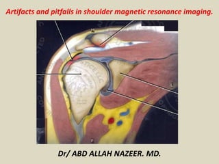
  • 1. Artifacts and pitfalls in shoulder magnetic resonance imaging. Dr/ ABD ALLAH NAZEER. MD.
  • 2. TENDONS: Long head of the biceps brachii The vincula of the biceps tendon (synovial bands attaching to the biceps within the tendon sheath) may be seen within the bicipital groove. Blood vessels can also be depicted in the bicipital groove. Bifid or duplicated biceps brachii tendon constitutes a normal variation, it may be mistaken for longitudinal split tendon tears.
  • 3. Biceps vincula (mesotendon). The biceps tendon obtains its blood supply from a mesotendon (arrow), also called vinculatendinum
  • 4. Vessels adjacent to the long head of the biceps tendon (arrows).
  • 5. Coronal fat-suppressed T2-weighted image shows thin, high-signal- intensity linear structures (arrows) in supraspinatus tendon and muscle. These are blood vessels that should not be misinterpreted for tears.
  • 6. Sagittal fat-suppressed T2-weighted MR image shows dilated veins (arrow) in spinoglenoid notch. These should not be mistaken for paralabral cysts.
  • 8. Rotator cuff The supraspinatus tendon usually inserts into the greater tubercle of the humerus. However, rarely it may be ectopically inserted into the bicipital groove. Mochizuki et al. have demonstrated that the supraspinatus tendon may also insert into the lesser tuberosity of the humerus. Also, Clark et al. have demonstrated the difficulty in separating the fibers of the supraspinatus and infraspinatus tendons. In some cases, one just cannot say exactly which tendon has a tear, but this should not be a source of distress.
  • 9. Frequently, in the area of the insertion, one can hardly determine where the supraspinatus tendon ends and where the infraspinatus tendon starts.
  • 10. Coronal (A) and abduction external rotation (B) fat-suppressed T1-weighted MR arthrograms show rotator cable (arrow) underlying supraspinatus tendon.
  • 11. A magic angle artifact may also occur in the supraspinatus tendon at short TE spin echo sequences; likewise seen in the intracapsular long head of the biceps brachii. In such cases, T2-weighted sequences are very useful to distinguish between magic angle artifact and rotator cuff tendinosis or partial tendon tear). The area of bright signal within the tendon does not persist or is decreased on T2-weighted images with longer TE.
  • 12. Magic angle and rotator cuff. Increased signal in the supraspinatus tendon at short TE sequence.
  • 13. Magic angle and rotator cuff. Coronal PD and T2-weighted images of the supraspinatus insertion. At left, increased signal on the short TE sequence (arrow), which is not seen on the long TE sequence at right (arrow).
  • 14. Another pitfall involving the supraspinatus tendon is the small sulcus of uncovered bone located between the supraspinatus insertion and the joint cartilage. The presence of this sulcus is important because this small area of exposed bone should not be mistaken for a chondral articular surface injury or even a partial tear. A small sulcus between the osseous insertion of the supraspinatus and the articular cartilage (arrow) is a normal finding.
  • 15. Glenohumeral ligament and capsular variants The MGHL may be thickened (a cord-like pattern) or thinned. The association of thickened MGHL and absent anterosuperior labrum is called a Buford complex. Compensatory thickening of the MGHL is due to the absent labrum. The frequency of Buford complex approximates to 1.5% of shoulders. The Buford complex should not be mistaken for detachment of the anterior superior labrum.
  • 16. Thickened MGHL (arrow) and absent anterosuperior labrum (Buford complex).
  • 17. Axial fat-suppressed T1-weighted MR arthrogram shows cordlike middle glenohumeral ligament in front of superior glenoid rim with absent labrum, representing Buford complex (arrow).
  • 18. Sagittal fat-suppressed T2-weighted MR images show normal superior (black arrow) and middle (white arrow, A) glenohumeral ligaments (A) and anomalous bifid middle glenohumeral ligament (arrow, B).
  • 19. Axial fat-suppressed T2-weighted MR image shows corrugated appearance of attachment of inferior glenohumeral ligament on humeral neck (arrow).
  • 20. Axial fat-suppressed T2-weighted MR image shows thickened posterior glenohumeral ligament adjacent to normal labrum (arrow). This anomaly can be mistaken for labral tear.
  • 21. LABRAL VARIANTS Anatomic variations of labral form and contour are commonly found. Usually, such variations occur in the superior half of the labrum, close to the insertion of many of the stabilizing capsular structures, and may easily mistaken for labral or capsular pathology. The sublabral foramen is an anatomic variation located in the anterosuperior labrum, where it is detached from the anterior glenoid margin, allowing for communication between the glenohumeral joint and the subscapular recess. It is important to recognize such a variation at MR arthrography, as it may be misinterpreted as anterior extension of a SLAP tear.
  • 22. The sublabral foramen (arrows) is an anatomic variation and sometimes it may be not easily differentiated from labral tear.
  • 23. Coronal T2-weighted MR image shows smooth, thin defect at base of superior labrum consistent with sublabral recess (arrow).
  • 24. Axial fat-suppressed T1-weighted MR arthrogram shows defect (arrow) at base of anterior superior labrum, consistent with sublabral foramen.
  • 25. Axial fat-suppressed T1-weighted MR arthrogram shows normal gap (arrow) between superior labrum and middle glenohumeral ligament that should not be mistaken for tear.
  • 26. Axial fat-suppressed MR arthrogram shows normal intermediate-signal-intensity cartilage (arrow) at interface between anterior labrum and glenoid fossa.
  • 27. Coronal MR arthrography T1-weighted sequence showing a normal sulcus in the region of the biceps-labral complex (arrow), which is typically parallel to the glenoid (A). In SLAP tears, the signal alteration extends towards the substance of the superior labrum (B).
  • 28. JOINT RECESSES AND BURSAE The subscapular recess is an extension of the glenohumeral joint located on the posterior and anterosuperior aspects of the subscapularis tendon, typically just below the coracoid process. Therefore, it may be confused for the subcoracoid bursa. The subscapular recess freely communicates with the glenohumeral joint via various possible synovial foramina located between the glenohumeral ligaments. It is should be highlighted that, in healthy shoulders, there is no communication between the glenohumeral joint and the subcoracoid bursa. However, a communication between the subacromial-subdeltoid bursa and subcoracoid bursa might be present.
  • 29. Subscapular recess (A) versus subcoracoid bursa (B). The subscapular recess communicates with the glenohumeral joint (short arrow). The subcoracoid bursa is located anteriorly to the subscapular tendon and inferiorly to the coracoid process, but does not communicate with the joint cavity. Coracoid process (arrowheads).
  • 30. POSITIONAL VARIATIONS The supraspinatus and infraspinatus tendons are best imaged with the patient’s shoulder placed in external rotation. Such tendons remain out of the standard at coronal and sagittal imaging as the humerus is internally rotated or externally rotated in excess. In such a condition, the increased tendons overlap may preclude the diagnosis of possible injuries, because the supraspinatus tendon may appear discontinuous. Also, if there is an exaggerated internal rotation, there may be a false subscapularis tendon thickening or artifactual signal abnormality from the MGHL and capsular structures. Such positioning may also result in abnormal signal intensity of the supraspinatus and infraspinatus, probably due to a magic angle effect.
  • 31. Exaggerated internal rotation on the image A, simulating subscapularis tendon thickening (short arrow). Certainly, the tendon is normal (long arrow) with external rotation (B).
  • 32. ACROMION Based on shape, the acromion process has been classified into four types, namely, type 1 (straight or flat); type 2 (curved); type 3 (hooked); type 4 (inverted). This fourth type has lately been added and represents a convex or upward pointing undersurface. These morphological acromial variations may be viewed by using either scapular Y- view radiographs or sagittal oblique and coronal oblique MRI sequences.
  • 33. The figures represent each acromion type.
  • 34. The os acromiale forms a pseudoarticulation with the base of the acromion through the fibrous tissue, periosteum, cartilage or synovium. A second “acromioclavicular” joint may be seen on images, where the os acromiale is seen articulating with the clavicle. It is important to identify an os acromiale because it plays a role in the development of shoulder impingement symptoms due to inferior displacement at deltoid contraction. Degenerative changes in this pseudoarticulation may occur across the synchondrosis or in association with acromioclavicular degeneration
  • 36. Axial T1-weighted MR image demonstrates the acromioclavicular joint and the os acromiale (white arrow). Acromion (A) and clavicle (C).
  • 37. Coronal fat-suppressed T1-weighted MR arthrogram shows normal low- signal-intensity attachment of deltoid tendon along inferior lateral aspect of acromion (arrow). Subacromial enthesophyte can also occur in this location.
  • 38. HUMERUS A normal groove is frequently found at the posterior aspect of the humerus near the junction of the head and proximal diaphysis, representing a potential pitfall at axial MR imaging, and should not be mistaken for a Hill-Sachs lesion. Such a distinction becomes important in patients with a history of glenohumeral instability, considering that the presence of a Hill-Sachs lesion might warrant surgical treatment. Usually, Hill-Sachs defects are visible on the uppermost axial images of the humerus, superolaterally at 5 mm from the top of the humeral head. The normal anatomic groove of the humerus usually lies at 20 to 30 mm from the superior humeral head and is more medial and posteriorly positioned on axial images. Depth and width of the defect are not considered to be reliable indicators for its differentiation
  • 39. Normal groove in the posterior aspect of the humeral head (A). Hill- Sachs defects are visible on the uppermost axial sections (B).
  • 40. Normal humeral head versus Hill-Sachs lesion. (A) On the axial T2 gradient echo weighted MR image, there is a slight flattening of the posteroinferior surface of the humeral head (arrow), which is a normal finding. (B) Axial fat saturated T2-weighted MR image obtained at the level of the coracoid process (arrowhead, B) typically shows a Hill-Sachs defect (arrow) in a patient with history of anterior shoulder dislocation.
  • 42. GLENOID The shallow glenoid cavity provides increased range of motion of the glenohumeral joint at the cost of stability. This stability is achieved by the surrounding structures, particularly the glenohumeral ligaments, labrum, and rotator cuff. Some imaging abnormalities and variations of the glenoid may be present. At the central region of the glenoid, there is an area of thickened subchondral bone (tubercle of Ossaki) with thinned overlying cartilage. It should not be mistaken for an area of chondral damage or loss. On the other hand, in this equivalent area, a smooth focal full thickness cartilage defect without thickening of the underlying bone may be seen as a normal variant and should not be confused with chondromalacia
  • 43. Tubercle of Ossaki. Area of focal subchondral bone thickening with thinned overlying cartilage (arrows).
  • 44. Axial (A) and sagittal (B) fat-suppressed T1-weighted MR arthrograms show depression (arrow) in central glenoid cavity devoid of cartilage. Finding represents bare area of glenoid cavity, which is normal variant.
  • 45. Axial (A) and sagittal (B) fat-suppressed T1-weighted MR arthrograms show depression (arrow) in central glenoid cavity devoid of cartilage. Finding represents bare area of glenoid cavity, which is normal variant.
  • 46. Axial images at the level of the inferior glenoid demonstrate the normal triangular type glenoid rim shape (A), the rounded " Lazy J " type glenoid rim shape (B) and the deltoid glenoid rim shape (C) (white arrowheads).
  • 47. Axial MR image at the level of the middle glenoid shows a slight retroversion of the normal glenoid.
  • 48. BONE MARROW Heterogeneous signal intensity from the bone marrow heterogeneous signal is typically observed in the region of the humeral head and neck. In adult patients, red marrow may be physiologically seen below the physeal scar with slightly hypointense signal at T1- weighted images. Red marrow should not be confused with marrow infiltrative processes, nodules or tumors. In infiltrative processes, the marrow signal becomes markedly low, hypointense to the muscle.
  • 49. Normal bone marrow striations.