Endodontic Access Cavity Preparation
•Transferir como PPTX, PDF•
116 gostaram•67,910 visualizações

Endodontic Access Cavity Preparation

Recomendados
Mais conteúdo relacionado
Mais procurados
Mais procurados (20)
Semelhante a Endodontic Access Cavity Preparation
Semelhante a Endodontic Access Cavity Preparation (20)
Mais de Dr Aaron Sarwal
Mais de Dr Aaron Sarwal (15)
Último
Último (20)
Endodontic Access Cavity Preparation
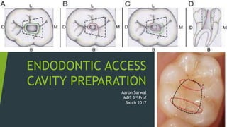
- 1. ENDODONTIC ACCESS CAVITY PREPARATION Aaron Sarwal MDS 3rd Prof Batch 2017
- 2. “...all the treatment that follows hinges on the accuracy and correctness of the entry” - FRANKLIN.S.WEINE
- 3. CONTENTS DEFINITION OBJECTIVES RULES FOR PROPER ACCESS PREPARATION PRINCIPLES OF ENDODONTIC ACCESS PREPARATION ARMAMENTARIUM GUIDELINES AIDS IN ACCESS PREPARATION ACCESS PREPARATION OF ANTERIOR AND POSTERIOR TEETH ERRORS IN ACCESS CAVITY PREPARATION CHALLENGES IN ACCESS PREAPARATIONS CONCLUSION
- 4. INTRODUCTION The hard tissue that encompasses the human dental pulp takes on numerous configurations and shapes. A thorough understanding of the complexity of the root canal system is essential for understanding the principles and problems of shaping and cleaning, for determining the apical limits and dimensions of canal preparations, and for performing successful microsurgical procedures.
- 5. DEFINITION Access cavity preparation is defined as endodontic coronal preparation which enables unobstructed access to the canal orifices, a straight line access to the apical foramen, complete control over instrumentation and accommodate obturation technique. According to Stephen Cohen, “it generally refers to the part of the preparation from the occlusal table to the root canal orifices.”
- 6. A PROPER CORONAL ACCESS FORMS THE FOUNDATION OF PYRAMID OF ENDODONTIC TREATMENT OBJECTIVES According to R.E Walton:- Straight line access Improved instrument control Improved obturation Decreased procedural errors
- 7. OBJECTIVES (ACC. TO RE WALTON) Conservation of tooth structure • Minimal weakening of tooth • Prevention of perforation Un-roofing the chamber and exposure of pulp horns • Maximum visibility • Location of canals
- 8. RULES FOR PROPER ACCESS PREPARATION The objective of entry is to give direct access to the apical foramina, not merely to the canal orifice. Access cavity preparations are different from typical operative occlusal preparations. The interior anatomy of the tooth under treatment must be determined. Rubber dam – when canals difficult to find the rubber dam should not be placed until correct location has been confirmed Endodontic entries are prepared through the occlusal or lingual surface never through the proximal or gingival surface. As a part of access preparation, the unsupported cusps of posterior teeth must be reduced. (Franklin S. Weine)
- 9. A PREOPERATIVE PERIAPICAL RADIOGRAPH IS A MUST, PRIOR TO ACCESS CAVITY PREPARATION RADIOGRAPHS HELP US GLEAN: • Morphology of tooth • Anatomy of root canal system • Number and length of canals • Branching of canal system • Position and depth of pulp chamber • Position of apical foramen • As well, calcifications, or resorption present, if any AN INTRA-ORAL PERIAPICAL RADIOGRAPH
- 10. DIVISIONS OF ACCESS CAVITY PREPARATION For sake of descriptive convenience Ingle has divided endodontic cavity preparation into CORONAL PREPARATION •0 RADICULAR PREPARATION
- 11. PHASES FOR ACCESS CAVITY PREPARATION Regardless of the tooth, there are three phases in the preparation of the access cavity: A. Penetration B. Enlarging C. Finishing
- 12. INSTRUMENTS FOR ACCESS CAVITY PREPARATION Access opening burs: They are round burs with 16mm bur shank (3mm longer than standard burs)
- 13. INSTRUMENTS FOR ACCESS CAVITY PREPARATION Access refining burs: These are coarse grit flame shaped, tapered round and diamonds for refining walls of access cavity preparation
- 14. INSTRUMENTS FOR ACCESS CAVITY PREPARATION Surgical length burs A. Munce Discovery Burs B. Muller Burs
- 15. INSTRUMENTS FOR ACCESS CAVITY PREPARATION: Endo Access Kit by Dentsply Mallifer
- 16. INSTRUMENTS FOR ACCESS CAVITY PREPARATION: Endo Access Kit by Dentsply Mallifer Cavity Access Kit Cavity Access Z Kit
- 17. INSTRUMENTS FOR ACCESS CAVITY PREPARATION: Endo Access Kit by Dentsply Mallifer
- 18. PRINCIPLES OF ENDODONTIC CAVITY PREPARATION I. Outline Form II. Convenience Form III. Removal of the remaining carious dentin and defective restorations. IV. Toilet of the cavity
- 19. PRINCIPLE I – OUTLINE FORM Must be correctly shaped and positioned. Establish complete access for instrumentation, from cavity margin to apical foramen. External outline form evolves from the internal anatomy of the tooth established by the pulp.
- 20. PRINCIPLE I: OUTLINE FORM Three factors of internal anatomy must be considered: 1.Size of the pulp chamber 2. Number of individual root canals, their curvature, and their position. 3.Shape of the pulp chamber
- 21. PRINCIPLE II: CONVENIENCE FORM Unobstructed access to the canal orifice Direct access to the apical foramen Cavity expansion to accommodate filling techniques Complete authority over the enlarging instrument.
- 22. UNOBSTRUCTED ACCESS TO CANAL ORIFICE Enough tooth structure must be removed to allow instruments to be placed easily. One should be able to see each orifice & easily reach it with instrument points. Entire wall need not be extended if instrument impingement occurs owing to severely curved root or an extra canal.
- 23. SHAMROCK PREPARATION: LEUBKE Leubke showed there is no need of extenstion of entire wall ,he recommended extension of only that portion of the wall were extra canal is present ,resulting in a clover leaf appearance in outline form- shamrock preparation
- 24. DIRECT ACCESS TO APICAL FORAMEN Enough tooth structure must be removed to allow endodontic instruments freedom within coronal cavity so that they can extend down the canal in unstrained position.
- 25. CAVITY EXPANSION TO ACCOMMODATE FILLING TECHNIQUES To make filling techniques more convenient or practical, outline form may have to be widely extended to accommodate these instruments
- 26. COMPLETE AUTHORITY OVER ENLARGING INSTRUMENT If the instrument is impinged at the canal orifice by tooth structure the intervening tooth structure will dictate the control of the instrument. If the tooth structure is removed from the orifice so that the instrument stands free in this area of the canal the instrument will then be controlled by only two factors: the clinician’s fingers on the handle of the instrument and the walls of the canal at the tip of the instrument.
- 27. PRINCIPLE III: REMOVAL OF THE REMAINING CARIOUS DENTIN AND DEFECTIVE RESTORATIONS To eliminate mechanically as many bacteria as possible from the interior of tooth To eliminate discoloration of tooth structure To eliminate the possibility of any bacteria-laden saliva leaking into the prepared cavity.
- 28. PRINCIPLE IV: TOILET OF THE CAVITY All of the caries, debris, and necrotic material must be removed (before the radicular preparation is begun). Use of hand instruments may be required along with copious irrigation.
- 29. LAWS OF ACCESS CAVITY PREPARATION (KRASNER AND RANKOW) LAW OF CENTRALITY LAW OF CEMENTO ENAMEL JUNCTION LAW OF CONCENTRICITY LAW OF COLOR CHANGE LAW OF SYMMETRY LAW OF ORIFICE LOCATION
- 30. LAW OF CONCENTRICITY The walls of the pulp chamber are always concentric to the external surface of the tooth at the level of the CEJ .
- 31. LAW OF SYMMETRY : 1 Except for maxillary molars, the orifices of the canals are equidistant from a line drawn in a mesial distal direction through the pulp chamber floor.
- 32. LAW OF SYMMETRY : 2 Except for the maxillary molars, the orifices of the canals lie on a line perpendicular to a line drawn in a mesial-distal direction across the centre of the floor of the pulp chamber.
- 33. LAW OF COLOR CHANGE The color of the pulp chamber floor is always darker than the walls.
- 34. LAW OF ORIFICE LOCATION Law of orifice location 1: the orifices of the root canals are always located at the junction of the walls and the floor. Law of orifice location 2: the orifices of the root canals are located at the angles in the floor wall junction. Law of orifice location 3: the orifices of the root canals are located at the terminus of the root development fusion lines.
- 37. ARMAMENTARIUM FOR ACCESS PREPARATION • Front surface mirror • Endodontic explorer • DG 16 • Endodontic excavator • Irrigating solutions • Cotton pliers • Broaches • Glass slab • Files & reamers • Burs • Rubber dam kit Armamentarium
- 38. THE ROUND BUR Three sizes of round burs, Nos. 2, 4, and 6, are routinely used. No. 2 Mandibular anterior teeth Maxillary premolar (narrow chambers & canals) Incisal pulp horn area (Maxillary anterior teeth) No. 4 Maxillary anterior teeth Maxillary and mandibular premolar teeth Maxillary and mandibular molars No. 2 & No. 4 Round bur
- 39. THE ROUND BUR No. 6 Only in large pulp chamber of molars Taurodontism No. 1 Used in the floor of pulp chamber to seek additional canal orifice. Eg MB2 Maillefer Endo-Z carbide fissure bur It is safe-ended and will not scar the pulpal floor. Moreover, it is longer bladed (9 mm) for sloping and funnelling the access cavity.
- 40. AIDS IN LOCATING ROOT CANAL ORIFICES Endodontic explorer Troughing of grooves with ultrasonic tips, Staining the chamber floor with 1% methylene blue dye, Performing the sodium hypochlorite ‘champagne bubble’ test and visualizing canal bleeding points are important aids in locating root canal orifices. Magnification and illumination
- 41. ACCESS PREPARATION GUIDELINES STEP 1- Diagnostic radiograph. Visualization of the location of the pulp space. Bucco-lingual angulations and coronal anatomy are judged visually.
- 42. ACCESS PREPARATION GUIDELINES STEP 2: Restorative material impinging on the straight- line access should be removed before pulp chamber is accessed to prevent lodging of debris in the canals. Caries is removed to prevent irrigating solutions from leaking past the rubber dam into the mouth and to prevent bacterial contamination of the canal system with saliva. Place an interim restoration. A 1 mm to 2 mm of occlusal adjustment of teeth may be done.
- 43. ACCESS PREPARATION GUIDELINES STEP 3: The roof of the pulp chamber is best perforated with a round bur. A no.2 bur ( anterior and premolar teeth) A no. 4 bur should be used in molar teeth. For teeth with porcelain crowns. The bur is best directed toward largest part of pulp chamber. In calcified, multi-rooted teeth, it is better to direct the access toward the largest canal.
- 44. ACCESS PREPARATION GUIDELINES STEP 4-Once the pulp chamber is located (with light upward pressure), the round bur is used to remove the roof of the pulp chamber from underneath; the “belly” of the bur should be used to cut on the outstroke. This should establish an initial outline form. The pulp chamber should be frequently flushed with sodium hypochlorite solution to remove debris and bacteria.
- 45. ACCESS PREPARATION GUIDELINES STEP 5- A sharp DG 16 double ended explorer is used to locate canal orifices. In heavily calcified teeth - transillumination, and the careful examination of internal dentin color. Once the canals are located, a no.10 or no. 15 K type of file is introduced into the canal to determine patency. Tooth length may be determined at this point.
- 46. ACCESS PREPARATION GUIDELINES STEP 6- Final outline form is established with a round tip, tapered, diamond bur after the canals have been located and the initial opening has been completed. This important outline form is dictated by the internal anatomy and modified to improve visibility, establish convenience form and conserve critical tooth structure.
- 47. Removal of caries/defects/restorations Direct round bur perpendicular to the lingual/occlusal surface at its centre and then parallel to long axis ,until a drop in effect - i.e. pulp chamber entry De-roofing of the chamber completed by working inside out Locate the canal orifices using endodontic explorer Remove the lingual shoulder using GG drills/Orifice enlargement
- 48. ACCESS CAVITY PREPARATION FOR : Maxillary Central Incisors Outline form-The inverted-triangular shaped access cavity is cut with its base at the cingulum to give straight line access. Width of base depends on distance between mesial and distal pulp horns. Shape may change from triangular to slightly oval due to less prominent pulp horns in older individuals.
- 51. ACCESS CAVITY PREPARATION FOR: Maxillary Lateral Incisors Shape of access cavity similar to maxillary central incisors,except that Smaller in size When pulp horns are present, shape of access cavity is rounded triangle If pulp horns are missing, shape is oval
- 53. ACCESS CAVITY PREPARATION FOR: Maxillary Canine Shape of access cavity No pulp horn Acess cavity is oval in shape with greater diameter labiopalatally
- 55. ACCESS CAVITY PREPARATION FOR: Mandibular Incisors Access cavity of mandibular central and lateral incisors is almost similar Shape is long oval with greater dimensions directed incisogingivally
- 57. ACCESS CAVITY PREPARATION FOR: Mandibular Canine Shape of access opening similar to maxillary canine- oval, but, Smaller in size Root canal outline narrower in mesiodistal dimension Two canals may be present
- 59. ERRORS IN ENDODONTIC ACCESS OPENING Errors occur when the operator fails to: to excavate and identify all caries and to remove unsupported, weak tooth structure or faulty restoration. to establish proper access to the pulp chamber space and root canal system to identify the angle of the crown to the root and angle of the tooth in the dental arch to recognize potential problems in access opening through crowned teeth.
- 60. Gouging at the labio cervical Gouging of labial wall Gouging of distal wall Ledge formationDiscoloration of crown - Failure to explore, debride or fill the second canal
- 61. ACCESS CAVITY PREPARATION FOR: Maxillary First Premolar Oval shaped acess cavity-The two horns are situated just within the peaks of their cusps. The orifices of the two canals are also slightly more within the horns. Thus, one can generally prepare a good access cavity without involving the cusps.
- 63. ACCESS CAVITY PREPARATION FOR: Maxillary Second Premolar
- 64. ACCESS CAVITY PREPARATION FOR: Mandibular First Pre-molar • Oval access cavity, wider mesiodistally • Presence of 30 degree lingual inclination of crown to root,hence starting point of bur should be half way up the lingual incline of buccal cusp.
- 66. ACCESS CAVITY PREPARATION FOR: Mandibular Second Pre-molar • Similar to mandibular first premolar • Enamel penetration initiated in central groove dueto small lingual tilt • Ovoid acess opening is wider mesiodistally
- 68. ERRORS OF ACCESS PREPARATION IN PRE-MOLARS
- 69. ACCESS CAVITY PREPARATION FOR: Maxillary First Molar Shape of pulp chamber –rhomboid; Palatal canal orifice located palatally, mesiobuccal canal orifice located under mesiobuccal cusp, distobuccal canal orifice located slightly distal and palatal to mesiobuccal orifice. A line drawn to connect all three orifices forms a triangle- molar triangle
- 71. THE SECOND MESIOBUCCAL CANAL In vivo, Stropko reported finding two canals in 73.2% of first molars before using the microscope and 93% after; 90% of the MB2s were negotiable to the apex. Gilles and Reader found that the mean distance of MB2 orifice from MB1 was 2.31mm (range 0.7 to 3.75 mm). Kulild and Peters found that the distance between MB1 and MB2 was on average 1.82 mm and the orifice was to the lingual of MB1 MB2 orifice can sometimes be found close to or even in the palatal canal orifice. While two mesiobuccal orifices are most common, three can also be present.
- 72. ACCESS CAVITY PREPARATION FOR: Maxillary Second Molar MB2 less likely to be present Three canals form a rounded triangle with base towards buccal side. Mesiobuccal orifice is located more towards mesial and buccal than first molar.
- 74. ACCESS CAVITY PREPARATION FOR: Maxillary Third Molar Alavi et al. found that: 50.9% of third maxillary molars had three separate roots of which 45.5% had two or more canals in the mesiobuccal root. About 45.7% had fused roots 2% had C-shaped canals 2% had four separate roots Modifications must be made in accessing these teeth compared to first and second molars to accommodate these anatomical variations
- 75. ERRORS OF ACCESS PREPARATION IN UPPER MOLARS
- 76. ACCESS CAVITY PREPARATION FOR: Mandibular First Molar This tooth most frequently requires endodontic treatment. The access cavity, which should not be triangular, rather trapezoidal or quadrangular with rounded corners. The classical triangular shape would hamper the identification of the second distal canal.
- 78. ACCESS CAVITY PREPARATION FOR: Mandibular Second Molar The access cavity of this tooth is started from the central fossa, and it is created according to the same rules used for the first molar. Because of the slight distal angulation of its roots, the access cavity can, however, be less extensive in this case. The shape of the access cavity depends on whether there is one, two, three, or four canals; it may be round to oval, triangular, or quadrangular
- 80. ACCESS CAVITY PREPARATION FOR: Mandibular Third Molar The lower third molar may require endodontic therapy for the same reasons as the upper third molar. When it is the last distal abutment, this tooth acquires great importance. The most varied and bizarre root morphology can correspond to an almost normal coronal appearance . Nonetheless, this tooth can also be treated successfully by endodontic means. The same rules that apply to the other lower molars also hold for its access cavity.
- 81. ERRORS OF ACCESS PREPARATION IN LOWER MOLARS Overextended Perforation into furcation Failure to find a second distal canal Ledge formation caused by faulty exploration and using too large of an instrument.
- 82. RECENT ADVANCES IN CONCEPTS OF ACCESS OPENING Many times straight line access leads to severe loss of strategic tooth structure which may be required for the strength of crown Atleast 2mm of of dentin thickness should be present between external tooth surface and the endodontic access at the finish line The dentin near the alveolar crest is irreplaceable An area of 4mm above and below crestal bone is important for ferrule, strength of tooth in cervical area, so it should be always conserved maximally GG drills are non end cutting and self centering ,so care must be taken to avoid strip perforation or overcutting at furcation area Pulp chamber should not be completely de-roofed; some of the roof is preserved all around the periphery of the tooth which is also called soffit to avoid damage to the lateral walls
- 86. But why? Are endodontically treated teeth stiffer due to loss of structure?
- 87. If not… Are endodontically treated teeth stiffer due to loss of moisture?
- 88. Then… There definitely must occur some changes in the dentin quality?
- 89. RADIX ENTOMOLARIS AND RADIX PARAMOLARIS Supernumerary roots in mandibular molarsRadix entomolaris: Presence of an additional disto lingual root in mandibular molars; extra root on the lingual side. Radix paramolaris: Presence of additional disto buccalroot in mandibular molars;extra root on buccal side.First reported by De Moor et al in 2004
- 90. CHALLENGING ACCESS CAVITY PREPARATION Teeth with minimal or no clinical crown:-depth of penetration needed to reach the pulp canal is measured on the preoperative radiograph. Clinician should study their root angulation on preoperative radiograph. Heaviliy restored teeth:-here crown / root angulation is altered. As these restorations block the passage of light ,poor visibility. Most cases, restorations are removed for better visibility.
- 91. CHALLENGING ACCESS ACVITY PREPARATION Teeth with calcified canals:- here use of magnification and transillumination, as well as color changes and pulp chamber shapes, dyes and champagne test should be used. Calcified dentin must be slowly removed using ultrasonic tips. Angled direction radiographs are used A small K file #6 or #8 coated with chelating agent is used.
- 92. CHALLENGING ACCESS ACVITY PREPARATION Crowded teeth:-conventional access preparation may not be possible instead buccal access preparation may be treatment of choice. Rotated teeth:- have altered crown to root relationships, so it poses additional problems:- 1. Failure to locate extra canals 2. Excessive gouging of the coronal or radicular tooth structure 3. Instrument separation in an attempt to locate the orifice 4. Failure to debride all pulp debris from the chamber.
- 93. CONCLUSION
- 94. REFERENCES: Pathways of pulp-stephen cohen(9th edition) British Dental Journal 197, 379 - 383 (2004) Sch. J. App. Med. Sci., 2014; 2(5B):1613-1617 Access Cavity and Endodontic Anatomy,by Arnaldo castellucci, m.d., d.d.s. Grossman endodontic practice-eleventh edition Dentsply Mallifer website
Notas do Editor
- 22.5mm 1 canal
- 21mm 1 canal
- 26mm 1 canal
- 21mm 1 c/r maybe 2c/1r labio lingual
- 24mm 1r/c 2r/c(14%)
- Errors in Anterior tooth
- 21mm 2c/r
- 21mm 2c/ 1r 1c/r
- 21.5mm 1c/r + 44% case w/ lat canal
- 22.5mm 1c/r 1c/2r 11% cases
- 21mm 3r/4c/5c
- 20MM 3C/R
- 21.9MM 2 R/ 3C/ 4C
- 22.5 MM 2R/ 3C/2C
- 20MM 2R/3C/2C
- If extensive restorations are marginally intact,then access cavity can be cut through them Porcelein restorations-Diamond burs Metal crowns-Fine cross cut metal carbide bur If possible ,complete removal of extensive restoration allows most favourable access
- Dyes can be used to locate sclerosed canals Precise dentin removal using ultrasonic tips advised Long shank low speed no2 round burs also used
- Access opening is an important step in root canal treatment and should not be neglected, as neglecting this step would lead to failure of the root canal treatment itself. Thus proper access opening will lay foundation for proper cleaning and shaping and obturation to be carried out successfully An error in access cavity preparation would compromise all subsequent work. This preliminary step permits localization, cleaning, shaping, disinfection, and three-dimensional obturation of the root canal system. Thus the success of the endodontic treatment depends entirely on precise, proper execution of this step