
Recomendados
Recomendados
Mais conteúdo relacionado
Semelhante a Conservative-presentation-group-B.pdf
Semelhante a Conservative-presentation-group-B.pdf (20)
Mais de ZohaaAljoubori
Último
Último (20)
Conservative-presentation-group-B.pdf
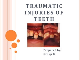
- 1. TRAUMATIC I N J U R I E S O F TEETH Prepared by: Group B
- 2. ➢Case History ✓Chief complaint ✓History of present illness ✓Medical History Traumatic Injuries of Teeth
- 3. ➢Clinical Examination ✓External Examination ✓Soft Tissues ✓Facial Skeleton ✓Teeth and Supporting Structures Traumatic Injuries of Teeth
- 5. ➢(1) Concussion ➢(2) Luxation ➢(3) Fracture Traumatic Injuries of Teeth
- 6. ➢tooth is not mobile ➢not displaced ➢periodontal ligament (PDL) absorbs injury + inflammed ✓leaves tooth tender to biting pressure + percussion Concussion
- 7. ➢Visual sign: ✓not displaced ➢Percussion test: ✓tender to touch or tapping ➢Mobility test: ✓no increased mobility Concussion
- 8. ➢Pulp Sensibility Test: ✓positive result ✓it is important in assessing future risk of healing complications ✓lack of response to the test indicates an increased risk of later pulp necrosis Concussion
- 9. ➢Radiographic findings: ✓no radiographic abnormalities ➢Radiographs: ✓occlusal ✓periapical ✓lateral view from mesial + distal aspect of tooth in question Concussion
- 10. ➢Treatment Objectives: ✓usually there is no treatment ➢Treatment: ✓monitor pulpal condition for at least 1 year Concussion
- 11. ➢Patient Instructions: ✓soft food for 1 week ✓brush with soft bristle ✓rinse with chlorhexidine 0.1% to prevent plaque accumulation Concussion
- 12. ➢tooth is displaced in a labial, lingual or lateral direction ➢PDL is usually torn ➢fractures of supporting alveolus may occur Luxation
- 13. ➢similar to extrusion injuries ✓partial or total separation of periodontal ligament Luxation
- 14. ➢Visual sign: ✓displaced, usually in a palatal/lingual or labial direction ➢Percussion test: ✓usually gives a metallic (ankylotic) sound ➢Mobility test: Luxation
- 15. ➢Pulp Sensibility Test: ✓likely give a lack of response except for teeth with minor displacement ✓test is important in assessing risk of healing complications ✓positive result at the initial examination indicates a reduced risk of future pulp necrosis Luxation
- 16. ➢Radiographic findings: ✓widened periapical ligament space best seen on occlusal or eccentric exposures ➢Radiographs: ✓occlusal ✓periapical ✓lateral view from mesial + distal aspect of tooth in question Luxation
- 17. ➢Treatment Objective: ✓reposition + splint a displaced tooth to facilitate pulp + periodontal ligament healing Luxation
- 18. ➢Treatment: ✓rinse the exposed part of root surface with saline before repositioning ✓apply local anesthesia ✓reposition tooth with forceps or with digital pressure to disengage it from its bony socket Luxation
- 19. ➢Treatment: ✓gently reposition it into its original position ✓stabilize the tooth for 4 weeks using a flexible splint ✓4 weeks is indicated due to associated bone fracture Luxation
- 20. ➢Patient Instructions: ✓soft food for 1 week ✓brush with soft bristle ✓rinse with chlorhexidine 0.1% to prevent plaque accumulation Luxation
- 21. ➢Ellis and Davey classification of crown fracture is useful in recording extent of damage to crown ✓ Class I – simple fracture of crown involving little or no dentin ✓Class II – extensive fracture of crown involving considerable Fracture
- 22. ✓ Class III – extensive fracture of crown with an exposure of dental pulp ✓Class IV – loss of entire crown Fracture
- 23. ➢Enamel Fracture ➢Enamel-Dentin Fracture ➢Enamel-Dentin-Pulp Fracture ➢Root Fracture Fracture
- 24. ➢fracture confined to the enamel with loss of tooth structure Enamel Fracture
- 25. ➢Visual sign: ✓visible loss of enamel ✓no visible sign of exposed dentin ➢Percussion test: ✓not tender ✓if tenderness is observed evaluate tooth for a possible luxation or root fracture injury Enamel Fracture
- 26. ➢Mobility test: ✓normal mobility ➢Sensibility test: ✓usually positive ✓test may be negative initially indicating transient pulpal damage Enamel Fracture
- 27. ➢Sensibility test: ✓monitor pulpal response until definitive pulpal diagnosis can be made ✓test is important in assessing risk of future healing complications ✓lack of response at initial examination indicates an increased risk of later pulpal necrosis Enamel Fracture
- 28. ➢Radiographic findings: ✓enamel lost is visible ➢Radiographs: ✓occlusal ✓periapical ✓recommended to rule out possible presence of root fracture or a luxation injury Enamel Fracture
- 29. ➢Treatment: ✓if tooth fragment is available, it can be bonded to the tooth ✓grinding or restoration with composite resin depending on extent + location of fracture Enamel Fracture
- 30. ➢fracture confined to enamel + dentin with loss of tooth structure, but not involving pulp Enamel-Dentin Fracture
- 31. ➢Visual sign: ✓visible loss of enamel + dentin ✓no visible sign of exposed pulp tissue ➢Percussion test: ✓not tender ✓if tenderness is observed evaluate tooth for a possible luxation or root fracture injury Enamel-Dentin Fracture
- 32. ➢Mobility test: ✓normal mobility ➢Sensibility test: ✓usually positive ✓test may be negative initially indicating transient pulpal damage Enamel-Dentin Fracture
- 33. ➢Sensibility test: ✓monitor pulpal response until definitive pulpal diagnosis can be made ✓test is important in assessing risk of future healing complications ✓lack of response at initial examination indicates an increased risk of later pulpal necrosis Enamel-Dentin Fracture
- 34. ➢Radiographic findings: ✓enamel-dentin lost is visible ➢Radiographs: ✓occlusal ✓periapical ✓recommended to rule out displacement or possible presence of root fracture Enamel-Dentin Fracture
- 35. ➢Treatment: ✓if tooth fragment is available, it can be bonded to the tooth ✓otherwise perform provisional treatment by covering exposed dentin with glass ionomer or a permanent restoration using a bonding agent + composite resin Enamel-Dentin Fracture
- 36. ➢(Complicated Crown Fracture) ➢a fracture involving enamel + dentin with loss of tooth structure + exposure of pulp Enamel-Dentin-Pulp Fracture
- 37. ➢Visual sign: ✓visible loss of enamel + dentin ✓exposed pulp tissue ➢Percussion test: ✓not tender ✓if tenderness is observed evaluate tooth for a possible luxation or root fracture injury Enamel-Dentin-Pulp Fracture
- 38. ➢Mobility test: ✓normal mobility ➢Sensibility test: ✓usually positive Enamel-Dentin-Pulp Fracture
- 39. ➢Sensibility test: ✓test is important in assessing risk of future healing complications ✓lack of response at initial examination indicates an increased risk of later pulpal necrosis Enamel-Dentin-Pulp Fracture
- 40. ➢Radiographic findings: ✓lost of tooth substance is visible ➢Radiographs: ✓occlusal ✓periapical ✓recommended to rule out displacement or possible presence of luxation or root fracture Enamel-Dentin-Pulp Fracture
- 41. ➢Treatment: ✓if young patients with open apices, it is very important to preserve pulp vitality by pulp capping or partial pulpotomy in order to secure further root development ✓this treatment is also treatment of choice in patients with closed apices Enamel-Dentin-Pulp Fracture
- 42. ➢Treatment: ✓Calcium hydroxide compunds + MTA are suitable materials for such procedures ✓in older patients with closed apices + luxation injury with displacement, root canal treatment is usually treatment of choice Enamel-Dentin-Pulp Fracture
- 43. ➢fracture involving: ✓enamel ✓dentin ✓cementum ✓with loss of tooth structure ✓but not exposing pulp Crown-Root Fracture without pulp involvement
- 44. ➢Visual sign: ✓crown fracture extending below gingival margin ➢Percussion test: ✓tender Crown-Root Fracture without pulp involvement
- 45. ➢Mobility test: ✓coronal fragment mobile ➢Sensibility test: ✓usually positive for apical fragment Crown-Root Fracture without pulp involvement
- 46. ➢Radiographic findings: ✓apical extension of fracture usually not visible ➢Radiographs: ✓occlusal ✓periapical ✓recommended to detect fracture lines in root ✓cone beam exposure can reveal whole fracture extension Crown-Root Fracture without pulp involvement
- 47. ➢Treatment: ✓Fragment removal only • removal of superficial coronal crown-root fragment • subsequent restoration of exposed dentin above gingival level Crown-Root Fracture without pulp involvement
- 48. ➢Treatment: ✓Fragment removal + gingivectomy (sometimes ostectomy) • removal of coronal segment with subsequent endodontic treatment + restoration with a post-retained crown Crown-Root Fracture without pulp involvement
- 49. ➢Treatment: ✓Orthodontic extrusion of apical fragment • removal of coronal segment with subsequent endodontic treatment + orthodontic extrusion of remaining root with sufficient length after extrusion to support a post- retained crown Crown-Root Fracture without pulp involvement
- 50. ➢Treatment: ✓Surgical extrusion • removal of mobile fractured fragment • subsequent surgical repositioning of root in a more coronal position Crown-Root Fracture without pulp involvement
- 51. ➢Treatment: ✓Decoronation (root submergence) • implant solution is planned, root fragment may be left in situ after in order to avoid alveolar bone resorption • thereby maintaining volume of alveolar process for later implant installation Crown-Root Fracture without pulp involvement
- 52. ➢Treatment: ✓Extraction • with immediate or delayed implant-retained crown restoration or a coventional bridge • fractures with severe apical extension, the extreme being a vertical fracture Crown-Root Fracture without pulp involvement
- 53. ➢fracture involving: ✓enamel ✓dentin ✓cementum ✓with loss of tooth structure ✓exposure of pulp Crown-Root Fracture with pulp involvement
- 54. ➢Visual sign: ✓crown fracture extending below gingival margin ➢Percussion test: ✓tender Crown-Root Fracture with pulp involvement
- 55. ➢Mobility test: ✓coronal fragment mobile ➢Sensibility test: ✓usually positive for apical fragment Crown-Root Fracture with pulp involvement
- 56. ➢Radiographic findings: ✓apical extension of fracture usually not visible ➢Radiographs: ✓occlusal ✓periapical ✓cone beam exposure can reveal whole fracture extension Crown-Root Fracture without pulp involvement
- 57. ➢Treatment: ✓Fragment removal + gingivectomy (sometimes ostectomy) • removal of coronal segment with subsequent endodontic treatment + restoration with a post-retained crown Crown-Root Fracture with pulp involvement
- 58. ➢Treatment: ✓Orthodontic extrusion of apical fragment • removal of coronal segment with subsequent endodontic treatment + orthodontic extrusion of remaining root with sufficient length after extrusion to support a post- retained crown Crown-Root Fracture with pulp involvement
- 59. ➢Treatment: ✓Surgical extrusion • removal of mobile fractured fragment • subsequent surgical repositioning of root in a more coronal position Crown-Root Fracture with pulp involvement
- 60. ➢Treatment: ✓Decoronation (root submergence) • implant solution is planned, root fragment may be left in situ after in order to avoid alveolar bone resorption • thereby maintaining volume of alveolar process for later implant installation Crown-Root Fracture with pulp involvement
- 61. ➢Treatment: ✓Extraction • with immediate or delayed implant-retained crown restoration or a coventional bridge • fractures with severe apical extension, the extreme being a vertical fracture Crown-Root Fracture with pulp involvement
- 62. ➢fracture confined to the root of tooth involving: ✓cementum ✓dentin ✓pulp Root Fracture
- 63. ➢Visual sign: ✓coronal segment may be mobile ✓some cases displaced ✓transient crown discoloration (red or gray) may occur ✓bleeding from gingival sulcus may be noted Root Fracture
- 64. ➢Percussion test: ✓tooth may be tender ➢Mobility test: ✓coronal segment may be mobile Root Fracture
- 65. ➢Sensibility test: ✓the test is important in assessing risk of healing complications ✓a positive sensibility test at the initial examination indicates a significantly reduced risk of later pulpal necrosis Root Fracture
- 66. ➢Sensibility test: ✓may give negative results initially ✓indicating transient or permanent neural damage ✓pulp sensibility test is usually negative for root fractures except for teeth with minor displacements Root Fracture
- 67. ➢Radiographic findings: ✓root fracture line is usually visible ✓fracture involves root of the tooth in a horizontal or diagonal plane Root Fracture
- 68. ➢Treatment: ✓rinse exposed root surface with saline before repositioning ✓if displaced, reposition the coronal segment of the tooth as soon as possible ✓check that correct position has been reached radiographically Root Fracture
- 69. ➢Treatment: ✓stabilize the tooth with flexible splint for 4 weeks ✓if the root fracture is near cervical area of the tooth stabilization is beneficial for a longer period of time (upto 4 months) Root Fracture
- 70. ➢Treatment: ✓monitor healing for at least 1 year to determine pulpal status ✓if pulp necrosis develops, then root canal treatment of the coronal tooth segment to the fracture is indicated Root Fracture
- 71. References: ❖ Books ➢ McDonald, Avery et al: Dentistry for the Child and Adolescent • (pages 458-459) ❖ Internet ➢http://www.dentaltraumaguide.org