SlideShare uma empresa Scribd logo
1 de 120
Embryology of Spine
n spinal dysraphism
By Dr Viralkumar Vasani
Moderator: Dr Satish S
• Formation and separation of the germ layers
• Dorsal and ventral induction phases, and
• Phases of neurogenesis,
• Migration,
• Organization and
• Myelination.
• During week 1 (stages 2–4) the blastocyst is formed,
• During week 2 (stages 5 and 6) implantation occurs
and the primitive streak is formed,
• Formation of the notochordal process and the
beginning of neurulation (stages 7– 10).
• Somites first appear at stage 9. The neural folds
begin to fuse at stage 10,
• Rostral and caudal neuropores close at stages11 and
12, respectively
Some Embryological fact
• first four embryonic weeks are also described as the period
of blastogenesis,
• Fifth to eighth weeks as the period of organogenesis
• At the junction of trimesters 1 and 2, the fetus of about 90
days has a greatest length of 90 mm, whereas at the
junction of trimesters 2 and 3, the fetus is
about 250 mm in length and weighs
approximately 1,000 g
• Newborn brain weighs 300–400g at full term. Male brains
weigh slightly more than those of females but, in either
case, the brain constitutes 10% of the body weight
Beginning of NS
• Gastrulation - birthday of the nervous system. Time
when
• 1)bilateral symmetry
• 2) three axes are established in the body of all
vertebrates
• 3) Neuroepithelium can first be identified and
distinguished from primitive germinal tissues
• Brain and spinal cord arise from an area of the ectoderm
known as the neural plate.
• The folding of the neural plate, leading to successively
the neural groove and the neural tube, is called primary
neurulation.
• The caudal part of the neural tube does not arise by
fusion of the neural folds but develops from the so-called
caudal eminence. This process is called secondary
neurulation
Stage Length Days External features Internal Feature related to CNS
Primary Gastrulation
• primitive streak (PS), midline structure, at the caudal end of the embryo
• Hensen’s node - cranial end of the primitive streak
• primitive groove, runs in the midline within the PS; the cranial end of the
primitive groove is the primitive pit.
• cells of the epiblast migrate toward the PS and invaginate through the primitive
groove. The first cells to ingress (while the PS is still elongating) are endodermal
cells, which displace the hypoblast cells laterally and form prospective
endoderm
• Displaced hypoblast cells form extraembryonic tissues.
• As the PS regresses, mesodermal cells ingress through the PS between the
epiblast and newly formed endoderm and become the mesoderm.
• The remaining epiblast cells spread out to replace the cells that have ingressed
through the primitive groove and thereby form both the neuroectoderm (the
neural tube) and the cutaneous ectoderm (skin
Developing spinal cord
•
• Neural tube closure
• Occurs craniocaudally from initial point of
contact
• Initiated in region of cervical spinal cord (5
somite stage )
• Posterior neuropore closes at day 25-27 (day 25 -
27)
• Posterior neuropore approximately located at S2
level
• Whole process known as neurulation (primary
neurulation )
Forces for neural tube
formation
• Mesoderm appears to be important for orien-
tation but not for closure of the neural tube.
• Expansion of the surface epithelium of the
embryo is the principal extrinsic force for folding
of the neuroepithelium to form the neural tube
• Intrinsic forces of the neuroepithelium, the cells of
the floor plate have a wedge shape—narrow at
the apex and broad at the base—that facilitates
bending.
• The ependymal cells that form the floor plate are the
first neural cells to differentiate, and they induce
growth of the parenchyma of the ventral zone more
than the dorsal regions
• mechanical effect may also facilitate curving of the
neural placode (Flat plate).
• The direction of proliferation of new cells in the
mitotic cycle, determined in part by the orientation of
the mitotic spindle, becomes another mechanical
force shaping the neural tube that is rostrocaudal
orientation of most mitotic spindles
• Adhesion molecules
Role of Hensen’s Node
• as the “organizer” of the embryo.
• As the PS elongates, prospective endodermal cells
within Hensen’s node migrate through the primitive pit.
• As the PS regresses, specialized mesodermal cells,
prospective notochordal cells, migrate through the
primitive pit and form the notochordal process in the
midline between the neuroectoderm and endoderm.
• Notochord plays an imp role in directing subsequent
neurulation.
• Between PODs 18 and 20, the notochordal process fuses
(intercalates) with the underlying endoderm to form the
notochordal plate. The notochordal plate is therefore incorporated
into the roof of the yolk sac, with the notochordal canal becoming
continuous with the yolk sac.
• Intercalation results in a direct communication, the primitive
neurenteric canal, that connects the amnionic and yolk sacs at the
level of Hensen’s node.
• The neurenteric canal persists for about 3 days, at the end of which
the notochordal plate folds dorsoventrally and separates
(excalates) from the endoderm and the neurenteric canal is
obliterated.
• Thereafter, the true notochord exists as a solid rod of notochordal
cells
Defination
Formation of neural tube
• Human neuroectoderm visible by day 16
• Pseudo stratified epithelium overlying the notochord
• Neural groove visible by day 17-19
• Neural folds (day 19 -21)
• Separate from the overlying ectoderm (dysjunction) and fuse to form the
neural tube.
• The neural folds separate from the cutaneous ectoderm and fuse to form a
closed neural tube between POD 21 and 23.
• Closure generally involves apposition and fusion of first the cutaneous ectoderm
and then the neuroectoderm.
• The first part of the human neural tube to close is the region of the caudal
rhombencephalon or cranial spinal cord, usually when five pairs of somites are
present.
• Cranial neural tube closure may involve the
coordinated interaction of as many as four
waves of discontinuous neural tube closure.
• The spinal cord closes craniocaudally in a linear
manner from the point of initial closure to the
caudal neuropore.
• Cranial neuropore closes between POD 23 and
25, whereas the caudal neuropore closes
between POD 25 and 27.
• Formation of neural crest
• Neural crest cells originate at the junction of
surface ectoderm and neuroectoderm
• First visible on day 19-21
• Continue forming till day 32
• Cranial neural crest cells contribute to the
branchial arches and the arachnoid and pia
mater of the cranium.
• spinal neural crest arises only after closure of the
neural tube.
• Spinal neural crest cells undergo terminal
differentiation into
• melanocytes of the body wall and limbs,
• Schwann cells investing the peripheral nerves,
• spinal cord meninges,
• dorsal root and autonomic ganglion cells of the
spinal nerves
• adrenal medulla.
• Secondary neurulation
• Caudal cell mass extends from the posterior
neuropore to the cloacal membrane (day 25-27)
• Pluripotent cells derived from the primitive
streak.
• Contains neurons, neural crest cells and glial
cells and ependymal cells
• The secondary neural tube is initially solid and
subsequently undergoes cavitation, eventually
forming the tip of the conus medullaris and
filum terminale by a process called retrogressive
differentiation.
• Three processes are responsible for further
development of the CCM
• Condensation
• Canalisation
• Retrogressive differentiation
• Final derivatives
• Distal sacral nerve roots,conus
medullaris,terminal ventricle,filum terminale
and sacrococcygeal remnant
• Secondary neurulation continues till day 52
• Ascent of the conus medullaris
• Process beginning day 43-48 and continuing into
post natal life probably
• 2 distinct processes
• Retrogressive differentiation of the caudal neural
tube (prior to day 54)
• Disparity between growth of spinal cord and
vertebral column.
• Most rapid ascent between 8 to 25 weeks of
gestation.
• At birth conus is at adult level of L1-L2 level.
• Low lying conus- below mid body of L2.
NTDs
• neural tube defects (NTDs), or localized failure of
primary neurulation, that can arise through one of
two mechanisms.
• The “nonclosure theory” proposes that NTDs
represent primary failure of neural tube closure.
• The “overdistention theory,” introduced in 1769
by Morgagni and popularized by Gardner
proposes that NTDs arise through overdistention
and rupture of a previously closed neural tube.
Embryological classification Of Cong. dis
Spinal Dysraphism
• Spinal dysraphism refers to a spectrum of disorders in
which there is defective midline closure of neural, bony,
or other mesenchymal tissues.
• The “open” dysraphic states include myelocele,
myelomeningocele, hydromyelia, Chiari II malformations,
hemimyelocele, and myeloschisis.
• The closed dysraphic states include entities such as
dermal sinus, lipomyelomeningocele, tight filum
terminale, meningocele, myelocystocele, diastemato-
myelia, neurenteric cyst, slit notochord, and
developmental tumors such as spinal lipomas
Classification of
Dysraphism
Spinal Dysraphism
Open
Myelomeningocele
Myelocele
Hemi-
MY/MYM
E
Closed With S/C
Mass
Lipomyelomeningocele
Meningocele
Lipomyelocele
Myelocys
tocele
Terminal
Myelocystocele
Closed without S/C
Mass Simple Dysraphic
state
Dermal
Sinus
Intradural Lipoma
Complex
Dysraphic State
Neuroenteric
cyst
Caudal
Agenesis
Segmental Vertebral
anamolies
Myelomeningoceles
• Disorder resulting from defective primary
neurulation
• 98% of all Spinal dysraphism
• Incidence
• 0.4 per 1000 live births
• Racially variable
• 85% caudal thoraco lumbar spine, 10 % in the torax
and the rest cervical
• 80-90 % associated with hydrocephalus and Chiari
• Trisomy 13 and trisomy 18
Associated defects
• Brain stem defect includes
• Medullary kinking, tectal beaking, and intrinsic nuclei
abnormalities
• Supratentorial abnormalities include
• partial or complete dysgenesis of the corpus callosum,
• polymicrogyria, a large massa intermedia, and gray matter
heterotopia.
• Mesodermal development of the skull
• small posterior fossa, short clivus,
• low-lying tentorium and torcular Herophili, wide incisura, and
• enlarged foramen magnum.
• Lückenschädel, or craniolacunia (scalloping of the skull
bones)
Cause of neuro-deterioration
• symptomatic hydrocephalus,
• syringomyelia
• Retethering of cord.
• Chiari II malformation, Risk factors
• mostly due to shunt malfunction resulting in
hydrocephalus
Patho Anatomy
• Failure of neural tube closure results in an exposed
neural placode. The groove in the center of the placode is
the remnant of the central canal.
• The spinal roots exit from the anterior surface of the
placode such that the ventral roots lie medially and the
dorsal roots lie laterally.
• The dura fuses with the defect in the fascia laterally.
Functional neural tissue + either caudal to the placode or
in the nerve roots exiting from the placode.
1. Axial schematic of myelomeningocele shows neural placode (star) protruding above skin
surface due to expansion of underlying subarachnoid space (arrow).
2. Axial T2-weighted MR image in 1-day-old boy shows neural placode (black arrow)
extending above skin surface due to expansion of underlying subarachnoid space (white
arrow), which is characteristic of myelomeningocele.
MR Image of
myelomeningocele
• Sagittal T2-weighted
MR image from same
patient with
myelomeningocele
shows neural placode
(white arrow)
protruding above skin
surface due to
expansion of
underlying
subarachnoid space
(black arrow).
Clinical Examination
• signs of myelopathy, including hyperreflexia and clonus, because they often
have an incomplete functional spinal cord transection in 2/3rd
• sensory level by stimulus - distally to proximally until the infant grimaces.
• A stimulus is applied above the sensory level, and the distal-most voluntary
motion seen for motor level determination
• With an L1-3 level, the infant has hip flexion with extended knees and clubfeet.
• The presence of intact hip adduction, hip flexion, and knee extension with
inverted feet is indicative of an L2-4 level.
• With an L5-S2 level, the infant has hip adduction, knee extension, and knee
flexion with dorsiflexed feet.
• Infants with a sacral level may appear intact except for weakness of plantar
flexion and rocker-bottom feet.
• A flaccid pelvic floor and patulous anus are often present
• Prenatal diagnosis
• Maternal serum Alpha feto protein : initial
screening test
• High resolution fetal ultrasonography.
• Can also demonstrate hydrocephalus and Chiari II
abnormality (lemon and banana sign)
• Amniocentesis : if MSAFP and USG are
suggestive
• Ach esterase levels along with AFP
• AFP can increase in other developmental
anomalies of the gut and kidneys.
“lemon sign” Normal fetal head
D/D
• At least 22 other fetal abnormalities besides
myelomeningocele increase MSAFP levels.
• Abdominal abnormalities such as omphalocele,
cloacal exstrophy, esophageal atresia, annular
pan- creas, duodenal atresia, and gastroschisis and
• urologic abnormalities such as congenital
nephrosis, polycystic kidneys, urinary tract
obstruction, and renal
• sacrococcygeal cystic teratoma
Preop evaluation- Clinical
• General
• Repaired within 72 hrs
• Enteral feeding avoided to prevent fecal soiling
of placode
• Prone position ,saline dressings
• Neurosurgical
• Sensory level determined
• Motor evaluation – distal most voluntary motion
evaluated. Limb abnormalities documented.
• Anal tone and anal reflex evaluated
• Ventricular size documented with preop USG and NCCT head.
• Imaging study shows that differentiating feature between a
myelomeningocele and myelocele is the position of the neural placode
relative to the skin surface
• The neural placode protrudes above the skin surface with a
myelomeningocele and is flush with the skin surface with a
myelocele
• Observe for symptoms of Chiari II
• Renal evaluation
• 90 % have neurogenic bladder.
• All should have preop Renal ultrasound for detecting severe
anomalies.
• CIC if fails to void.
Myelocele
1. Axial schematic of myelocele shows neural placode (arrow) flush with skin surface.
2. Axial T2-weighted MR image in 1-day-old girl shows exposed neural placode (arrow) that
is flush with skin surface, consistent with myelocele. There is no expansion of underlying
subarachnoid space.
Repair
• Timing of repair:
• Myelomeningocele repair can be performed safely up to 72
hours after birth
• Delayed repair – Increases chance of ventriculitis by 5 times,
• shunt infection developed in about 75%, and the mortality was
13%
• In case Delay
• 1) Cultures from the neural placode – No growth – go ahead n
repair
• If infection +, -external ventricular drainage and appropriate
antibiotics until the infection clears – Then repair
• Shunt before repair – High chance of shunt inf./ Meningitits – IQ
impairment (due to inf)
• PREPARATION
• Intraoperatively avoid hypothermia, hypovolemia, and hypoglycemia.
• A doughnut-shaped sponge - to protect the myelomeningocele while
intubation.
• If severe Hydrocephalus - CSF diversion before closure of the
myelomeningocele - to minimize pressure on the
myelomeningocele dural closure
• entire back and flanks are prepared and draped to facilitate
extensive closure if needed.
• Contact between povidone- iodine solution and the neural placode
should be avoided
• purposes of myelomeningocele repair are to protect the
functional spinal cord tissue, prevent loss of CSF, and
minimize the risk for meningitis by reconstructing the neural
tube and its coverings.
• The margin between the arachnoid of the neural placode and
the dystrophic epidermis, or the junctional zone, is the site of
the initial incision.
• The goal is to free the neural placode from the surrounding
junctional zone circumferentially.
• Duraplasty with thoracolumbar fascia or another dural
substitute is performed when necessary to prevent leakage
of CSF.
Post op care
• Post op antibiotics
• Prevention of fecal contamination of wound
• Nurse in Trendlenberg’s
• Observe for Hydrocephalus – shunt if HCP present
• Complications
• Superficial wound dehiscence
• Meningitis
• Symptomatic chiari
• Ensure functioning shunt
• Hindbrain decompression
Prognosis
• 10 to 15% succumb <6yr of age even with Multi specialty
aggressive approach
• >95% Lives >2 years
• 8 to 17 % will have urinary control rest on Drugs / CIC
• >87% will have social fecal incontinence
• L3 function allows one to stand erect, and L4 and L5 function
allows ambulation – During the first decade, approximately
60% of children with spina bifida are community ambulators,
without or with assistive devices (including wheelchairs) –
Reduces to 17% in teenagers
• IQ stays N if no inf. – only <10% economically independent
Occult spinal dysraphism
• Aka Closed dysraphism
• Of 2 types ; with/without s/c mass
• Congenital spinal defects covered by intact skin
• Causative lesions
• Fatty filum terminale
• Lipomyelomeningocele
• Split cord malformations type I and II
• Inclusion lesions (dermoid, dermal sinus tract)
• Neurenteric cyst
• Myelocystocele
Closed / Occult type
• Closed With S/C Mass
• Lipomyelomeningocele
• Meningocele
• Lipomyelocele
• Myelocystocele
• Terminal Myelocystocele
• Closed without S/C Mass
• Simple Dysraphic state
• Dermal Sinus
• Intradural Lipoma
• Complex Dysraphic State
• Diastometamyelia
• Neuroenteric cyst
• Caudal Agenesis
• Closed Spinal Dysraphisms With a Subcutaneous Mass
1. Lipomas with a dural defect—
• include both lipomyeloceles and lipomyelomeningoceles.
• result from a defect in primary neurulation whereby mesenchymal tissue enters the
neural tube and forms lipomatous tissue.
• characterized clinically by the presence of a subcutaneous fatty mass above the
intergluteal crease.
• The main differentiating is the position of the placode–lipoma interface. With a
lipomyelocele, the placode–lipoma interface lies within the spinal canal where as it is
outside of the spinal canal in case of lipomyelomeningocele, it lies due to expansion of
the sub arachnoid space .
2. Meningocele—Herniation of a CSF-filled sac lined by dura and arachnoid mater
is referred to as a meningocele. The spinal cord is not located within a
meningocele but may be tethered to the neck of the CSF-filled sac.
• Posterior meningoceles herniate through a posterior spina bifida (osseous defect of
posterior spinal elements) and are usually lumbar or sacral in location but also can
occur in the occipital and cervical regions.
• Anterior meningoceles are usually presacral in location but also can occur elsewhere
Manifestations of occult
spinal dysraphism
Cutaneous stigmata Orthopedic deformities Urologic problems
Asymmetric gluteal cleft Foot or leg deformities Neurogenic bladder
Capillary hemangioma Scoliosis UTIs
Subcutaneous lipomas Sacral agenesis Incontinence
Hypertrichosis Delay in toilet training
Dermal sinus tract
Cutis aplasia
Neurological signs and symptoms in
different age groups
Infants Toddler Older children Young adults
Decreased
spontaneous leg
movements
Delayed walking Asymmetric motor/
sensory
development
Back pain
Absent reflexes Abnormal gait Back/leg pain Leg cramping/pain
Leg atrophy UMN signs Spasticity
Foot asymmetry Painless ulceration Hyperreflexia
Decreased urinary
stream
Bowel/bladder
incontinence.
Plain radiological findings
Structure Findings
Lamina Fusion defects,midline defects,abnormal
spinous processes
Vertebral bodies Hemivertebrae, Butterfly vertebrae, Block
vertebrae, Midline cleft defects, canal
stenosis
Disk space Congenital narrowing
Pedicles Flattening, thinning
Widening of spinal canal Interpedicular widening, scalloping of
posterior border, Midline bony spur.
Failure of development Reduced number of vertebral bodies,
Absence of parts of vertebrae, sacral
dysjunction
Spinal curvature Scoliosis, kyphosis, lordosis.
Lipomyelomeningoceles
• Lipoma tethering the cord to the
subcutaneous tissue
• Fascial, spinous and dural defect
• Lipoma cord interface distracted out of the
spinal canal by traction created by tethering
LMMC
Axial schematic of
lipomyelomeningocele shows
placode–lipoma interface
(arrow) lies outside of spinal
canal due to expansion of
subarachnoid space.
Axial T1-weighted MR image
in 18-month-old boy shows
lipomyelomeningocele
(arrow) that is differentiated
from lipomyelocele by
location of placode–lipoma
interface outside of spinal
canal due to expansion of
subarachnoid space.
LMC- Lipomyelocele
• Axial schematic of lipomyelocele shows placode–lipoma interface lies within
spinal canal.
• Axial T2-weighted MR image in 3-year-old girl shows placode–lipoma
interface within spinal canal, characteristic for lipomyelocele.
Lipomyelocele
Sagittal T1-weighted MR image in 3-
year-old girl with lipomyelocele
shows subcutaneous fatty mass
(black arrow) and placode–lipoma
interface (white arrow) within spinal
canal.
Chapman’s classification
of LMM
• Classification
• Type I (dorsal lipoma)
• Type II (transitional lipoma)
• Type III ( terminal lipoma)
• Dorsal lipoma (Type I)
• Fibrolipomatous stalk tethering cord proximal to
conus
• Usually at middle lumbar to lumbo sacral level
• Dorsal spinal cord dysraphic at site of attachment
of lipoma
• Site of attachment medial to the dorsal root entry
• Normal spinal cord distal to myeloschisis.
• Roots lie within the subarachnoid space.
Dorsal lipoma
• Caudal or terminal lipomas (type III)
• Directly from conus medullaris or filum
terminale
• Largely or wholly intradural
• Nerve roots entangled in the lipoma
• Lipoma cord interface caudal to the dorsal
root entry zone.
• Filum may be fatty, thickened and sometimes
attached to subcuatneous tissue ( sacral
dimple).
Terminal lipomas
• Transitional lipomas
• Share the charcteristics of both Type 1 and
type 2.
• No normal spinal cord distal to lipoma
attachment
• Initially dorsal roots may be separate but
caudally become enmeshed into the lipoma.
• Frequently assymmetric attachment to cord.
Transitional lipomas
Abnormal embryology
LMM
• Usually a disjunction in timing of neural
tube closure and cutaneous ectoderm
closure
• Elements of the ectoderm become
incorporated into the incompletely closed
neural tube.
Clinical features
• Subcutaneous masses over the back
• Stigmata of occult dysraphism
• Hypertrichosis
• Hemangioma
• Hypo/ hyperpigmented patch
• Dermal pit or sinus
• Atretic meningocele
• Assymmetric gluteal cleft
• Inexorable symptomatic progression - in
untreated cases
• Risk of precipitous neurologic deterioration
• Orthopedic syndrome
• Limb length discrepancy, high pedal arches,
hammer toes, calcaneovarus/ valgus foot
deformity.
• Urologic syndrome
• Urinary incontinence, post void dribbling, urgency,
frequency
• Intractable pain in the legs, back, pelvis or
perineum.
Indication for surgical
repair
• Asymptomatic infant older than 2 months
• Presence of orthopedic, pain or urologic
syndrome
• Neurological symptoms
• Prior to corrective spinal surgery.
Principal Goal of surgery
• Detethering of spinal cord
• Decompression of the cord by removing as much
lipoma as possible
• Reconstruct the spinal cord and dural sac
• Preservation of the functional tissue
• Surgical principles
• Relationship between the lipoma-cord interface and
dorsal roots to be established
• Conservative excision of the lipoma to avoid injury to
the cord/ exiting roots.
• Complications of surgical repair
• Early – CSF leak/ pseudo meningoceles & New
Neurological deficit
• Late – retethering of the cord
• Mostly presenting between 3-8, 11-22 months after
surgery
• Upto 20% cases may demonstrate retethering
• Diagnosis primarily clinical.
• Aseptic meningitis from host-graft inflammation
• meningitis,
• Intradural abscess,
• wound infection, and wound breakdown
Lipoma of the terminal
filum
 Less severe form of Occult SD
 More than 2 mm thickness of the filum on MR imaging
 Frequently assosciated with sacral/gluteal cleft dimples.
 May be associated with VATER association, imperforate
anus, cloacal extrophy and other urogenital
abnormalities.
 Sometimes a/w sacral agenesis
 Reflects defective secondary neurulation
 Clinical presentation
 Orthopedic
 Urologic
 Pain
 All asymptomatic infants and symptomatic adults are
surgical candidates.
 Surgical procedure is the exposure of filum through
lumbosacral laminectomy or interlaminar approach
 The filum identified separated from nerve roots and
cut.
Closed SD without S/C Mass
• Closed without S/C Mass
• Simple Dysraphic state
• Intradural Lipoma
• Filar lipoma,
• Tight filum terminale,
• Persistent terminal ventricle
• Dermal Sinus
• Complex Dysraphic State
• Diastometamyelia
• Neuroenteric cyst
• Caudal Agenesis
• Segmental Vertebral anamolies
• An intradural lipoma - a lipoma located along the dorsal midline that is contained within
the dural sac. No open spinal dysraphism is present.
• M.C site: lumbosacral - present with tetheredcord syndrome, a clinical syndrome of
progressive neurologic abnormalities in the setting of traction on a lowlying conus
medullaris.
• Fibrolipomatous thickening of the filum terminale is referred to as a filar lipoma. On
imaging, a filar lipoma _ - Normal variant if there is no clinical evidence of tetheredcord
syndrome
• Tight filum terminale is characterized by hypertrophy and shortening of the filum
terminale causing tethering of the spinal cord and impaired ascent of the conus
medullaris. The conus medullaris is low lying relative to its normal position that is above
the L2–L3 disc level.
• Persistence of a small, ependymal lined cavity within the conus medullaris is referred to
as a persistent terminal ventricle.
• Location immediately above the filum terminale and lack of contrast enhancement,
which differentiate this entity from other cystic lesions of the conus medullaris.
Intra dural Lipoma
Complex dysraphic states
divided into two categories:
• disorders of midline notochordal integration
• dorsal enteric fistula
• neurenteric cyst
• Diastematomyelia
• disorders of notochordal formation, which include
• caudal agenesis and
• segmental spinal dysgenesis
• Diastematomyelia—Separation of the spinal cord into two
hemicords. The two hemi cords are usually symmetric,
although the length of separation is variable.
• two types of diastematomyelia.
• In type 1, the two hemicords are located within individual
dural tubes separated by an osseous or cartilaginous
septum.
• In type 2, there is a single dural tube containing two hemi
cords, sometimes with an intervening fibrous septum.
Diastematomyelia can present clinically with scoliosis and
Tethering
Split cord malformation
• Longitudinal developmental splitting of the cord over
one or multiple levels
• Two types:
• Both types may be present simultaneously at different
levels
Type I malformations
(formerly diastematomyelia)
are characterized by a bony
septum that cleaves the spinal
canal in the sagittal plane and
a duplicated thecal sac
Type II malformations
(formerly diplomyelia) are
characterized by a cleft cord
within a single dural sac, often
tethered by a fibrous midline
septum to the adjacent dura
(single dural sheath,
hemicords separated by
fibrotic bands
Split cord Malformation
• Exceedingly rare
• Represent 3.8% to 5% of all congenital spinal anomalies.
• prevalence of SCM to be 1 in 5000 (0.02%) live births.
• slight female preponderance, approximately 1.3:1.7
• The peak age is 4 to 7 years,
• second peak between 12 and 16 years - post pubescent
growth spurt.
• Type I SCMs > type II lesions
Pang’s theory of
embryogenesis
Unifide theory
• Pang - unified theory - type I and type II SCM to a single
error in embryogenesis:
• Formation of abnormal fistula through midline embryonic
disc that maintains communication b/w Yolk sac &
amniotic cavity contact b/w ecto + Endoderm
• This fistula causes splitting of Notochord & overlying
neural plate- mostly rostral to henson’s node
Associated Anomaly
• Tethered/low-lying cord (>50%),
• Kyphoscoliosis (44% to 60%),
• Syringomyelia (27.5% to 44%), and
• Spina bifida (11% to 26%)
Clinical signs - symptoms
• “faun’s tail,” consists of a patch of unusually coarse, raised
hair - strong association with type I SCM.
• Capillary hemangioma underlying these hairy patches.
• Nearly 50% of patients have gross (i.e., structural)
asymmetry of the lower extremities-- neuro-orthopedic
syndrome.
• characterized by a triad of
1. limb length discrepancy,
2. muscular atrophy (resulting in secondary weakness), and
3. clubfoot deformity (talipes equinovarus).
• The smaller limb is often ipsilateral to a smaller hemicord.
MRI Findings
• MRI Spine shows
1. presence of two hemi cords, on T1-w sequences.
2. demonstrating the presence of an associated fatty filum, affect one
or both hemicords, a dermal sinus tract, or a terminal lipoma.
• Associated tethering anomalies are present in approximately half
of all patients and in more than 90% of those with a low-lying
conus.
• T2-weighted MRI for
1. categorization of the malformation as type I or type II .
2. hypointense fibrous band in type II SCM.
3. To demonstrate syringomyelia proximal to the cleft, which may
extend into one or both hemicords
Type 1 & Type 2 SCM MRI
MGTMNT
• 85% of patients without intervention suffer
from a progressive neurological deficit versus
only 4.5% after surgical treatment
• Pang too suported prophylactic surgery in
Type 1 malformation.. Whereas in Type 2 both-
some – W&W policy
• Surgery – careful – incision 1 to 2 level more
exposure
Operative
• Operative management
• Surgical detethering of cord by excision of the bony
spur/ division of the fibrous bands
• Avoid damage to the hemi cords during excision of the
spur.
• For type I SCM, the initial laminectomies should be
limited to adjacent levels while initially avoiding
exposure of dura at the level of the midline bony spur.
• Rongeurs or a high-speed drill (or both) - to perform
bilateral paramedian laminectomies ( preserve the
midline lamina and spinous process to prevent any
torque or lateral force from disrupting the bony spur
prematurely.)
• Careful when removing lamina at the level of the SCM in type II
malformations because of the frequent presence of transdural
adhesions - most commonly attached dorsally but ventrally
too. Often, the dura at this level grossly abnormal.
• All non-neural and non- functional adhesive bands should be
transected, beginning dorsally and then gently rolling the
hemicords to one side and transecting any ventral attachments
• Hemicords are typically closely approximated with no clear
intervening plane. Resection of the fibrous band within the
split spinal cord itself is not indicated.
• Any associated tethering lesion (sinus tract, fatty filum, or
terminal lipoma) should also be addressed
Outcome & Complication
Of SCM
Secondary repair of an SCM
in a patient with a
previously closed
myelomeningocele at the
same level - failed wound
healing as high as 10%
Neurenteric cysts
• Persistent neurenteric canal communicating between yolk sac and
amniotic cavity
• Intradural, extramedullary mucosa lined cysts
• Formed from persistents tracts communicating with respiratory and
gut epithelia.
• Neurenteric cysts represent a more localized form of dorsal enteric
fistula.
• These cysts are lined with mucin secreting epithelium similar to the GIT
and located in the cervicothoracic spine anterior to the spinal cord
• Associated with vertebral anomalies
• MRI- demonstrates non- contrast enhancing intradural
extramedullary cyst
• Presentation usually in late years (50-60
years)
• May also present in pediatric age group
• Most common location is cervico- thoracic
• Usually postero-lateral surgical approach
• Complete excision of cyst – long term
symptom free survival.
Dermal sinus tracts
 Abnormal tracts communicating between the skin
and intraspinal compartment.
 Most common- lumbosacral location
 May occur anywhere from nasion to coccyx in
midline
 May be accompanied by other cutaneous stigmata.
 Tract terminates within thecal sac mostly
 Half may have associated dermoids, epidermoids,
teratoma at termination.
• Potential pathway for spread of infection
• Repeated episodes of meningitis with atypical
organisms
• Operative repair consists of complete excision of
the track under prophylactic antibiotic cover.
• Gram positive and gram negative coverage
Meningoceles
• Distinguished from MMCs by absence of
hydrocephalus , chiari malformation or lower
limb abnormalities
• Dural defect through which CSF space and
meninges herniate.
• Concomitant neurocutaneous lesions such as
lipomas, dermal sinus tracts may cause
tethering
• Surgical repair of defect at 4-6 months of age.
Posterior meningocele
Anterior meningocele
• Herniation of meninges and CSF in ventral
location
• Commonly in presacral and lumbosacral region
• Female predominance
• Currarino’s triad- anorectal abnormalities,
presacral mass, sacral bony abnormalities.
• Presacral tumour may be epidermoid, dermoid or
teratoma
• Meningitis by atypical organisms may also occur.
• Repair the dural defect and detether the cord.
Anterior meningoceles
myelocystoceles
• Terminal dilatation of the central canal that
herniates through defective posterior elements
• Expanded spinal cord, CSF, fibrous bands and
meninges and lipomatous elements
• Result from disordered development of the
caudal cell mass
• Associated anomalies of the anorectal system,
lower GI tract and spinal column
Myelocystocele
Herniation of dilated
central canal through
posterior spinal defect.
Terminal Myelocystocele
Terminal myelocystocele—
Herniation of large terminal
syrinx (syringocele) into a
posterior meningocele
through a posterior spinal
defect. The terminal syrinx
component communicates
with the central canal, and
the meningocele
component communicates
with the subarachnoid
space. The terminal syrinx
and meningocele
components do not usually
communicate with each
other.
• Surgical repair attempted because of the
resultant tethering of spinal cord.
• Per-op trumpet shaped distended conus
often adheres to the superficial fat
• Detethering may be difficult.
Thank you

Mais conteúdo relacionado

Mais procurados

Imaging of spinal dysraphism
Imaging of spinal dysraphismImaging of spinal dysraphism
Imaging of spinal dysraphismMuthu Magesh
 
Dandy walker malformation - Hội chứng Dandy Walker
Dandy walker malformation - Hội chứng Dandy WalkerDandy walker malformation - Hội chứng Dandy Walker
Dandy walker malformation - Hội chứng Dandy WalkerVõ Tá Sơn
 
Spinal dysraphism and its management
Spinal dysraphism and its managementSpinal dysraphism and its management
Spinal dysraphism and its managementMukhtar Khan
 
Dandy-Walker Malformations
Dandy-Walker Malformations Dandy-Walker Malformations
Dandy-Walker Malformations Ade Wijaya
 
Presentation1, radiological imaging of pediatric leukodystrophy.
Presentation1, radiological imaging of pediatric leukodystrophy.Presentation1, radiological imaging of pediatric leukodystrophy.
Presentation1, radiological imaging of pediatric leukodystrophy.Abdellah Nazeer
 
Presentation1.pptx, radiological imaging of spinal dysraphism.
Presentation1.pptx, radiological imaging of spinal dysraphism.Presentation1.pptx, radiological imaging of spinal dysraphism.
Presentation1.pptx, radiological imaging of spinal dysraphism.Abdellah Nazeer
 
Presentation1.pptx, radiological imaging of hydrocephalus.
Presentation1.pptx, radiological imaging of hydrocephalus.Presentation1.pptx, radiological imaging of hydrocephalus.
Presentation1.pptx, radiological imaging of hydrocephalus.Abdellah Nazeer
 
Dandy-Walker Malformation: Classification and Management
Dandy-Walker Malformation: Classification and ManagementDandy-Walker Malformation: Classification and Management
Dandy-Walker Malformation: Classification and ManagementDr. Shahnawaz Alam
 
Presentation1.pptx, radiological imaging of congenital anomalies of the spine...
Presentation1.pptx, radiological imaging of congenital anomalies of the spine...Presentation1.pptx, radiological imaging of congenital anomalies of the spine...
Presentation1.pptx, radiological imaging of congenital anomalies of the spine...Abdellah Nazeer
 
Intradural extramedullary mass - a case on MRI
Intradural extramedullary mass - a case on  MRIIntradural extramedullary mass - a case on  MRI
Intradural extramedullary mass - a case on MRIREKHAKHARE
 
Congenital malformations of brain
Congenital malformations of brainCongenital malformations of brain
Congenital malformations of brainabinash66
 
Presentation2.pptx intra cranial cyst
Presentation2.pptx intra cranial cystPresentation2.pptx intra cranial cyst
Presentation2.pptx intra cranial cystAbdellah Nazeer
 
Craniovertebral JUNCTION ANOMALIES
Craniovertebral JUNCTION ANOMALIESCraniovertebral JUNCTION ANOMALIES
Craniovertebral JUNCTION ANOMALIESUphar Gupta
 
congenital brain anomalies
congenital brain anomalies congenital brain anomalies
congenital brain anomalies Dev Lakhera
 

Mais procurados (20)

Imaging of spinal dysraphism
Imaging of spinal dysraphismImaging of spinal dysraphism
Imaging of spinal dysraphism
 
Dandy walker malformation - Hội chứng Dandy Walker
Dandy walker malformation - Hội chứng Dandy WalkerDandy walker malformation - Hội chứng Dandy Walker
Dandy walker malformation - Hội chứng Dandy Walker
 
Spinal dysraphism and its management
Spinal dysraphism and its managementSpinal dysraphism and its management
Spinal dysraphism and its management
 
Craniovertebral anomalies
Craniovertebral anomaliesCraniovertebral anomalies
Craniovertebral anomalies
 
intracerebral calcification
intracerebral calcificationintracerebral calcification
intracerebral calcification
 
Dandy-Walker Malformations
Dandy-Walker Malformations Dandy-Walker Malformations
Dandy-Walker Malformations
 
Chiari malformation
Chiari malformationChiari malformation
Chiari malformation
 
Presentation1, radiological imaging of pediatric leukodystrophy.
Presentation1, radiological imaging of pediatric leukodystrophy.Presentation1, radiological imaging of pediatric leukodystrophy.
Presentation1, radiological imaging of pediatric leukodystrophy.
 
Dandy Walker syndrome
Dandy Walker syndromeDandy Walker syndrome
Dandy Walker syndrome
 
Presentation1.pptx, radiological imaging of spinal dysraphism.
Presentation1.pptx, radiological imaging of spinal dysraphism.Presentation1.pptx, radiological imaging of spinal dysraphism.
Presentation1.pptx, radiological imaging of spinal dysraphism.
 
Presentation1.pptx, radiological imaging of hydrocephalus.
Presentation1.pptx, radiological imaging of hydrocephalus.Presentation1.pptx, radiological imaging of hydrocephalus.
Presentation1.pptx, radiological imaging of hydrocephalus.
 
Dandy-Walker Malformation: Classification and Management
Dandy-Walker Malformation: Classification and ManagementDandy-Walker Malformation: Classification and Management
Dandy-Walker Malformation: Classification and Management
 
Presentation1.pptx, radiological imaging of congenital anomalies of the spine...
Presentation1.pptx, radiological imaging of congenital anomalies of the spine...Presentation1.pptx, radiological imaging of congenital anomalies of the spine...
Presentation1.pptx, radiological imaging of congenital anomalies of the spine...
 
Intradural extramedullary mass - a case on MRI
Intradural extramedullary mass - a case on  MRIIntradural extramedullary mass - a case on  MRI
Intradural extramedullary mass - a case on MRI
 
Congenital malformations of brain
Congenital malformations of brainCongenital malformations of brain
Congenital malformations of brain
 
Presentation2.pptx intra cranial cyst
Presentation2.pptx intra cranial cystPresentation2.pptx intra cranial cyst
Presentation2.pptx intra cranial cyst
 
Craniovertebral JUNCTION ANOMALIES
Craniovertebral JUNCTION ANOMALIESCraniovertebral JUNCTION ANOMALIES
Craniovertebral JUNCTION ANOMALIES
 
Cervical myelopathy
Cervical myelopathyCervical myelopathy
Cervical myelopathy
 
congenital brain anomalies
congenital brain anomalies congenital brain anomalies
congenital brain anomalies
 
Craniosynostosis
Craniosynostosis Craniosynostosis
Craniosynostosis
 

Destaque

Destaque (6)

Hydrocephalus presentation
Hydrocephalus presentationHydrocephalus presentation
Hydrocephalus presentation
 
Hydrocephalus
HydrocephalusHydrocephalus
Hydrocephalus
 
SPiNAL INJURY AND IT'S CURRENT MANAGEMENT : CME -
SPiNAL INJURY AND IT'S CURRENT MANAGEMENT : CME  -SPiNAL INJURY AND IT'S CURRENT MANAGEMENT : CME  -
SPiNAL INJURY AND IT'S CURRENT MANAGEMENT : CME -
 
Hydrocephalus
HydrocephalusHydrocephalus
Hydrocephalus
 
Spinal cord injury
Spinal cord injurySpinal cord injury
Spinal cord injury
 
Spinal injury ppt
Spinal injury pptSpinal injury ppt
Spinal injury ppt
 

Semelhante a Spinal dysraphism

Basics of Embryology and short review of congenital errors of CNS in radiolog...
Basics of Embryology and short review of congenital errors of CNS in radiolog...Basics of Embryology and short review of congenital errors of CNS in radiolog...
Basics of Embryology and short review of congenital errors of CNS in radiolog...Dr. Dipesh Yadav
 
Fate of embryonic layers and structures develope from them
Fate of embryonic layers and structures develope from themFate of embryonic layers and structures develope from them
Fate of embryonic layers and structures develope from themNizadSultana
 
Embryology developmentofcentralnervoussystem-100708132545-phpapp02
Embryology developmentofcentralnervoussystem-100708132545-phpapp02Embryology developmentofcentralnervoussystem-100708132545-phpapp02
Embryology developmentofcentralnervoussystem-100708132545-phpapp02PTCHANDAN221
 
embryology_developmentofcentralnervoussystem_100708132545_phpapp02.pdf
embryology_developmentofcentralnervoussystem_100708132545_phpapp02.pdfembryology_developmentofcentralnervoussystem_100708132545_phpapp02.pdf
embryology_developmentofcentralnervoussystem_100708132545_phpapp02.pdfJoharseid1
 
Embryological development of the nervous system and special
Embryological development of the nervous system and specialEmbryological development of the nervous system and special
Embryological development of the nervous system and specialVernon Pashi
 
Embryology of the Central Nevous system and anomalies
Embryology of the Central Nevous system and anomaliesEmbryology of the Central Nevous system and anomalies
Embryology of the Central Nevous system and anomaliesNyombi Eric
 
Development of central nervous system
Development of central nervous systemDevelopment of central nervous system
Development of central nervous systemDikshya upreti
 
Development of oral cavity and face .ppt by dr. samidha arora
Development of oral cavity and face .ppt by dr. samidha aroraDevelopment of oral cavity and face .ppt by dr. samidha arora
Development of oral cavity and face .ppt by dr. samidha aroraSamidha Arora
 
Development nervous system
Development nervous systemDevelopment nervous system
Development nervous systemFarhan Ali
 
Neurulation developmental biology
Neurulation developmental biologyNeurulation developmental biology
Neurulation developmental biologyKashif Manzoor
 
embroyogenesis of CNS
embroyogenesis of CNSembroyogenesis of CNS
embroyogenesis of CNSirfan malik
 
Lecture fourteen neuroanatomy, Embryology of the CNS.pdf
Lecture fourteen neuroanatomy, Embryology of the CNS.pdfLecture fourteen neuroanatomy, Embryology of the CNS.pdf
Lecture fourteen neuroanatomy, Embryology of the CNS.pdfNatungaRonald1
 
GENERAL EMBRYOLOGY third wk DEVELOPMENT.pptx
GENERAL  EMBRYOLOGY third wk DEVELOPMENT.pptxGENERAL  EMBRYOLOGY third wk DEVELOPMENT.pptx
GENERAL EMBRYOLOGY third wk DEVELOPMENT.pptxAkhilaV16
 

Semelhante a Spinal dysraphism (20)

Basics of Embryology and short review of congenital errors of CNS in radiolog...
Basics of Embryology and short review of congenital errors of CNS in radiolog...Basics of Embryology and short review of congenital errors of CNS in radiolog...
Basics of Embryology and short review of congenital errors of CNS in radiolog...
 
Fate of embryonic layers and structures develope from them
Fate of embryonic layers and structures develope from themFate of embryonic layers and structures develope from them
Fate of embryonic layers and structures develope from them
 
Embryology developmentofcentralnervoussystem-100708132545-phpapp02
Embryology developmentofcentralnervoussystem-100708132545-phpapp02Embryology developmentofcentralnervoussystem-100708132545-phpapp02
Embryology developmentofcentralnervoussystem-100708132545-phpapp02
 
embryology_developmentofcentralnervoussystem_100708132545_phpapp02.pdf
embryology_developmentofcentralnervoussystem_100708132545_phpapp02.pdfembryology_developmentofcentralnervoussystem_100708132545_phpapp02.pdf
embryology_developmentofcentralnervoussystem_100708132545_phpapp02.pdf
 
Embryological development of the nervous system and special
Embryological development of the nervous system and specialEmbryological development of the nervous system and special
Embryological development of the nervous system and special
 
Embryology of the Central Nevous system and anomalies
Embryology of the Central Nevous system and anomaliesEmbryology of the Central Nevous system and anomalies
Embryology of the Central Nevous system and anomalies
 
NS1 EMBRYOLOGY.pptx
NS1 EMBRYOLOGY.pptxNS1 EMBRYOLOGY.pptx
NS1 EMBRYOLOGY.pptx
 
Development of nervous system
Development of nervous systemDevelopment of nervous system
Development of nervous system
 
CNS Development
CNS DevelopmentCNS Development
CNS Development
 
Third week of development.pdf
Third week of development.pdfThird week of development.pdf
Third week of development.pdf
 
Development of central nervous system
Development of central nervous systemDevelopment of central nervous system
Development of central nervous system
 
Development of oral cavity and face .ppt by dr. samidha arora
Development of oral cavity and face .ppt by dr. samidha aroraDevelopment of oral cavity and face .ppt by dr. samidha arora
Development of oral cavity and face .ppt by dr. samidha arora
 
Development nervous system
Development nervous systemDevelopment nervous system
Development nervous system
 
Embryonic stage
Embryonic stageEmbryonic stage
Embryonic stage
 
CNS System.pptx
CNS System.pptxCNS System.pptx
CNS System.pptx
 
Neurulation developmental biology
Neurulation developmental biologyNeurulation developmental biology
Neurulation developmental biology
 
embroyogenesis of CNS
embroyogenesis of CNSembroyogenesis of CNS
embroyogenesis of CNS
 
Lecture fourteen neuroanatomy, Embryology of the CNS.pdf
Lecture fourteen neuroanatomy, Embryology of the CNS.pdfLecture fourteen neuroanatomy, Embryology of the CNS.pdf
Lecture fourteen neuroanatomy, Embryology of the CNS.pdf
 
GENERAL EMBRYOLOGY third wk DEVELOPMENT.pptx
GENERAL  EMBRYOLOGY third wk DEVELOPMENT.pptxGENERAL  EMBRYOLOGY third wk DEVELOPMENT.pptx
GENERAL EMBRYOLOGY third wk DEVELOPMENT.pptx
 
4. third week 1
4. third week 14. third week 1
4. third week 1
 

Último

Call Girl Bangalore Nandini 7001305949 Independent Escort Service Bangalore
Call Girl Bangalore Nandini 7001305949 Independent Escort Service BangaloreCall Girl Bangalore Nandini 7001305949 Independent Escort Service Bangalore
Call Girl Bangalore Nandini 7001305949 Independent Escort Service Bangalorenarwatsonia7
 
Book Call Girls in Kasavanahalli - 7001305949 with real photos and phone numbers
Book Call Girls in Kasavanahalli - 7001305949 with real photos and phone numbersBook Call Girls in Kasavanahalli - 7001305949 with real photos and phone numbers
Book Call Girls in Kasavanahalli - 7001305949 with real photos and phone numbersnarwatsonia7
 
Call Girls Service Nandiambakkam | 7001305949 At Low Cost Cash Payment Booking
Call Girls Service Nandiambakkam | 7001305949 At Low Cost Cash Payment BookingCall Girls Service Nandiambakkam | 7001305949 At Low Cost Cash Payment Booking
Call Girls Service Nandiambakkam | 7001305949 At Low Cost Cash Payment BookingNehru place Escorts
 
Call Girls Whitefield Just Call 7001305949 Top Class Call Girl Service Available
Call Girls Whitefield Just Call 7001305949 Top Class Call Girl Service AvailableCall Girls Whitefield Just Call 7001305949 Top Class Call Girl Service Available
Call Girls Whitefield Just Call 7001305949 Top Class Call Girl Service Availablenarwatsonia7
 
Call Girls Jp Nagar Just Call 7001305949 Top Class Call Girl Service Available
Call Girls Jp Nagar Just Call 7001305949 Top Class Call Girl Service AvailableCall Girls Jp Nagar Just Call 7001305949 Top Class Call Girl Service Available
Call Girls Jp Nagar Just Call 7001305949 Top Class Call Girl Service Availablenarwatsonia7
 
Sonagachi Call Girls Services 9907093804 @24x7 High Class Babes Here Call Now
Sonagachi Call Girls Services 9907093804 @24x7 High Class Babes Here Call NowSonagachi Call Girls Services 9907093804 @24x7 High Class Babes Here Call Now
Sonagachi Call Girls Services 9907093804 @24x7 High Class Babes Here Call NowRiya Pathan
 
Kolkata Call Girls Services 9907093804 @24x7 High Class Babes Here Call Now
Kolkata Call Girls Services 9907093804 @24x7 High Class Babes Here Call NowKolkata Call Girls Services 9907093804 @24x7 High Class Babes Here Call Now
Kolkata Call Girls Services 9907093804 @24x7 High Class Babes Here Call NowNehru place Escorts
 
VIP Call Girls Lucknow Nandini 7001305949 Independent Escort Service Lucknow
VIP Call Girls Lucknow Nandini 7001305949 Independent Escort Service LucknowVIP Call Girls Lucknow Nandini 7001305949 Independent Escort Service Lucknow
VIP Call Girls Lucknow Nandini 7001305949 Independent Escort Service Lucknownarwatsonia7
 
Call Girl Koramangala | 7001305949 At Low Cost Cash Payment Booking
Call Girl Koramangala | 7001305949 At Low Cost Cash Payment BookingCall Girl Koramangala | 7001305949 At Low Cost Cash Payment Booking
Call Girl Koramangala | 7001305949 At Low Cost Cash Payment Bookingnarwatsonia7
 
Low Rate Call Girls Pune Esha 9907093804 Short 1500 Night 6000 Best call girl...
Low Rate Call Girls Pune Esha 9907093804 Short 1500 Night 6000 Best call girl...Low Rate Call Girls Pune Esha 9907093804 Short 1500 Night 6000 Best call girl...
Low Rate Call Girls Pune Esha 9907093804 Short 1500 Night 6000 Best call girl...Miss joya
 
Asthma Review - GINA guidelines summary 2024
Asthma Review - GINA guidelines summary 2024Asthma Review - GINA guidelines summary 2024
Asthma Review - GINA guidelines summary 2024Gabriel Guevara MD
 
Call Girl Lucknow Mallika 7001305949 Independent Escort Service Lucknow
Call Girl Lucknow Mallika 7001305949 Independent Escort Service LucknowCall Girl Lucknow Mallika 7001305949 Independent Escort Service Lucknow
Call Girl Lucknow Mallika 7001305949 Independent Escort Service Lucknownarwatsonia7
 
Housewife Call Girls Hoskote | 7001305949 At Low Cost Cash Payment Booking
Housewife Call Girls Hoskote | 7001305949 At Low Cost Cash Payment BookingHousewife Call Girls Hoskote | 7001305949 At Low Cost Cash Payment Booking
Housewife Call Girls Hoskote | 7001305949 At Low Cost Cash Payment Bookingnarwatsonia7
 
Call Girls Hosur Just Call 7001305949 Top Class Call Girl Service Available
Call Girls Hosur Just Call 7001305949 Top Class Call Girl Service AvailableCall Girls Hosur Just Call 7001305949 Top Class Call Girl Service Available
Call Girls Hosur Just Call 7001305949 Top Class Call Girl Service Availablenarwatsonia7
 
Call Girls In Andheri East Call 9920874524 Book Hot And Sexy Girls
Call Girls In Andheri East Call 9920874524 Book Hot And Sexy GirlsCall Girls In Andheri East Call 9920874524 Book Hot And Sexy Girls
Call Girls In Andheri East Call 9920874524 Book Hot And Sexy Girlsnehamumbai
 
Book Call Girls in Yelahanka - For 7001305949 Cheap & Best with original Photos
Book Call Girls in Yelahanka - For 7001305949 Cheap & Best with original PhotosBook Call Girls in Yelahanka - For 7001305949 Cheap & Best with original Photos
Book Call Girls in Yelahanka - For 7001305949 Cheap & Best with original Photosnarwatsonia7
 
Artifacts in Nuclear Medicine with Identifying and resolving artifacts.
Artifacts in Nuclear Medicine with Identifying and resolving artifacts.Artifacts in Nuclear Medicine with Identifying and resolving artifacts.
Artifacts in Nuclear Medicine with Identifying and resolving artifacts.MiadAlsulami
 
VIP Call Girls Pune Vrinda 9907093804 Short 1500 Night 6000 Best call girls S...
VIP Call Girls Pune Vrinda 9907093804 Short 1500 Night 6000 Best call girls S...VIP Call Girls Pune Vrinda 9907093804 Short 1500 Night 6000 Best call girls S...
VIP Call Girls Pune Vrinda 9907093804 Short 1500 Night 6000 Best call girls S...Miss joya
 
High Profile Call Girls Jaipur Vani 8445551418 Independent Escort Service Jaipur
High Profile Call Girls Jaipur Vani 8445551418 Independent Escort Service JaipurHigh Profile Call Girls Jaipur Vani 8445551418 Independent Escort Service Jaipur
High Profile Call Girls Jaipur Vani 8445551418 Independent Escort Service Jaipurparulsinha
 

Último (20)

Call Girl Bangalore Nandini 7001305949 Independent Escort Service Bangalore
Call Girl Bangalore Nandini 7001305949 Independent Escort Service BangaloreCall Girl Bangalore Nandini 7001305949 Independent Escort Service Bangalore
Call Girl Bangalore Nandini 7001305949 Independent Escort Service Bangalore
 
Book Call Girls in Kasavanahalli - 7001305949 with real photos and phone numbers
Book Call Girls in Kasavanahalli - 7001305949 with real photos and phone numbersBook Call Girls in Kasavanahalli - 7001305949 with real photos and phone numbers
Book Call Girls in Kasavanahalli - 7001305949 with real photos and phone numbers
 
Call Girls Service Nandiambakkam | 7001305949 At Low Cost Cash Payment Booking
Call Girls Service Nandiambakkam | 7001305949 At Low Cost Cash Payment BookingCall Girls Service Nandiambakkam | 7001305949 At Low Cost Cash Payment Booking
Call Girls Service Nandiambakkam | 7001305949 At Low Cost Cash Payment Booking
 
Escort Service Call Girls In Sarita Vihar,, 99530°56974 Delhi NCR
Escort Service Call Girls In Sarita Vihar,, 99530°56974 Delhi NCREscort Service Call Girls In Sarita Vihar,, 99530°56974 Delhi NCR
Escort Service Call Girls In Sarita Vihar,, 99530°56974 Delhi NCR
 
Call Girls Whitefield Just Call 7001305949 Top Class Call Girl Service Available
Call Girls Whitefield Just Call 7001305949 Top Class Call Girl Service AvailableCall Girls Whitefield Just Call 7001305949 Top Class Call Girl Service Available
Call Girls Whitefield Just Call 7001305949 Top Class Call Girl Service Available
 
Call Girls Jp Nagar Just Call 7001305949 Top Class Call Girl Service Available
Call Girls Jp Nagar Just Call 7001305949 Top Class Call Girl Service AvailableCall Girls Jp Nagar Just Call 7001305949 Top Class Call Girl Service Available
Call Girls Jp Nagar Just Call 7001305949 Top Class Call Girl Service Available
 
Sonagachi Call Girls Services 9907093804 @24x7 High Class Babes Here Call Now
Sonagachi Call Girls Services 9907093804 @24x7 High Class Babes Here Call NowSonagachi Call Girls Services 9907093804 @24x7 High Class Babes Here Call Now
Sonagachi Call Girls Services 9907093804 @24x7 High Class Babes Here Call Now
 
Kolkata Call Girls Services 9907093804 @24x7 High Class Babes Here Call Now
Kolkata Call Girls Services 9907093804 @24x7 High Class Babes Here Call NowKolkata Call Girls Services 9907093804 @24x7 High Class Babes Here Call Now
Kolkata Call Girls Services 9907093804 @24x7 High Class Babes Here Call Now
 
VIP Call Girls Lucknow Nandini 7001305949 Independent Escort Service Lucknow
VIP Call Girls Lucknow Nandini 7001305949 Independent Escort Service LucknowVIP Call Girls Lucknow Nandini 7001305949 Independent Escort Service Lucknow
VIP Call Girls Lucknow Nandini 7001305949 Independent Escort Service Lucknow
 
Call Girl Koramangala | 7001305949 At Low Cost Cash Payment Booking
Call Girl Koramangala | 7001305949 At Low Cost Cash Payment BookingCall Girl Koramangala | 7001305949 At Low Cost Cash Payment Booking
Call Girl Koramangala | 7001305949 At Low Cost Cash Payment Booking
 
Low Rate Call Girls Pune Esha 9907093804 Short 1500 Night 6000 Best call girl...
Low Rate Call Girls Pune Esha 9907093804 Short 1500 Night 6000 Best call girl...Low Rate Call Girls Pune Esha 9907093804 Short 1500 Night 6000 Best call girl...
Low Rate Call Girls Pune Esha 9907093804 Short 1500 Night 6000 Best call girl...
 
Asthma Review - GINA guidelines summary 2024
Asthma Review - GINA guidelines summary 2024Asthma Review - GINA guidelines summary 2024
Asthma Review - GINA guidelines summary 2024
 
Call Girl Lucknow Mallika 7001305949 Independent Escort Service Lucknow
Call Girl Lucknow Mallika 7001305949 Independent Escort Service LucknowCall Girl Lucknow Mallika 7001305949 Independent Escort Service Lucknow
Call Girl Lucknow Mallika 7001305949 Independent Escort Service Lucknow
 
Housewife Call Girls Hoskote | 7001305949 At Low Cost Cash Payment Booking
Housewife Call Girls Hoskote | 7001305949 At Low Cost Cash Payment BookingHousewife Call Girls Hoskote | 7001305949 At Low Cost Cash Payment Booking
Housewife Call Girls Hoskote | 7001305949 At Low Cost Cash Payment Booking
 
Call Girls Hosur Just Call 7001305949 Top Class Call Girl Service Available
Call Girls Hosur Just Call 7001305949 Top Class Call Girl Service AvailableCall Girls Hosur Just Call 7001305949 Top Class Call Girl Service Available
Call Girls Hosur Just Call 7001305949 Top Class Call Girl Service Available
 
Call Girls In Andheri East Call 9920874524 Book Hot And Sexy Girls
Call Girls In Andheri East Call 9920874524 Book Hot And Sexy GirlsCall Girls In Andheri East Call 9920874524 Book Hot And Sexy Girls
Call Girls In Andheri East Call 9920874524 Book Hot And Sexy Girls
 
Book Call Girls in Yelahanka - For 7001305949 Cheap & Best with original Photos
Book Call Girls in Yelahanka - For 7001305949 Cheap & Best with original PhotosBook Call Girls in Yelahanka - For 7001305949 Cheap & Best with original Photos
Book Call Girls in Yelahanka - For 7001305949 Cheap & Best with original Photos
 
Artifacts in Nuclear Medicine with Identifying and resolving artifacts.
Artifacts in Nuclear Medicine with Identifying and resolving artifacts.Artifacts in Nuclear Medicine with Identifying and resolving artifacts.
Artifacts in Nuclear Medicine with Identifying and resolving artifacts.
 
VIP Call Girls Pune Vrinda 9907093804 Short 1500 Night 6000 Best call girls S...
VIP Call Girls Pune Vrinda 9907093804 Short 1500 Night 6000 Best call girls S...VIP Call Girls Pune Vrinda 9907093804 Short 1500 Night 6000 Best call girls S...
VIP Call Girls Pune Vrinda 9907093804 Short 1500 Night 6000 Best call girls S...
 
High Profile Call Girls Jaipur Vani 8445551418 Independent Escort Service Jaipur
High Profile Call Girls Jaipur Vani 8445551418 Independent Escort Service JaipurHigh Profile Call Girls Jaipur Vani 8445551418 Independent Escort Service Jaipur
High Profile Call Girls Jaipur Vani 8445551418 Independent Escort Service Jaipur
 

Spinal dysraphism

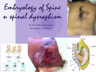
  • 1. Embryology of Spine n spinal dysraphism By Dr Viralkumar Vasani Moderator: Dr Satish S
  • 2.
  • 3. • Formation and separation of the germ layers • Dorsal and ventral induction phases, and • Phases of neurogenesis, • Migration, • Organization and • Myelination.
  • 4. • During week 1 (stages 2–4) the blastocyst is formed, • During week 2 (stages 5 and 6) implantation occurs and the primitive streak is formed, • Formation of the notochordal process and the beginning of neurulation (stages 7– 10). • Somites first appear at stage 9. The neural folds begin to fuse at stage 10, • Rostral and caudal neuropores close at stages11 and 12, respectively
  • 5. Some Embryological fact • first four embryonic weeks are also described as the period of blastogenesis, • Fifth to eighth weeks as the period of organogenesis • At the junction of trimesters 1 and 2, the fetus of about 90 days has a greatest length of 90 mm, whereas at the junction of trimesters 2 and 3, the fetus is about 250 mm in length and weighs approximately 1,000 g • Newborn brain weighs 300–400g at full term. Male brains weigh slightly more than those of females but, in either case, the brain constitutes 10% of the body weight
  • 6. Beginning of NS • Gastrulation - birthday of the nervous system. Time when • 1)bilateral symmetry • 2) three axes are established in the body of all vertebrates • 3) Neuroepithelium can first be identified and distinguished from primitive germinal tissues
  • 7. • Brain and spinal cord arise from an area of the ectoderm known as the neural plate. • The folding of the neural plate, leading to successively the neural groove and the neural tube, is called primary neurulation. • The caudal part of the neural tube does not arise by fusion of the neural folds but develops from the so-called caudal eminence. This process is called secondary neurulation
  • 8. Stage Length Days External features Internal Feature related to CNS
  • 9. Stage Length Days External features Internal Feature related to CNS
  • 10.
  • 11.
  • 12. Primary Gastrulation • primitive streak (PS), midline structure, at the caudal end of the embryo • Hensen’s node - cranial end of the primitive streak • primitive groove, runs in the midline within the PS; the cranial end of the primitive groove is the primitive pit. • cells of the epiblast migrate toward the PS and invaginate through the primitive groove. The first cells to ingress (while the PS is still elongating) are endodermal cells, which displace the hypoblast cells laterally and form prospective endoderm • Displaced hypoblast cells form extraembryonic tissues. • As the PS regresses, mesodermal cells ingress through the PS between the epiblast and newly formed endoderm and become the mesoderm. • The remaining epiblast cells spread out to replace the cells that have ingressed through the primitive groove and thereby form both the neuroectoderm (the neural tube) and the cutaneous ectoderm (skin
  • 13.
  • 15. • Neural tube closure • Occurs craniocaudally from initial point of contact • Initiated in region of cervical spinal cord (5 somite stage ) • Posterior neuropore closes at day 25-27 (day 25 - 27) • Posterior neuropore approximately located at S2 level • Whole process known as neurulation (primary neurulation )
  • 16. Forces for neural tube formation • Mesoderm appears to be important for orien- tation but not for closure of the neural tube. • Expansion of the surface epithelium of the embryo is the principal extrinsic force for folding of the neuroepithelium to form the neural tube • Intrinsic forces of the neuroepithelium, the cells of the floor plate have a wedge shape—narrow at the apex and broad at the base—that facilitates bending.
  • 17. • The ependymal cells that form the floor plate are the first neural cells to differentiate, and they induce growth of the parenchyma of the ventral zone more than the dorsal regions • mechanical effect may also facilitate curving of the neural placode (Flat plate). • The direction of proliferation of new cells in the mitotic cycle, determined in part by the orientation of the mitotic spindle, becomes another mechanical force shaping the neural tube that is rostrocaudal orientation of most mitotic spindles • Adhesion molecules
  • 18. Role of Hensen’s Node • as the “organizer” of the embryo. • As the PS elongates, prospective endodermal cells within Hensen’s node migrate through the primitive pit. • As the PS regresses, specialized mesodermal cells, prospective notochordal cells, migrate through the primitive pit and form the notochordal process in the midline between the neuroectoderm and endoderm. • Notochord plays an imp role in directing subsequent neurulation.
  • 19. • Between PODs 18 and 20, the notochordal process fuses (intercalates) with the underlying endoderm to form the notochordal plate. The notochordal plate is therefore incorporated into the roof of the yolk sac, with the notochordal canal becoming continuous with the yolk sac. • Intercalation results in a direct communication, the primitive neurenteric canal, that connects the amnionic and yolk sacs at the level of Hensen’s node. • The neurenteric canal persists for about 3 days, at the end of which the notochordal plate folds dorsoventrally and separates (excalates) from the endoderm and the neurenteric canal is obliterated. • Thereafter, the true notochord exists as a solid rod of notochordal cells
  • 21. Formation of neural tube • Human neuroectoderm visible by day 16 • Pseudo stratified epithelium overlying the notochord • Neural groove visible by day 17-19 • Neural folds (day 19 -21) • Separate from the overlying ectoderm (dysjunction) and fuse to form the neural tube. • The neural folds separate from the cutaneous ectoderm and fuse to form a closed neural tube between POD 21 and 23. • Closure generally involves apposition and fusion of first the cutaneous ectoderm and then the neuroectoderm. • The first part of the human neural tube to close is the region of the caudal rhombencephalon or cranial spinal cord, usually when five pairs of somites are present.
  • 22. • Cranial neural tube closure may involve the coordinated interaction of as many as four waves of discontinuous neural tube closure. • The spinal cord closes craniocaudally in a linear manner from the point of initial closure to the caudal neuropore. • Cranial neuropore closes between POD 23 and 25, whereas the caudal neuropore closes between POD 25 and 27.
  • 23. • Formation of neural crest • Neural crest cells originate at the junction of surface ectoderm and neuroectoderm • First visible on day 19-21 • Continue forming till day 32 • Cranial neural crest cells contribute to the branchial arches and the arachnoid and pia mater of the cranium.
  • 24. • spinal neural crest arises only after closure of the neural tube. • Spinal neural crest cells undergo terminal differentiation into • melanocytes of the body wall and limbs, • Schwann cells investing the peripheral nerves, • spinal cord meninges, • dorsal root and autonomic ganglion cells of the spinal nerves • adrenal medulla.
  • 25. • Secondary neurulation • Caudal cell mass extends from the posterior neuropore to the cloacal membrane (day 25-27) • Pluripotent cells derived from the primitive streak. • Contains neurons, neural crest cells and glial cells and ependymal cells • The secondary neural tube is initially solid and subsequently undergoes cavitation, eventually forming the tip of the conus medullaris and filum terminale by a process called retrogressive differentiation.
  • 26.
  • 27. • Three processes are responsible for further development of the CCM • Condensation • Canalisation • Retrogressive differentiation • Final derivatives • Distal sacral nerve roots,conus medullaris,terminal ventricle,filum terminale and sacrococcygeal remnant • Secondary neurulation continues till day 52
  • 28. • Ascent of the conus medullaris • Process beginning day 43-48 and continuing into post natal life probably • 2 distinct processes • Retrogressive differentiation of the caudal neural tube (prior to day 54) • Disparity between growth of spinal cord and vertebral column. • Most rapid ascent between 8 to 25 weeks of gestation. • At birth conus is at adult level of L1-L2 level. • Low lying conus- below mid body of L2.
  • 29. NTDs • neural tube defects (NTDs), or localized failure of primary neurulation, that can arise through one of two mechanisms. • The “nonclosure theory” proposes that NTDs represent primary failure of neural tube closure. • The “overdistention theory,” introduced in 1769 by Morgagni and popularized by Gardner proposes that NTDs arise through overdistention and rupture of a previously closed neural tube.
  • 31. Spinal Dysraphism • Spinal dysraphism refers to a spectrum of disorders in which there is defective midline closure of neural, bony, or other mesenchymal tissues. • The “open” dysraphic states include myelocele, myelomeningocele, hydromyelia, Chiari II malformations, hemimyelocele, and myeloschisis. • The closed dysraphic states include entities such as dermal sinus, lipomyelomeningocele, tight filum terminale, meningocele, myelocystocele, diastemato- myelia, neurenteric cyst, slit notochord, and developmental tumors such as spinal lipomas
  • 32. Classification of Dysraphism Spinal Dysraphism Open Myelomeningocele Myelocele Hemi- MY/MYM E Closed With S/C Mass Lipomyelomeningocele Meningocele Lipomyelocele Myelocys tocele Terminal Myelocystocele Closed without S/C Mass Simple Dysraphic state Dermal Sinus Intradural Lipoma Complex Dysraphic State Neuroenteric cyst Caudal Agenesis Segmental Vertebral anamolies
  • 33. Myelomeningoceles • Disorder resulting from defective primary neurulation • 98% of all Spinal dysraphism • Incidence • 0.4 per 1000 live births • Racially variable • 85% caudal thoraco lumbar spine, 10 % in the torax and the rest cervical • 80-90 % associated with hydrocephalus and Chiari • Trisomy 13 and trisomy 18
  • 34.
  • 35. Associated defects • Brain stem defect includes • Medullary kinking, tectal beaking, and intrinsic nuclei abnormalities • Supratentorial abnormalities include • partial or complete dysgenesis of the corpus callosum, • polymicrogyria, a large massa intermedia, and gray matter heterotopia. • Mesodermal development of the skull • small posterior fossa, short clivus, • low-lying tentorium and torcular Herophili, wide incisura, and • enlarged foramen magnum. • Lückenschädel, or craniolacunia (scalloping of the skull bones)
  • 36. Cause of neuro-deterioration • symptomatic hydrocephalus, • syringomyelia • Retethering of cord. • Chiari II malformation, Risk factors • mostly due to shunt malfunction resulting in hydrocephalus
  • 37. Patho Anatomy • Failure of neural tube closure results in an exposed neural placode. The groove in the center of the placode is the remnant of the central canal. • The spinal roots exit from the anterior surface of the placode such that the ventral roots lie medially and the dorsal roots lie laterally. • The dura fuses with the defect in the fascia laterally. Functional neural tissue + either caudal to the placode or in the nerve roots exiting from the placode.
  • 38. 1. Axial schematic of myelomeningocele shows neural placode (star) protruding above skin surface due to expansion of underlying subarachnoid space (arrow). 2. Axial T2-weighted MR image in 1-day-old boy shows neural placode (black arrow) extending above skin surface due to expansion of underlying subarachnoid space (white arrow), which is characteristic of myelomeningocele.
  • 39. MR Image of myelomeningocele • Sagittal T2-weighted MR image from same patient with myelomeningocele shows neural placode (white arrow) protruding above skin surface due to expansion of underlying subarachnoid space (black arrow).
  • 40. Clinical Examination • signs of myelopathy, including hyperreflexia and clonus, because they often have an incomplete functional spinal cord transection in 2/3rd • sensory level by stimulus - distally to proximally until the infant grimaces. • A stimulus is applied above the sensory level, and the distal-most voluntary motion seen for motor level determination • With an L1-3 level, the infant has hip flexion with extended knees and clubfeet. • The presence of intact hip adduction, hip flexion, and knee extension with inverted feet is indicative of an L2-4 level. • With an L5-S2 level, the infant has hip adduction, knee extension, and knee flexion with dorsiflexed feet. • Infants with a sacral level may appear intact except for weakness of plantar flexion and rocker-bottom feet. • A flaccid pelvic floor and patulous anus are often present
  • 41. • Prenatal diagnosis • Maternal serum Alpha feto protein : initial screening test • High resolution fetal ultrasonography. • Can also demonstrate hydrocephalus and Chiari II abnormality (lemon and banana sign) • Amniocentesis : if MSAFP and USG are suggestive • Ach esterase levels along with AFP • AFP can increase in other developmental anomalies of the gut and kidneys.
  • 43.
  • 44. D/D • At least 22 other fetal abnormalities besides myelomeningocele increase MSAFP levels. • Abdominal abnormalities such as omphalocele, cloacal exstrophy, esophageal atresia, annular pan- creas, duodenal atresia, and gastroschisis and • urologic abnormalities such as congenital nephrosis, polycystic kidneys, urinary tract obstruction, and renal • sacrococcygeal cystic teratoma
  • 45. Preop evaluation- Clinical • General • Repaired within 72 hrs • Enteral feeding avoided to prevent fecal soiling of placode • Prone position ,saline dressings • Neurosurgical • Sensory level determined • Motor evaluation – distal most voluntary motion evaluated. Limb abnormalities documented. • Anal tone and anal reflex evaluated
  • 46. • Ventricular size documented with preop USG and NCCT head. • Imaging study shows that differentiating feature between a myelomeningocele and myelocele is the position of the neural placode relative to the skin surface • The neural placode protrudes above the skin surface with a myelomeningocele and is flush with the skin surface with a myelocele • Observe for symptoms of Chiari II • Renal evaluation • 90 % have neurogenic bladder. • All should have preop Renal ultrasound for detecting severe anomalies. • CIC if fails to void.
  • 47. Myelocele 1. Axial schematic of myelocele shows neural placode (arrow) flush with skin surface. 2. Axial T2-weighted MR image in 1-day-old girl shows exposed neural placode (arrow) that is flush with skin surface, consistent with myelocele. There is no expansion of underlying subarachnoid space.
  • 48. Repair • Timing of repair: • Myelomeningocele repair can be performed safely up to 72 hours after birth • Delayed repair – Increases chance of ventriculitis by 5 times, • shunt infection developed in about 75%, and the mortality was 13% • In case Delay • 1) Cultures from the neural placode – No growth – go ahead n repair • If infection +, -external ventricular drainage and appropriate antibiotics until the infection clears – Then repair
  • 49. • Shunt before repair – High chance of shunt inf./ Meningitits – IQ impairment (due to inf) • PREPARATION • Intraoperatively avoid hypothermia, hypovolemia, and hypoglycemia. • A doughnut-shaped sponge - to protect the myelomeningocele while intubation. • If severe Hydrocephalus - CSF diversion before closure of the myelomeningocele - to minimize pressure on the myelomeningocele dural closure • entire back and flanks are prepared and draped to facilitate extensive closure if needed. • Contact between povidone- iodine solution and the neural placode should be avoided
  • 50. • purposes of myelomeningocele repair are to protect the functional spinal cord tissue, prevent loss of CSF, and minimize the risk for meningitis by reconstructing the neural tube and its coverings. • The margin between the arachnoid of the neural placode and the dystrophic epidermis, or the junctional zone, is the site of the initial incision. • The goal is to free the neural placode from the surrounding junctional zone circumferentially. • Duraplasty with thoracolumbar fascia or another dural substitute is performed when necessary to prevent leakage of CSF.
  • 51.
  • 52.
  • 53. Post op care • Post op antibiotics • Prevention of fecal contamination of wound • Nurse in Trendlenberg’s • Observe for Hydrocephalus – shunt if HCP present • Complications • Superficial wound dehiscence • Meningitis • Symptomatic chiari • Ensure functioning shunt • Hindbrain decompression
  • 54. Prognosis • 10 to 15% succumb <6yr of age even with Multi specialty aggressive approach • >95% Lives >2 years • 8 to 17 % will have urinary control rest on Drugs / CIC • >87% will have social fecal incontinence • L3 function allows one to stand erect, and L4 and L5 function allows ambulation – During the first decade, approximately 60% of children with spina bifida are community ambulators, without or with assistive devices (including wheelchairs) – Reduces to 17% in teenagers • IQ stays N if no inf. – only <10% economically independent
  • 55. Occult spinal dysraphism • Aka Closed dysraphism • Of 2 types ; with/without s/c mass • Congenital spinal defects covered by intact skin • Causative lesions • Fatty filum terminale • Lipomyelomeningocele • Split cord malformations type I and II • Inclusion lesions (dermoid, dermal sinus tract) • Neurenteric cyst • Myelocystocele
  • 56. Closed / Occult type • Closed With S/C Mass • Lipomyelomeningocele • Meningocele • Lipomyelocele • Myelocystocele • Terminal Myelocystocele • Closed without S/C Mass • Simple Dysraphic state • Dermal Sinus • Intradural Lipoma • Complex Dysraphic State • Diastometamyelia • Neuroenteric cyst • Caudal Agenesis
  • 57. • Closed Spinal Dysraphisms With a Subcutaneous Mass 1. Lipomas with a dural defect— • include both lipomyeloceles and lipomyelomeningoceles. • result from a defect in primary neurulation whereby mesenchymal tissue enters the neural tube and forms lipomatous tissue. • characterized clinically by the presence of a subcutaneous fatty mass above the intergluteal crease. • The main differentiating is the position of the placode–lipoma interface. With a lipomyelocele, the placode–lipoma interface lies within the spinal canal where as it is outside of the spinal canal in case of lipomyelomeningocele, it lies due to expansion of the sub arachnoid space . 2. Meningocele—Herniation of a CSF-filled sac lined by dura and arachnoid mater is referred to as a meningocele. The spinal cord is not located within a meningocele but may be tethered to the neck of the CSF-filled sac. • Posterior meningoceles herniate through a posterior spina bifida (osseous defect of posterior spinal elements) and are usually lumbar or sacral in location but also can occur in the occipital and cervical regions. • Anterior meningoceles are usually presacral in location but also can occur elsewhere
  • 58. Manifestations of occult spinal dysraphism Cutaneous stigmata Orthopedic deformities Urologic problems Asymmetric gluteal cleft Foot or leg deformities Neurogenic bladder Capillary hemangioma Scoliosis UTIs Subcutaneous lipomas Sacral agenesis Incontinence Hypertrichosis Delay in toilet training Dermal sinus tract Cutis aplasia
  • 59. Neurological signs and symptoms in different age groups Infants Toddler Older children Young adults Decreased spontaneous leg movements Delayed walking Asymmetric motor/ sensory development Back pain Absent reflexes Abnormal gait Back/leg pain Leg cramping/pain Leg atrophy UMN signs Spasticity Foot asymmetry Painless ulceration Hyperreflexia Decreased urinary stream Bowel/bladder incontinence.
  • 60. Plain radiological findings Structure Findings Lamina Fusion defects,midline defects,abnormal spinous processes Vertebral bodies Hemivertebrae, Butterfly vertebrae, Block vertebrae, Midline cleft defects, canal stenosis Disk space Congenital narrowing Pedicles Flattening, thinning Widening of spinal canal Interpedicular widening, scalloping of posterior border, Midline bony spur. Failure of development Reduced number of vertebral bodies, Absence of parts of vertebrae, sacral dysjunction Spinal curvature Scoliosis, kyphosis, lordosis.
  • 61.
  • 62. Lipomyelomeningoceles • Lipoma tethering the cord to the subcutaneous tissue • Fascial, spinous and dural defect • Lipoma cord interface distracted out of the spinal canal by traction created by tethering
  • 63. LMMC Axial schematic of lipomyelomeningocele shows placode–lipoma interface (arrow) lies outside of spinal canal due to expansion of subarachnoid space. Axial T1-weighted MR image in 18-month-old boy shows lipomyelomeningocele (arrow) that is differentiated from lipomyelocele by location of placode–lipoma interface outside of spinal canal due to expansion of subarachnoid space.
  • 64. LMC- Lipomyelocele • Axial schematic of lipomyelocele shows placode–lipoma interface lies within spinal canal. • Axial T2-weighted MR image in 3-year-old girl shows placode–lipoma interface within spinal canal, characteristic for lipomyelocele.
  • 65. Lipomyelocele Sagittal T1-weighted MR image in 3- year-old girl with lipomyelocele shows subcutaneous fatty mass (black arrow) and placode–lipoma interface (white arrow) within spinal canal.
  • 66.
  • 67. Chapman’s classification of LMM • Classification • Type I (dorsal lipoma) • Type II (transitional lipoma) • Type III ( terminal lipoma)
  • 68. • Dorsal lipoma (Type I) • Fibrolipomatous stalk tethering cord proximal to conus • Usually at middle lumbar to lumbo sacral level • Dorsal spinal cord dysraphic at site of attachment of lipoma • Site of attachment medial to the dorsal root entry • Normal spinal cord distal to myeloschisis. • Roots lie within the subarachnoid space.
  • 70. • Caudal or terminal lipomas (type III) • Directly from conus medullaris or filum terminale • Largely or wholly intradural • Nerve roots entangled in the lipoma • Lipoma cord interface caudal to the dorsal root entry zone. • Filum may be fatty, thickened and sometimes attached to subcuatneous tissue ( sacral dimple).
  • 72. • Transitional lipomas • Share the charcteristics of both Type 1 and type 2. • No normal spinal cord distal to lipoma attachment • Initially dorsal roots may be separate but caudally become enmeshed into the lipoma. • Frequently assymmetric attachment to cord.
  • 74. Abnormal embryology LMM • Usually a disjunction in timing of neural tube closure and cutaneous ectoderm closure • Elements of the ectoderm become incorporated into the incompletely closed neural tube.
  • 75. Clinical features • Subcutaneous masses over the back • Stigmata of occult dysraphism • Hypertrichosis • Hemangioma • Hypo/ hyperpigmented patch • Dermal pit or sinus • Atretic meningocele • Assymmetric gluteal cleft
  • 76.
  • 77.
  • 78. • Inexorable symptomatic progression - in untreated cases • Risk of precipitous neurologic deterioration • Orthopedic syndrome • Limb length discrepancy, high pedal arches, hammer toes, calcaneovarus/ valgus foot deformity. • Urologic syndrome • Urinary incontinence, post void dribbling, urgency, frequency • Intractable pain in the legs, back, pelvis or perineum.
  • 79. Indication for surgical repair • Asymptomatic infant older than 2 months • Presence of orthopedic, pain or urologic syndrome • Neurological symptoms • Prior to corrective spinal surgery.
  • 80. Principal Goal of surgery • Detethering of spinal cord • Decompression of the cord by removing as much lipoma as possible • Reconstruct the spinal cord and dural sac • Preservation of the functional tissue • Surgical principles • Relationship between the lipoma-cord interface and dorsal roots to be established • Conservative excision of the lipoma to avoid injury to the cord/ exiting roots.
  • 81. • Complications of surgical repair • Early – CSF leak/ pseudo meningoceles & New Neurological deficit • Late – retethering of the cord • Mostly presenting between 3-8, 11-22 months after surgery • Upto 20% cases may demonstrate retethering • Diagnosis primarily clinical. • Aseptic meningitis from host-graft inflammation • meningitis, • Intradural abscess, • wound infection, and wound breakdown
  • 82. Lipoma of the terminal filum  Less severe form of Occult SD  More than 2 mm thickness of the filum on MR imaging  Frequently assosciated with sacral/gluteal cleft dimples.  May be associated with VATER association, imperforate anus, cloacal extrophy and other urogenital abnormalities.  Sometimes a/w sacral agenesis  Reflects defective secondary neurulation
  • 83.  Clinical presentation  Orthopedic  Urologic  Pain  All asymptomatic infants and symptomatic adults are surgical candidates.  Surgical procedure is the exposure of filum through lumbosacral laminectomy or interlaminar approach  The filum identified separated from nerve roots and cut.
  • 84. Closed SD without S/C Mass • Closed without S/C Mass • Simple Dysraphic state • Intradural Lipoma • Filar lipoma, • Tight filum terminale, • Persistent terminal ventricle • Dermal Sinus • Complex Dysraphic State • Diastometamyelia • Neuroenteric cyst • Caudal Agenesis • Segmental Vertebral anamolies
  • 85. • An intradural lipoma - a lipoma located along the dorsal midline that is contained within the dural sac. No open spinal dysraphism is present. • M.C site: lumbosacral - present with tetheredcord syndrome, a clinical syndrome of progressive neurologic abnormalities in the setting of traction on a lowlying conus medullaris. • Fibrolipomatous thickening of the filum terminale is referred to as a filar lipoma. On imaging, a filar lipoma _ - Normal variant if there is no clinical evidence of tetheredcord syndrome • Tight filum terminale is characterized by hypertrophy and shortening of the filum terminale causing tethering of the spinal cord and impaired ascent of the conus medullaris. The conus medullaris is low lying relative to its normal position that is above the L2–L3 disc level. • Persistence of a small, ependymal lined cavity within the conus medullaris is referred to as a persistent terminal ventricle. • Location immediately above the filum terminale and lack of contrast enhancement, which differentiate this entity from other cystic lesions of the conus medullaris.
  • 87. Complex dysraphic states divided into two categories: • disorders of midline notochordal integration • dorsal enteric fistula • neurenteric cyst • Diastematomyelia • disorders of notochordal formation, which include • caudal agenesis and • segmental spinal dysgenesis
  • 88. • Diastematomyelia—Separation of the spinal cord into two hemicords. The two hemi cords are usually symmetric, although the length of separation is variable. • two types of diastematomyelia. • In type 1, the two hemicords are located within individual dural tubes separated by an osseous or cartilaginous septum. • In type 2, there is a single dural tube containing two hemi cords, sometimes with an intervening fibrous septum. Diastematomyelia can present clinically with scoliosis and Tethering
  • 89. Split cord malformation • Longitudinal developmental splitting of the cord over one or multiple levels • Two types: • Both types may be present simultaneously at different levels
  • 90. Type I malformations (formerly diastematomyelia) are characterized by a bony septum that cleaves the spinal canal in the sagittal plane and a duplicated thecal sac Type II malformations (formerly diplomyelia) are characterized by a cleft cord within a single dural sac, often tethered by a fibrous midline septum to the adjacent dura (single dural sheath, hemicords separated by fibrotic bands
  • 91. Split cord Malformation • Exceedingly rare • Represent 3.8% to 5% of all congenital spinal anomalies. • prevalence of SCM to be 1 in 5000 (0.02%) live births. • slight female preponderance, approximately 1.3:1.7 • The peak age is 4 to 7 years, • second peak between 12 and 16 years - post pubescent growth spurt. • Type I SCMs > type II lesions
  • 93. Unifide theory • Pang - unified theory - type I and type II SCM to a single error in embryogenesis: • Formation of abnormal fistula through midline embryonic disc that maintains communication b/w Yolk sac & amniotic cavity contact b/w ecto + Endoderm • This fistula causes splitting of Notochord & overlying neural plate- mostly rostral to henson’s node
  • 94. Associated Anomaly • Tethered/low-lying cord (>50%), • Kyphoscoliosis (44% to 60%), • Syringomyelia (27.5% to 44%), and • Spina bifida (11% to 26%)
  • 95. Clinical signs - symptoms
  • 96. • “faun’s tail,” consists of a patch of unusually coarse, raised hair - strong association with type I SCM. • Capillary hemangioma underlying these hairy patches. • Nearly 50% of patients have gross (i.e., structural) asymmetry of the lower extremities-- neuro-orthopedic syndrome. • characterized by a triad of 1. limb length discrepancy, 2. muscular atrophy (resulting in secondary weakness), and 3. clubfoot deformity (talipes equinovarus). • The smaller limb is often ipsilateral to a smaller hemicord.
  • 97. MRI Findings • MRI Spine shows 1. presence of two hemi cords, on T1-w sequences. 2. demonstrating the presence of an associated fatty filum, affect one or both hemicords, a dermal sinus tract, or a terminal lipoma. • Associated tethering anomalies are present in approximately half of all patients and in more than 90% of those with a low-lying conus. • T2-weighted MRI for 1. categorization of the malformation as type I or type II . 2. hypointense fibrous band in type II SCM. 3. To demonstrate syringomyelia proximal to the cleft, which may extend into one or both hemicords
  • 98. Type 1 & Type 2 SCM MRI
  • 99.
  • 100.
  • 101.
  • 102. MGTMNT • 85% of patients without intervention suffer from a progressive neurological deficit versus only 4.5% after surgical treatment • Pang too suported prophylactic surgery in Type 1 malformation.. Whereas in Type 2 both- some – W&W policy • Surgery – careful – incision 1 to 2 level more exposure
  • 103. Operative • Operative management • Surgical detethering of cord by excision of the bony spur/ division of the fibrous bands • Avoid damage to the hemi cords during excision of the spur. • For type I SCM, the initial laminectomies should be limited to adjacent levels while initially avoiding exposure of dura at the level of the midline bony spur. • Rongeurs or a high-speed drill (or both) - to perform bilateral paramedian laminectomies ( preserve the midline lamina and spinous process to prevent any torque or lateral force from disrupting the bony spur prematurely.)
  • 104. • Careful when removing lamina at the level of the SCM in type II malformations because of the frequent presence of transdural adhesions - most commonly attached dorsally but ventrally too. Often, the dura at this level grossly abnormal. • All non-neural and non- functional adhesive bands should be transected, beginning dorsally and then gently rolling the hemicords to one side and transecting any ventral attachments • Hemicords are typically closely approximated with no clear intervening plane. Resection of the fibrous band within the split spinal cord itself is not indicated. • Any associated tethering lesion (sinus tract, fatty filum, or terminal lipoma) should also be addressed
  • 105. Outcome & Complication Of SCM Secondary repair of an SCM in a patient with a previously closed myelomeningocele at the same level - failed wound healing as high as 10%
  • 106. Neurenteric cysts • Persistent neurenteric canal communicating between yolk sac and amniotic cavity • Intradural, extramedullary mucosa lined cysts • Formed from persistents tracts communicating with respiratory and gut epithelia. • Neurenteric cysts represent a more localized form of dorsal enteric fistula. • These cysts are lined with mucin secreting epithelium similar to the GIT and located in the cervicothoracic spine anterior to the spinal cord • Associated with vertebral anomalies • MRI- demonstrates non- contrast enhancing intradural extramedullary cyst
  • 107.
  • 108. • Presentation usually in late years (50-60 years) • May also present in pediatric age group • Most common location is cervico- thoracic • Usually postero-lateral surgical approach • Complete excision of cyst – long term symptom free survival.
  • 109. Dermal sinus tracts  Abnormal tracts communicating between the skin and intraspinal compartment.  Most common- lumbosacral location  May occur anywhere from nasion to coccyx in midline  May be accompanied by other cutaneous stigmata.  Tract terminates within thecal sac mostly  Half may have associated dermoids, epidermoids, teratoma at termination.
  • 110. • Potential pathway for spread of infection • Repeated episodes of meningitis with atypical organisms • Operative repair consists of complete excision of the track under prophylactic antibiotic cover. • Gram positive and gram negative coverage
  • 111.
  • 112. Meningoceles • Distinguished from MMCs by absence of hydrocephalus , chiari malformation or lower limb abnormalities • Dural defect through which CSF space and meninges herniate. • Concomitant neurocutaneous lesions such as lipomas, dermal sinus tracts may cause tethering • Surgical repair of defect at 4-6 months of age.
  • 115. • Herniation of meninges and CSF in ventral location • Commonly in presacral and lumbosacral region • Female predominance • Currarino’s triad- anorectal abnormalities, presacral mass, sacral bony abnormalities. • Presacral tumour may be epidermoid, dermoid or teratoma • Meningitis by atypical organisms may also occur. • Repair the dural defect and detether the cord. Anterior meningoceles
  • 116. myelocystoceles • Terminal dilatation of the central canal that herniates through defective posterior elements • Expanded spinal cord, CSF, fibrous bands and meninges and lipomatous elements • Result from disordered development of the caudal cell mass • Associated anomalies of the anorectal system, lower GI tract and spinal column
  • 117. Myelocystocele Herniation of dilated central canal through posterior spinal defect.
  • 118. Terminal Myelocystocele Terminal myelocystocele— Herniation of large terminal syrinx (syringocele) into a posterior meningocele through a posterior spinal defect. The terminal syrinx component communicates with the central canal, and the meningocele component communicates with the subarachnoid space. The terminal syrinx and meningocele components do not usually communicate with each other.
  • 119. • Surgical repair attempted because of the resultant tethering of spinal cord. • Per-op trumpet shaped distended conus often adheres to the superficial fat • Detethering may be difficult.

Notas do Editor

  1. Myelocystocele—A nonterminal myelocystocele occurs when a dilated central canal herniates through a posterior spina bifida defect (Fig. 9). Myelocystoceles are covered with skin and can occur anywhere but are most commonly seen in the cervical or cervicothoracic regions