anomalous RCA stenting
•
2 gostaram•760 visualizações

The document reports on two cases of successful percutaneous coronary intervention on anomalous right coronary arteries originating from the left coronary sinus. In both cases, the Voda guiding catheter was able to selectively engage the anomalous arteries, allowing for stenting of significant lesions. The Voda catheter provided stable support due to its shape and location opposite the left coronary ostium. Percutaneous intervention of anomalous coronary arteries requires careful planning and catheter selection. In these cases, the Voda catheter facilitated successful revascularization.

Recomendados
Mais conteúdo relacionado
Mais procurados
Mais procurados (20)
Semelhante a anomalous RCA stenting
Semelhante a anomalous RCA stenting (20)
Último
Último (20)
anomalous RCA stenting
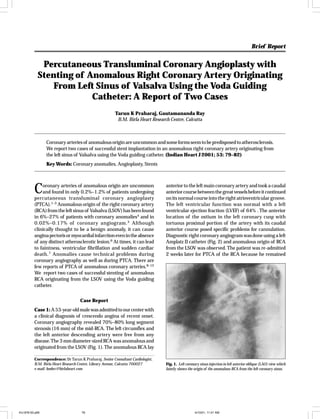
- 1. Brief Report Indian Heart J 2001; 53: 000–000 Percutaneous Transluminal Coronary Angioplasty with Stenting of Anomalous Right Coronary Artery Originating From Left Sinus of Valsalva Using the Voda Guiding Catheter: A Report of Two Cases Tarun K Praharaj, Gautamananda Ray B.M. Birla Heart Research Centre, Calcutta Coronary arteries of anomalous origin are uncommon and some forms seem to be predisposed to atherosclerosis. We report two cases of successful stent implantation in an anomalous right coronary artery originating from the left sinus of Valsalva using the Voda guiding catheter. (Indian Heart J 2001; 53: 79–82) Key Words: Coronary anomalies, Angioplasty, Stents C oronary arteries of anomalous origin are uncommon and found in only 0.2%–1.2% of patients undergoing percutaneous transluminal coronary angioplasty (PTCA).1–3 Anomalous origin of the right coronary artery (RCA) from the left sinus of Valsalva (LSOV) has been found in 6%–27% of patients with coronary anomalies4 and in 0.02%–0.17% of coronary angiogram. 5 Although clinically thought to be a benign anomaly, it can cause angina pectoris or myocardial infarction even in the absence of any distinct atherosclerotic lesion.6 At times, it can lead to faintness, ventricular fibrillation and sudden cardiac death. 7 Anomalies cause technical problems during coronary angiography as well as during PTCA. There are few reports of PTCA of anomalous coronary arteries.8–12 We report two cases of successful stenting of anomalous RCA originating from the LSOV using the Voda guiding catheter. anterior to the left main coronary artery and took a caudal anterior course between the great vessels before it continued on its normal course into the right atrioventricular groove. The left ventricular function was normal with a left ventricular ejection fraction (LVEF) of 64% . The anterior location of the ostium in the left coronary cusp with tortuous proximal portion of the artery with its caudal anterior course posed specific problems for cannulation. Diagnostic right coronary angiogram was done using a left Amplatz II catheter (Fig. 2) and anomalous origin of RCA from the LSOV was observed. The patient was re-admitted 2 weeks later for PTCA of the RCA because he remained Case Report Case 1: A 53-year-old male was admitted to our center with a clinical diagnosis of crescendo angina of recent onset. Coronary angiography revealed 70%–80% long segment stenosis (16 mm) of the mid-RCA. The left circumflex and the left anterior descending artery were free from any disease. The 3 mm diameter-sized RCA was anomalous and originated from the LSOV (Fig. 1). The anomalous RCA lay Correspondence: Dr Tarun K Praharaj, Senior Consultant Cardiologist, B.M. Birla Heart Research Centre, Library Avenue, Calcutta 700027 e-mail: bmbrc@birlaheart.com IHJ-876-00.p65 79 Fig. 1. Left coronary sinus injection in left anterior oblique (LAO) view which faintly shows the origin of the anomalous RCA from the left coronary sinus. 4/10/01, 11:01 AM
- 2. 80 Praharaj et al. Indian Heart J 2001; 53: 79–82 Stenting of Anomalous Right Coronary Fig. 2. Right coronary angiogram was done using a left Amplatz Catheter in LAO view showing severe long segment stenosis in the mid-RCA. Fig. 4. Right coronary angiogram in LAO view showing the inflated balloon. Fig. 3. Right coronary angiogram in LAO view showing left Voda guiding catheter deeply engaged in the RCA with its tip well seated in the mid ostium. Fig. 5. Final diagnostic angiogram following stenting. symptomatic despite medical treatment. The artery could not be cannulated even after using different catheters including the Amplatz catheter. Finally, the RCA was selectively cannulated by Voda Left 8 F guide catheter (inner lumen diameter 0.080", Boston Scientific Corporation, Minnesota) (Fig. 3). The RCA stenotic lesion was successfully crossed with a 0.014" Hi-torque intermediate wire (Advanced Cardiovascular System, Califorina), and dilated with a 3×20 mm Rocket balloon (Advanced Cardiovascular system, California) (Fig. 4). After predilatation of the narrowed segment, a 3×18 mm MultiLink stent (Advanced Cardiovascular System, California) was deployed at 16 atm. A final diagnostic angiogram showed an excellent angiographic result (Fig. 5). The immediate post-procedure IHJ-876-00.p65 80 stay of the patient was uneventful and he was discharged three days later on regular calcium channel blockers, aspirin and ticlopidine (for 6 weeks). The patient continued to remain asymptomatic with a good quality of life on oneyear follow-up. Case 2: A 56-year-old male presented with a clinical diagnosis of effort angina of recent onset with diabetes and hypertension. His coronary angiography revealed a normal left main coronary artery and left anterior descending artery. However, the left circumflex artery was a small caliber vessel, totally occluded in its mid-segment and filled through collaterals from the left anterior descending artery. Mid-RCA had a 70%–80% long segment narrowing with subtotal occlusion in its distal segment. The distal RCA was 4/10/01, 11:01 AM
- 3. Indian Heart J 2001; 53: 79–82 Praharaj et al. Stenting of Anomalous Right Coronary 81 Fig. 6. Left coronary sinus injection in left anterior oblique view showing the origin of the anomalous RCA from the LSOV. Fig. 8. Right coronary angiogram in LAO view showing the inflated balloon. Fig. 7. Right coronary angiogram done using left Voda guiding catheter in LAO view showing long segment stenosis in the mid-RCA and subtotal occlusion of the distal RCA. Fig. 9. Right coronary angiogram in LAO view showing the final diagnostic angiogram. seen to be filled through collaterals from the left anterior descending artery. The RCA (3 mm diameter) originated from the LSOV (Fig. 6) and passed between the pulmonary artery and aorta to reach the right atrioventricular groove. Thereafter it followed a normal course. The LVEF by twodimensional echocardiography was 52%. There was mild hypokinesia of the inferior wall. The anomalous RCA had an ostium located anteriorly and superiorly and could not be cannulated with Judkin, Multipurpose and Amplatz catheters. However, cannulation was easily possible with a Voda 8 F guiding catheter (Fig. 7). The long segment narrowing in mid-RCA and the subtotally occluded distal RCA were successfully crossed with a 0.014" Hi-torque intermediate wire and dilated with a 3×20 mm Rocket balloon. The lesion in the mid-RCA was successfully dilated at 10 atm and the distal segment was stented after predilatation, using a 3×15 mm MultiLink stent at 16 atm (Fig. 8). The final diagnostic angiogram revealed excellent result (Fig. 9). A totally occluded left circumflex artery was successfully crossed with a 0.014" Hi-torque intermediate wire and predilated using a 2.5×20 mm balloon and a 2.5× 15 mm MultiLink stent was deployed at 12 atm with good angiographic result. The immediate post-procedure stay of the patient was uneventful and the patient was discharged on regular medications. He remained asymptomatic at follow-up after 7 months. IHJ-876-00.p65 81 4/10/01, 11:01 AM
- 4. 82 Praharaj et al. Discussion PTCA in patients with an anomalous RCA is technically challenging. It demands a high degree of awareness, and complete evaluation of the coronary artery anatomy and distribution in order to avoid complications. The complication rate of coronary arteriography and PTCA is related to the duration of the procedure. Topaz et al.11 have described various aspects of orifice configuration, anatomy of the artery, location of atherosclerotic lesions and also guiding catheter selection. Proper guiding catheter selection decreases procedure time in PTCA involving anomalous coronary arteries and thus increases success rate. In both cases, we were able to cannulate the anomalous RCA using the Voda guiding catheter. In the first case, the initial angiography was done using the left Amplatz catheter. However, during PTCA, the cannulation was not possible with the Amplatz catheter. Use of the Voda guiding catheter in both cases provided easy cannulation with enough backup support. The choice of the Voda guiding catheter was based on its curvature, large area of support and location of the artery just opposite to the left ostium. It provided the maximum stable support required for the smooth passage of the balloon as well as the stent. The tip of the catheter sits well in the anomalous vessel and the secondary curve rests stably against the opposite aortic wall. The anatomical course of the anomalous RCA in both our cases corresponds to the course described by Ilia.13 Usually, the anomalous RCA originating from the LSOV almost invariably follows a similar course.5 Thus, it appears that the Voda guiding catheter may be the best for PTCA of a coronary artery with similar anomaly. Several techniques have been reported for PTCA in an anomalous RCA.5, 11–14 However, we could cannulate the anomalous RCA in both our cases with relative ease and got good back-up support using the Voda guiding catheter. Thus, after careful study of the course of the anomalous artery, location of the lesion and selective use of the Voda guiding catheter, angioplasty and stenting can be performed IHJ-876-00.p65 Indian Heart J 2001; 53: 79–82 Stenting of Anomalous Right Coronary 82 in patients with an anomalous RCA originating from the LSOV with excellent results. References 1. Engel HJ, Torres C, Page HL Jr. Major variations in anatomical origin of the coronary arteries: angiographic observations in 4250 patients without associated congenital heart disease. Cathet Cardiovasc Diagn 1975; 1: 157–169 2. Kimbiris D, Iskandrian AS, Segal BL, Bemis CE. Anomalous aortic origin of coronary arteries. Circulation 1978; 58: 606–615 3. Ogden JA. Congenital anomalies of the coronary arteries. Am J Cardiol 1970; 25: 474–479 4. Taylor AJ, Rogan KM, Virmani R. Sudden cardiac death associated with isolated congenital coronary artery anomalies. J Am Coll Cardiol 1992; 20: 640–647 5. Douglas JS, French RH, King SB. Coronary artery anomalies. In: King SB, Douglas JS (eds): Coronary angiography and angioplasty. New York: McGraw-Hill, 1985, p. 33–85 6. Benge W, Martins JB, Funk DC. Morbidity associated with anomalous origin of the right coronary artery from the left sinus of Valsalva. Am Heart J 1980; 99: 96–100 7. Roberts WC, Siegel RJ, Zipes DP. Origin of the right coronary artery from the left sinus of Valsalva with associated chest pain: report of two cases. Cathet Cardiovasc Diagn 1976; 2: 397 8. Stauffer J, Sigwart U, Vogt P, Aymon D, Kappenberger L. Transluminal angioplasty of a single coronary artery. Am Heart J 1991; 122: 569– 571 9. Kimbiris D, Lo E, Iskandrian AS. Percutaneous transluminal angioplasty of anomalous left circumflex coronary artery. Cathet Cardiovasc Diagn 1987; 13: 407–410 10. Mooss A, Hentz M. Percutaneous transluminal angioplasty of anomalous right coronary artery. Cathet Cardiovasc Diagn 1989; 16: 16–18 11. Topaz O, Di Sciascio G, Goudreau E, Cowley MJ, Nath A, Kohli RS, et al. Coronary angioplasty of anomalous coronary arteries. Notes on technical aspects. Cathet Cardiovasc Diagn 1990; 21: 106–111 12. Sohara H, Tsurukawa T, Kawabata K, Kawano R, Amitani S, Kurose M, et al. Pitfalls of intervention therapy in a patient with anomalous origin of the right coronary artery from the left sinus of Valsalva associated with organic stenosis. J Cardiol 1997; 29: 111–115 13. Ilia R. Percutaneous transluminal angioplasty of coronary arteries with anomalous origin. Cathet Cardiovasc Diagn 1995; 35: 36–41 14. Das GS, Wysham DG. Double wire technique for additional guiding catheter support in anomalous left circumflex coronary artery angioplasty. Cathet Cardiovasc Diagn 1991; 24: 102–104 4/10/01, 11:01 AM