Enteral feeding is a narrow feeding tube is place through nose down it to stomach. This tube is used to give fluid, medication and liquid food complete with nutrients directly in to stomach.
#ppt on Enteral Feeding, #Enteral Feeding
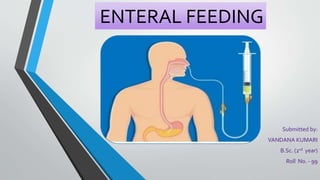
2. DEFINITION
•Enteral feeding is a narrow feeding tube is place
through nose down it to stomach.This tube is used
to give fluid, medication and liquid food complete
with nutrients directly in to stomach.
3. PURPOSE
• To provide nourishment who cannot feed them self or through mouth, If
patient having mouth surgery, coma patients, unconscious etc.
• To administer medication.
• To administer supplemental fluid.
• To monitor gastrointestinal bleeding.
4. TYPES OF NASOGASTRIC
FEEDING
• Continuous Feeding : - Small amount of feed given to long time. It is also
called regular feeding. Person place in semi flower's position during this
type of feeding.
• Bolus Feeding : - This type of feeding also know as gravity feeding. In this
large amount of feed given in short time (with in 20 min) during this feeding
patient placed in flower position.
5. Articles
• A tray containing : -
Formula of feed
Graduated container
Large syringe (30 to 60 ml)
Water in container
Stethoscope
6. PROCEDURE OF ENTERAL
FEEDING
• Wash hand
• Take the tube and check whether it is in good order, Expel the water from
the tube and check the tube for patency.
• Measure distance on the tube from the tip of the nose to the ear lobe plus
the distance from ear lobe to the tip of xiphoid process of the sternum. Mark
the distance of the tube.
• Lubricate the tube for about 6 to 8 inches with lubricant using rag piece or
paper square. Lubrication should be applied to the minimum.
7. • Hold the tube coiled in the right hand and introduce the tip into left nostril.
• Pass the tube gently but quickly backward and downward. Momentary
resistance may occur as the tube is passed into nasopharynx. Have the client
to flex head with draw the table about one inch, rotate it side to side gently
advance the tube.
• When the tube reaches the pharynx the client may gag. Allow him to rest for
movement. Ask him to take panting.
• Have the client take sips of water and swallow on command.Advance the
tube 3 to 4 inches each time client swallows continue to advance the tube
until it reaches the previously designated mark.
8. • Check the placement of the tube in stomach –
i. Aspirate for gastric content with a syringe.
ii. Place the end of the tube with syringe barrel or funnel into bowel of water
and note the rhythm of escaping bubbles.
iii. Ask client to speak.
• After inserting tube place tape it to the side of the faces and wait for
sometime before giving the feeding.
• Before giving the feed, pour some water through the funnel and lower the
funnel slowly, So as to expel the air then give the feed and medication kept
ready for client. When the feed is finished, pour a little water and camp the
tube firmly to prevent leakage of fluid.
9. SIZE AND COLOUR OF
NASOGASTRIC
SIZE COLOUR
8 BLUE
10 BLACK
12 WHITE
14 GREEN
16 ORANGE
18 RED
20 YELLOW
• For pediatrics, the size of nasogastric tube is 8 to 14 French.
• For adult, the size of nasogastric tube is 12 to 18 French
10. TYPES OF NASOGASTRIC
TUBE
• Levine’sTube : - It is also called single Lumen tube. Levine tube are
nasogastric tube that has one opening and used administering medication
used for feeding.
• Salem sump : - It is double Lumen tube which is a large bore nasogastric
tube with double Lumen (2 opening)
1) Most common nasogastric tube.
2) It is used for irrigation of stomach and tube feeding.
12. NURSING MANAGEMENT
• Monitor drainage output, confirm that a x-ray was used for nasogastric
placement, all ordered procedures, irrigation, how often to perform
suctioning . Care is typically performed every 4 hours or less as needed.
• Check tube placement for signs of migration: take note to where it is
marked on the patient and measure external tube length and compare to
what is documented in the chart.
• Check tape and change as needed.
• Use a pen light to assess skin and observe for signs of irritation and redness.
• Clean area around the tube every 4 hours.
13. • Apply a lubricate around nose in order to maintain skin integrity and
reduce the risk of infection.
• Frequent mouth care to keep the mucous membranes moist: brush teeth
every 8 hours (or use an oral sponge) and offer lip balm in order to
maintain skin integrity and reduce the risk of infection.
• Report complaints and signs of nose or throat irritation (excessive mucus,
sore throat, or hoarseness).
• Document all nursing actions taken and the patient’s response to
intervention.
14. HOWTO REMOVE
NGTUBE
• Before removing a decompression tube, the nurse may intermittently
clamp it for a trial period of several hours to ensure that the patient does not
experience nausea, vomiting, or distention.
• Before any tube is removed, it is flushed with 10 ml of water or normal saline
to ensure that is free of debris and away from the gastric lining.
• Gloves are worn when removing the tube.The tube is withdrawn gently and
slowly for 15 to 20 cm (6 to 8 inches) until the tip reaches the esophagus; the
remainder is withdrawn rapidly from the nostril.
• If the tube does not come out easily, force should not be used, and the
problem should be reported to the physician.
• Provides oral hygiene after the tube is removed.
15. SUMMARY
• Enteral feeding is given when oral intake is impossible.
• Food is given in modified form.
• The normal health of intestinal mucosa is well maintained.
• Satisfaction of taking food is felt by the patients.
• Biochemical monitoring is required.
• Comparatively less technical skill is required.
• Calculation of food intake is less complicated.
• Less expensive.