
Recomendados
Mais conteúdo relacionado
Mais procurados
Mais procurados (20)
Semelhante a Nucleus structure and function
Semelhante a Nucleus structure and function (20)
Mais de Thippeswamy M
Último
Último (20)
Nucleus structure and function
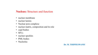
- 1. Nucleus: Structure and function • nuclear membrane • nuclear lamins • Nuclear pore complexe • nuclear matrix, composition and its role • cajal bodies • SFCs • nuclear speckles • PML bodies • Nucleolus Dr. M. THIPPESWAMY
- 2. • Nucleus is the structure where Eukaryotes store their genetic material while nucleoid is the place where Prokaryotes store their genetic material. • Nucleus is large and well organized, whereas nucleoid is small and poorly organized. • Nucleus is surrounded by a double layered membrane called “nuclear membrane” and separates from other cell organelles. • Such membrane cannot be found in nucleoid. • Nucleus contains many chromosomes while nucleoid generally has only one circular DNA molecule. • Nucleolus and nucloeplasm are present inside the nucleus, whereas they are absent in nucleiod. Difference between Nucleus and Nucleoid
- 3. • The nucleus is the genetic control center of a eukaryotic cell. • In most cells, there is only one nucleus. • It is spherical, and the most prominent part of the cell, making up 10% of the cell’s volume. • It has a unique structure and function that is essential for the cell. • It appears as a dense, roughly spherical or irregular organelle. • The composition by dry weight of the nucleus is approximately: DNA 9%, RNA 1%, Histone Protein 11%, Residual Protein 14%, Acidic Proteins 65%. The Nucleus
- 4. The structures are: 1. Nuclear Envelope or Karyotheca 2. Nuclear Matrix 3. Nucleoplasm 4. Nucleolus • The viscous liquid within it is called nucleoplasm. It contains numerous sub-nuclear bodies, including nucleoli, Splicing speckles, Cajal bodies (CB), gems, and Promyelocytic leukemia (PML) bodies in addition to chromosomes
- 5. • Under electron microscope the nuclear envelope in the interphase or prophase stage appears to consist of two concentric membranes, viz., inner nuclear membrane and outer nuclear membrane separated by a space called perinuclear space. • At the outer surfaces of the outer membrane, continuity with endoplasmic reticulum, many ribosomes are found attached and they are actively engaged in protein synthesis. The Nuclear Envelope • In fact, the outer membrane continuously produces numerous finger shaped outgrowths, which expand into flat membranous sacs. This process is called blebbing, which is very common in some protozoan nuclei • The inner membrane is associated with intermediate fibers called lamins, they form a kind of reticular network and provide mechanical support to the membrane. Chromosomes are found bound to the inner surface of the membrane.
- 6. The Nuclear Lamins • Nuclear Lamina, a fibrous network of proteins; at inner surface of the Nuclear membrane consists of three principal extrinsic membrane proteins, lamins A, B and C which together forms a fibrous network at the inner surface provides support and strength • The lamins bind to specific proteins present in the inner nuclear envelope, such as emerin and lamin B receptors. The lamins provide skeletal support to nuclear envelope. • These also provide the site for attachment to chromatin fibers. Lamins also help in dissolution of nuclear envelope at the time of cell division and its reorganization after the cell division is over. • When phosphate groups are not attached to lamins, lamins are assembled to form an ordered structure, which help in organizing nuclear envelope. • Moreover, at the time of cell division, lamins are phosphorylated resulting in their depolymerization so that nuclear envelope also disappears. The phosphorylation and dephosphorylation of lamins is triggered by a specific protein kinase.
- 7. The Nuclear Pore Complex (NPC) • Nuclear pore complexes allow the transport of molecules across the nuclear envelope. • This transport includes RNA and ribosomal proteins moving from nucleus to the cytoplasm and proteins (such as DNA polymerase and lamins), carbohydrates, signaling molecules and lipids moving into the nucleus. • The proteins that make up the nuclear pore complex are known as nucleoporins. Each NPC contains at least 456 individual protein molecules and is composed of 30 distinct proteins (nucleoporins). • Nucleoporin-mediated transport is not directly energy requiring, but depends on concentrations gradients associated with the RAN cycle (RAs-related Nuclear protein). • Each of the eight protein subunits surrounding the actual pore (the outer ring) projects a spoke-shaped protein over the pore channel. • The center of the pore often appears to contain a plug-like structure. Nuclear pore complex is molecular machine acts as a basket
- 8. • The entry and exit of large molecules from the cell nucleus is tightly controlled by the nuclear pore complexes (NPCs). • Although small molecules can enter the nucleus without regulation. • Macromolecules such as RNA and proteins require association with transport factors like karyopherins called importins (Cytoplasm to nucleus) to enter the nucleus and exportins to exit (Nucleus to cytoplasm). • The ability of both importins and exportins to transport their cargo is regulated by the small Ras related GTPase, Ran. The Nuclear Transport
- 9. Step 1. NLS – attached protein binds soluble NLS receptor (importin a/b) Step 2. NLS transports complex to cytoplasmic filaments Step 3. Cytoplasmic filaments bend toward nucleus Step 4. Change in conformation of transporter Step 5. Importin – NLS protein complex binds to Ran-GTP and importin dissociates Step 6. Ran-GTP-importin b shuttled back to cytoplasm Step 7. Importin a subunit transported by an exportin The Nuclear import Nuclear Localization Signals (NLS)
- 10. Export – RNAs • Move as ribonucleoproteins (RNPs) (Except t-RNA – direct transport by exportin-t) • Protein component contains nuclear export signal (NES) • Exportins recognize NES • Binds Ran-GTP – stabilizes complex • Carried to cytoplasm • Ran-GAP1 converts Ran-GTP to Ran-GDP The Nuclear Export
- 11. • The amorphous liquid found within the nucleus is often referred to as karyolymph. Chromatin material, nucleoli and other enzymatic components are suspended in this liquid. • In addition, the fluid at the inner nuclear membrane is saturated with another network of fibrils are lamins, they are a kind of intermediate filaments. • The nuclear sap also contains another kind of network of proteins, unique to the nuclear sap called nuclear matrix a heterogeneous kind of proteins. • Within such a network, chromatin material, various species of RNAs and enzyme components are found embedded. • Apart from the network, the fluid has the enzymatic components required for chromosomal duplication, transcription and processing and repair activities. • During meiosis, specialized structures like synaptonemal complex, appearance at zygotene stage and disappearance at diplotene stage, very dramatic. • Nuclear matrix also posses certain enzymatic activities like kinases, methylases, acetylases and few others, but respiratory activities like glycolysis, is totally absent. Nuclear sap
- 12. • The nuclear matrix is the structural framework of nucleus that consists of the peripheral lamins and pore complexes, an internal ribonucleic protein network, and residual nucleoli. • The nuclear matrix contains proteins that contribute to the preservation of nuclear shape and organization. • The nuclear matrix enables spatial organization of DNA replication, transcription and repair processes; it harbors numerous enzymes and transcription factors. • The nucleoplasm contains numerous sub- nuclear bodies, including nucleoli, Splicing speckles, Cajal bodies (CB), gems, and Promyelocytic leukemia (PML) bodies The Nuclear matrix The nuclear matrix is represented in red.
- 13. Splicing speckles • Nuclear speckles, also known as interchromatin granule clusters, are nuclear domains enriched in pre-mRNA splicing factors, located in the interchromatin regions of the nucleoplasm of mammalian cells. • When observed by immunofluorescence microscopy, they usually appear as 20–50 irregularly shaped structures that vary in size. • Speckles are dynamic structures, and theirconstituents can exchange continuously with the nucleoplasm and other nuclear locations, including active transcription sites. • Studies on the composition, structure, and dynamics of speckles have provided an important paradigm for understanding the functional organization of the nucleus and the dynamics of the gene expression machinery.
- 14. Cajal bodies snRNPs: small nuclear ribonucleo proteins snoRNPs: small nucleolar ribonucleo protein • They were first reported by Santiago Ramón y Cajal in 1903, who called them nucleolar accessory bodies due to their association with the nucleoli in neuronal cells. • Numerous Cajal bodies, or coiled bodies are found in many cell types and are typically 0.2-1 um in diameter. • These structures appear as a tangle of coiled threads and are characterized by the presence of the p80 coilin protein. • Cajal bodies are thought to play a role in snRNP biogenesis and in the trafficking of snRNPs and small nucleolar RNPs (snoRNPs). • Cajal bodies are rich in spliceosomal U1, U2, U4/U6 and U5 snRNPs as well as U7 snRNP involved in histone 3’-end processing (most histone transcripts are not polyadenylated rather their 3’ ends are produced by an endonucleolytic cleavage) and U3 and U8 snoRNPs involved in processing of pre-rRNA. • It is believed that snRNPs and snoRNPs move through Cajal bodies then on to nuclear speckles or nucleoli respectively. Coilin-dependent snRNP assembly is essential for zebrafish embryogenesis
- 15. SUMOylation is a post-translational modification involved in various cellular processes, such as nuclear-cytosolic transport, transcriptional regulation, apoptosis, protein stability, response to stress, and progression through the cell cycle. Promyelocytic leukaemia (PML) nuclear bodies • PML bodies vary in size from 0.3-1 um in diameter and a nucleus typically contains 10-30 of these structures. • The primary role of PML bodies remains unclear; but they may play a role in transcriptional regulation and in anti-viral responses. • Individuals suffering from acute promyelocytic leukaemia (APL) have a translocation in which the PML gene is fused to the gene encoding the alpha-retinoic acid receptor, resulting in the production of a fusion protein. • Cells from these individuals exhibit fragmented PML bodies. However, treatment of APL patients with retinoic acid results in cancer remission and the restoration of normal PML body structure. • Acute promyelocytic leukemia (APL), the M3 subtype of acute myeloid leukemia (AML), is important because patients with APL can develop serious blood-clotting or bleeding problems.
- 16. • Gems are found adjacent to Cajal bodies and are characterized by the presence of the survival of motor neurons gene product (SMN) and Gemin 2. • Cytoplasmic SMN and Gemin 2 are involved in the assembly of snRNPs and therefore the nuclear pool may play a role in snRNP maturation. • Spinal muscular atrophy, a motor neuron disorder, results from reduced levels or a mutation in SMN proteins. Gems (Gemini of Cajal Bodies)
- 17. • The nucleolus, a large nuclear domain, is the ribosome factory of the cells. • Ribosomal RNAs are synthesized, processed and assembled with ribosomal proteins in the nucleolus, and the ribosome subunits are then transported to the cytoplasm. • The position of the nucleolus in the nucleus is eccentric (peculiar). The nucleolus • A nucleolus is often associated with the nuclear organization region (NOR) which represents the secondary constriction of the chromosomes. • The NOR region codes for 18S, 5.8S and 28S RNAs. The genes for the forth type of rRNA occur outside the nuclear organizer. • The nucleolus is not bounded by any limiting membrane. Calcium ions are supposed to maintain its intact organization.
- 18. The nucleolus active in synthesis of ribosomes typically exhibits three regions under electron microscope. 1. Fibrillar centre: This pale – staining part represents the inner most region of the nucleolus. It comprises of 5 filaments having 5-10 nm (50-100 A) diameter and several 100 A long. These fibrils formed of rDNA (ribosanal DNA), rRNA and ribonucleoprotein particles (RNP). These particles gradually modify into granules. 2. Granular cortex: This is the outmost region of the nucleolus where processing and maturation of pre-ribosomal particles taken place. 3. Amorphous matrix: This is proteinacious ground substance in which granules and fibrils remain suspended. • The function of the nucleolus is to synthesize rRNA and assemble ribosomal subunits. • The rRNA genes are transcribed by RNA polymerase I as a large pre-rRNA precursor that is cleaved to produce 5.8S, 18S, and 28S rRNAs found in ribosomes. • rRNA is post-transcriptionally modified and assembled with ribosomal proteins, which are synthesized in the cytoplasm and imported into the nucleus through the nuclear pores. • This results in the formation of the large and small ribosomal sub-units, which are subsequently transported from the nucleus to the cytoplasm where they mediate mRNA translation. • Nucleolus place significant role in cell division. It disappears during prophase and at the telophase, tiny nucleoli reappear at the chromosaomal locations of the ribosomal RNA genes (NOs).
- 19. THANK YOU