2. FASCIOLA GENERAL CHARACTERS
Distribution
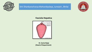
F.hepatica has a thin, dorso ventrally flattened, leaf-shaped, elongated and
oval body. The maximum width is at about anterior third of the body from where
the body tapers anteriorly as well as posteriorly, however, the anterior end is
somewhat rounded, while it is bluntly pointed posteriorly. F. indica has its greatest
width at about the middle of the body, and the posterior end is rounded. It is
usually pinkish in color but it appears brownish due to ingested bile of the host.
This parasite is cosmopolitan in distribution throughout all the sheep and
cattle rearing areas of the world.
Habit and habitat-
Fasciola is digenetic endoparasites. It completes its life cycle in two hosts. Its
primary host is sheep and secondary host is a freshwater gastropod either Limnea
truncate or some species of Planorbis or Bulinus. Fasciola hepatica is also known
as sheep liver fluke as it resides in the liver and bile ducts of sheep. It can also
occur in other vertebrate hosts like goat, horse, dog, ass monkey, man , elephant
etc. A single sheep may accommodate around 200 adult flukes in its liver
and as a result the liver may stop to function. Consequently liver rot occurs.
3. External features
Size and shape of the body:
This parasite may measure about 1.8 to 3 cm long and 0.4 to
1.5 cm in width at the middle region. From the middle region
the body tapers both anteriorly and posteriorly. Anterior
end is a bit rounded whereas posterior end is pointed. It is a
soft, oval, dorso-ventrally flattened leaf like animal.
Color of the body: The color of this animal is usually
pinkish. The transparency of the body wall enables the view
of internal organs like vitelline glands along the lateral
margins, alimentary canals in the center.
Oral cone: The anterior rounded end of this animal is
prominently projected. This part is also called as oral cone or
head lobe. At the tip of this oral cone a triangular aperture
called mouth is located.
4. Suckers:
This animal does not have any hooks or spines but has two
small suckers namely, anterior sucker and the ventral sucker.
Anterior sucker is situated at the bottom of the cup shaped
muscular mouth. It is also called as oral sucker. The
muscles of this oral sucker radiate from the margins of the
mouth to the periphery of the sucker. These oral suckers
help in adhesion and ingestion.
Ventral sucker is situated mid ventrally behind the oral
sucker. It is also called as acetabulum and it does not have
any aperture.
Apertures: Along with mouth there are two more apertures
namely gonopore and excretory pore. The gonopore is a small
aperture lying in front of the acetabulum. A single excretory
pore is situated at the posterior end of the body. Because of the
presence of an incomplete alimentary canal, there is no anus in
this animal.
Scales: The body of Fasciola is covered by tegument from
which numerous minute backwardly directed spicules or scales
arise. These spicules help in locomotion, protection and in
anchorage of the parasite body in the bile ducts of the host.
5. FASCIOLA
DIGESTIVE-SYSTEM
Fasciola has an incomplete alimentary canal and
thus it do not have anus.
The mouth is situated at the anterior end surrounded
by oral sucker.
The mouth leads into an ovoid pharynx.
Pharynx has a small lumen and thick walls provided
with radial muscles and pharyngeal glands. The
pharynx leads into oesophagus.
Oesophagus is lined by single layer of epithelial cells
and it in turn opens into large intestine.
The intestine has numerous branches both on the
right and left side. Each branch again gives out
numerous irregular side branches called caeca.
Outer caeca are large and further branched while the
inner caeca are short and simple.
Pharynx and oesophagus are internally lined with
cuticle and intestine is lined by endodermal columnar
epithelial cells. Caecal epithelium has secretory
gland cells.
6. Mechanism of feeding and digestion
The flukes when hungry migrate into smaller bile ducts and capillaries for
feeding. They suck the lymph, bile and tissue pieces with the help of the oral
sucker. The digestion in these flukes is extracellular type and it takes place in
the intestine.
The distribution of the digested food is done by diverticula of the intestine.
Monosaccharide's can directly diffuse through the body surface. Amino acids
are absorbed by the tegument. The food is reserved in the form of glycogen
and fats in the mesenchyme and muscles.
7. FASCIOLA EXCRETORY SYSTEM
The excretory system of Fasciola is also known as Protonephredial system.
It consists of following parts,
Flame cells
The flame cells are modified mesenchymal cells to perform the excretory
function. Each flame cell consists of intracellular lumen in which few long
cilia are present. Each of these cilia arises from a basal granule. As these
structures vibrate like a flickering of a flame they are named so.
Excretory ducts
The lumen of the flame cell is in continuation with the microscopic excretory or
capillary ducts. The capillary ducts from a few protonephridia open into narrow
collecting tubule. Several such tubules open into larger twigs which in turn open into
vessels. The excretory vessels of the anterior part of body open into four trunks.
Excretory canal
These four trunks unite posteriorly to form a single longitudinal excretory canal.
This longitudinal excretory canal extends up to posterior end of the body where it
opens out through single excretory pore. The excretory vessels of the posterior part,
open directly into the longitudinal excretory canal. All the ducts except the median
longitudinal canal are lined with cilia.
8. FASCIOLA NERVOUS SYSTEM
As flukes are parasitic in nature, they do not have sense organs.
They possess developed nervous system. Brain forms a cerebral
ring around pharynx and it bears a pair of lateral cerebral ganglia
and ventral ganglia. Fine nerves arise from the brain and supply
the anterior and posterior regions.
The suckers receive more supply. Three pairs of longitudinal nerve
cords extend posteriorly and they give out numerous peripheral
branches to various organs. Of these three pairs one is dorsal; one
ventral and last one is lateral. Out of these three lateral cords are
well developed and are connected by few transverse commissures.