Thrombosis- Dr. Shubhangi V. Agale
•Transferir como PPT, PDF•
14 gostaram•1,898 visualizações

Thrombosis Definition: Formation of solid plug any where in intact cardiovascular system from constituents of blood during life.

Recomendados
Mais conteúdo relacionado
Mais procurados
Mais procurados (20)
Destaque
Destaque (20)
Semelhante a Thrombosis- Dr. Shubhangi V. Agale
Semelhante a Thrombosis- Dr. Shubhangi V. Agale (20)
Último
Último (20)
Thrombosis- Dr. Shubhangi V. Agale
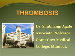
- 1. Dr. Shubhangi Agale Associate Professor Grant Govt Medical College, Mumbai.
- 2. Functions of Normal Hemostasis Maintain blood in a fluid and clot free state Induce a rapid and localised hemostatic plug at a site of vascular injury Hemostasis and thrombosis are regulated by: the vascular wall,
- 3. Normal flow of liquid blood is maintained by following properties of endothelial cells Antiplatelet properties Anticoagulant properties Endothelium
- 4. Thrombosis Definition: Formation of solid plug any where in intact cardiovascular system from constituents of blood during life.
- 5. Differential Diagnosis 1. Blood clot: A mass of coagulated blood formed in vitro e.g. in a test tube. 2. Haematoma: Extra vascular accumulation of blood clot e.g. into the tissues. 3. Haemostatic plug: Blood clots formed in a healthy individual at the site of injury.
- 6. Effects of Thrombosis : “ Life threatening” 1. Ischaemic injury: Thrombi may decrease/ stop the blood supply to part of an organ/ tissue and cause ischaemia which may subsequently result in infarction. 2. Thromboembolism: The thrombus or its part may get dislodged and be carried along in the blood stream as embolus to lodge in a distant vessel e.g. Pulmonary embolism.
- 7. Pathogenesis Three primary influences predispose to thrombus formation (Virchow triad) Endothelial injury Stasis or turbulence of blood flow Hypercoagulability of blood
- 10. 1. Endothelial Injury 1. Role of vessel wall. 2. Role of Platelets. 3. Role of coagulation system. Role of vessel wall:-Integrity of vessel wall is important to maintain normal blood flow.
- 11. Endothelial Injury Intact endothelium has following functions- 1. Protects the flowing blood from thrombogenic influence of subendothelium. 2. Elaborates few anti thrombotic factors like heparin like substance,thrombomodulin,inhibitors of platelet aggregation,fibrinolysis-TPA. 3. Release of prothrombotic factors- Thromboplastin,von Willibrand’s factor,platelet activating factor,inhibitor of TPA.
- 13. A leads to – Exposure of subendothelium ( collagen, elastin, fibronectin,laminin, glycosaminoglycans)which are Thrombogenic. Brief vasoconstriction of small blood vessels –to reduce the blood flow. Major significance in arterial thrombi/in heart. Vasculr injury::
- 14. Conditions where vascular injury predispose to formation of thrombi Endocardial injury in myocardial infarction, cardiac surgery, prosthetic valves. Ulcerated plaques in athrosclerosis. Hypertension, Diabetis Mellitus, cigarette smoking. Arterial diseases.
- 16. Endothelial dysfunction without endothelial loss Hemodynamic stresses of Hypertension Turbulent flow over scarred valves Bacterial toxins Homocystinuria Hypercholesterolemia Radiation Products absorbed from cigarette smoke
- 17. 2. Alteration in Normal Blood Flow Turbulence a) Causes endothelial injury or dysfunction, b) Forms counter currents and local pockets of stasis Stasis: Disrupts normal blood flow Normal blood flow (Laminar): Platelets flow centrally separated from the endothelium by plasma
- 18. Stasis and Turbulence Bring platelets into contact with endothelium Prevent dilution of clotting factors Retard inflow of clotting factor inhibitor and permit the build-up of thrombi Promote endothelial cell activation, predisposing to local thrombosis
- 20. 2. Alteration of blood flow Formation of arterial and cardiac thrombi is facilitated by turbulence in the blood flow. Stasis initiates the venous thrombi even without evidence of endothelial injury. In turbulence and stasis, the normal axial flow of blood is disturbed so that the platelets come into contact with the endothelium. Inhibitors of coagulation fail to reach the site of thrombus resulting in enlargement of the thrombus site.
- 22. 3. Hypercoagulability Any alteration of the coagulation pathways that predisposes to thrombosis. A) Primary or Genetic. 1. Mutation in factor V gene (Leiden) 2. Mutation in Prothrombin gene. 3. Mutation in Methyltetrahydrofolate gene.
- 23. 4. Rare a) Antithrombin III deficiency b) Protein C c) Protein S deficiency d) Fibrinolysis defects Hypercoagulability
- 24. Hypercoagulability Secondary (Acquired): A) High risk: 1. Prolonged bed rest/immobilisation 2. Myocardial infarction, 3. Atrial fibrillation 4. Tissue damage (surgery, fracture, burns, cancer) 5. Prosthetic cardiac valves, 6. DIC 7. Heparin induced thrombocytopenia 8. Antiphospholipid antibody syndrome
- 25. Gross appearance Arterial thrombi-White, mural, firm ,pale. Venous thrombi- red, occlusive, soft, gelatinous. Mixed or laminated- Alternate red & white layers –Lines of Zahn.
- 26. Types of thrombi Antemortem Thrombi. 1. Gross- Dry,granular,firm,friab le 2. Adherant to vessel wall. 3. Shape- May or may not fit their vascular contours. 4. Surface contains apparent lines of Zahn. Postmortem clots. 1. Gross-Gelatinous, soft, rubbery. 2. Weakly attached. 3. Take the shape of vessel or its bifurcation. 4. The surface is chicken fat yellow covering the underlying red currant jelly.
- 28. Microscopy Composition depends upon rate of flow of blood. Lines of Zahn are formed by light staining aggregated platelets admixed with fibrin and dark staining layer of red cells. Red thrombi have more abundant red cells leucocytes & platelets entrapped in fibrin meshwork.
- 30. Sites for Thrombi Any where in cardiovascular system Variable in size and shape Arterial: at ulcerated Atherosclerotic plaque Cardiac: MIAuricular appendage, Stenotic valve Vessel bifurcation due to turbulence Venous thrombi: At site of stasis Firmly attached at the point of origin Arterial: Thrombi grow retrograde Venous: Thrombi extend towards heart
- 31. Mitral valve stenosis Atrial fibrillation Stasis due to hyperviscosity syndrome (Polycythemia) Deformed red cells (Sickle cell anemia) Clinical settings contributing to thrombosis
- 32. Cardiac Thrombi Vegetations of infective endocarditis. Maccallum patch in RHD. Myocardial infarction-subendocardial. Ball valve thrombus. Atrial appendages.
- 36. Arterial Thrombi Usually occlusive Sites: Coronary, Cerebral, Femoral Thrombus on atherosclerotic plaque or sometimes vasculitis Firmly adherent to injured endothelium Gray-white, friable
- 37. Arterial thrombi Aorta:aneurysms,arteritis. Coronary arteries:atherosclerosis. Mesentric artery:atherosclerosis,arteritis. Arteries of limbs:atherosclerosis,diabetes mellitus, Buerger’s disease, Raynaud’s disease. Renal artery: atherosclerosis,arteritis. Cerebral artery:atherosclerosis,vasculitis.
- 41. Venous thrombi Veins of lower limbs:deep veins of legs,varicose veins. Popliteal,femoral and iliac veins:postoperative stage,postpartum. Pulmonary veins:CHF,pulmonary hypertension Hepatic and portal vein:portal hypertension Superior vena cava:infections in head and neck Mesentric veins:volvulus,intestinal obstruction
- 42. Arterial vs Venous Thrombus Grossly: Thrombi are friable, a mixture of red and gray in irregular layers, dull, and attached to the endothelium Arterial thrombus: Dry, friable gray masses composed of almost regularly arranged layers of platelets and fibrin, irregularly mixed with small amounts of darker red coagulated blood (White or conglutination thrombus) Venous thrombus: Red, gelatinous (Stasis or red coagulation thrombus)
- 43. Capillary Thrombi Vasculitis. Acute inflammatory lesions. Disseminated intravascular coagulation.
- 44. Fate of Thrombus Propagation: May accumulate more platelet and fibrin leading to fibrosis and inflammation Recanalisation: Reestablish vascular obstruction Embolisation: Thrombi may dislodge Dissolution: Removed by fibrinolytic activity Organisation: Thrombi may induce flow
- 48. Predisposing Factors Primary(Genetic)factors: 1. Defficiency of antithrombin,Protien C / S. 2. Defects in fibrinolysis. 3. Mutation in Factor V. Secondary (acquired) factors: 1. Prolonged bed rest / Immobilisation. 2. Use of oral contraceptives. 3. Cigarette smoking. 4. Tissue damage: trauma,fractures,burns.
- 49. 1. Heart diseases- MI, CHF, RHD, Cardiomyopathy. 2. Atherosclerosis. 3. Aneurysms of Aorta. 4. Varicose veins. 5. Nephrotic syndrome. 6. Dissseminated cancers. Clinical conditions predisposing to thrombosis
- 50. Clinical Effects of Thrombosis Cardiac thrombi- sudden death, thromboembolism Arterial thrombi- sudden death, thrombosis of coronary artery Venous thrombi-thromboembolism, skin, thrombophlebitis, Capillary thrombi- DIC
- 51. Clinical Correlation Arterial Thrombi: Obstruction of coronary arteries (Myocardial infarction), cerebral, Renal arteries, and arteries of spleen Venous Thrombi: Congestion and ed ema distal to obstruction, may embolise to lung (Pulmonary embolism) causing death Superficial venous Thrombi: Congestion, swelling, pain, tenderness (rarely embolise)
- 52. Clinical Correlation Deep Venous Thrombi: (Popliteal, femoral, Iliac) Occurs in cardiac failure due to stasis Immobalisation Release of procoagulant substances from tissues: Puerperium, Amniotic fluid infusion into circulation in delivery Hypercoagulability: late pregnancy, Postpartum period Release of tumor associated procoagulant
- 53. SUMMARY Definition Effects of thrombi Pathogenesis Virchow’s triad Antemortem / postmortem thrombi Gross appearance Microscopy Types of thrombi Fate of thrombus Predisposing Factors Clinical Effects of Thrombosis