SlideShare uma empresa Scribd logo
1 de 45
Prostheses
The principal lower limb prostheses are
• Partial foot prostheses
• Syme’s prosthesis
• Transtibial prostheses
• Transfemoral prostheses
• Knee disarticulation
• Hip disarticulation
Partial Foot Prostheses
• Purpose
1. Restore foot function particularly in walking
2. Simulate the shape of the missing foot
segment
• Indications
1. Loss of one or more toes
2. Transmetatarsal amputation
3. Amputation or disarticulation through tarsals
Partial Foot Prostheses
• Loss of one or more toes
 Pad the toe section to improve appearance of the upper
section of the shoe
 Arch support to maintain alignment of amputated foot.
• Transmetatarsal amputation
 Plastic socket to protect amputated ends of the metatarsals
 Rigid plate restores the foot length
 Toe filler
 Rocker bar at the bottom of the prosthesis to aid late stance.
Partial foot prostheses
• Amputation or disarticulation through tarsals
Prostheses used in transmetatarsal
amputation augmented with a plastic calf
shell which is strapped around the leg.
Trans-tibial Prostheses
Trans-tibial level refers to -
• Amputation in which tibia and fibula are
transected
• Retention of anatomical knee joint
• Intact motor and sensory functions.
• Indicated in vascular diseases.
Parts of Trans-tibial Prostheses
1. Foot-ankle assembly
2. Socket
3. Shank
4. Suspension component
Foot-ankle assembly
Uses
• Restores contour of the foot
• Absorbs shock at heel contact
• Plantarflexes in early stance
• Simulates metatarsophalangeal
hyperextension in late stance phase
• In neutral position during swing phase
Foot-ankle assembly
 Types
• Non-articulated feet
1. SACH foot (solid ankle cushion heel)
2. SAFE foot (stationary attachment flexible
endoskeleton)
• Articulated feet
1. Single-Axis feet
2. Multiple-Axis feet
SACH FOOT (Solid ankle Cushion Heel)
SAFE FOOT ( Solid Ankle Flexible Endoskeleton)
Shank
 Substitute for human leg
 Restores length and shape
 Located above foot-ankle assembly and
below socket in transtibial prostheses
 Types
• Exoskeleton shank
• Endoskeleton shank
ENDOSKELETON EXOSKELETON
Socket
Uses
• Maximum distribution of load
• Assist in venous blood circulation
• Provide tactile feedback
Patellar tendon bearing (PTB) socket has a
prominent indentation over patella tendon.
Socket has reliefs and build-ups
Structure of Socket
• Reliefs-concavities in the socket over areas
contacting sensitive structures such as bony
prominences.
• Located over
i. fibular head,
ii. tibial crest,
iii. tibial condyles and
iv. anterior-distal tibia.
• Posterior brim is trimmed to provide room for
hamstring tendons
Structure of Socket
• Build-ups- convexities n the socket over areas
contacting pressure-tolerant tissues.
• Located over
i. belly of gastrocnemius,
ii. patellar tendon,
iii. proximomedial tibia (pes anserinus) and
iv. tibial and fibular shafts.
Alignment of Socket
On the shank in slight flexion and in slight lateral
tilt.
• Flexion
i. enhances loading on patellar tendon,
ii. prevent genu recurvatum,
iii. resist tendency of amputated limb to slide
too deeply into socket,
iv. facilitates contraction of quadriceps muscle
• Lateral tilt reduces loading on fibular head
Types of socket
• Lined socket
i. Made of polyethylene foam liner, silicone
ii. Liner adds or reduces volume of the socket
iii. Adds to the bulk of the prosthesis
iv. Heat insulator
v. Reduces risk of abrasion between socket and skin
• Unlined socket
i. Made of thermoplastic material
ii. Usually given when stump has stabilized in volume
iii. Easier to clean
iv. Difficult to alter the shape of the socket.
Suspension
• Prosthesis requires some from suspension to hold
it in place while walking or climbing stairs or
jumping.
• Types of suspension
i. Cuff variants
ii. Distal attachment
iii. Brim variants
iv. Thigh corset
v. Vacuum-assisted socket system
PTB
Transtibial
prosthesis
PTB SC/SP
Trans-femoral prosthesis
Components
• Foot-ankle assembly
• Shank
• Knee unit
• Socket
• Suspension device
Knee Unit
Knee units have four features:
• Axis
• Friction mechanism
• Extension aid
• Mechanical stabilizer
Knee Unit: AXIS SYSYTEM
Two types of knee units
i. Single axis
ii. Polycentric linkage
a) 4 or more pivoting bars
b) Provide greater stability
c) center of knee rotation in posterior to
weight bearing line
Knee Unit: FRICTION MECHANISMS
• Change the knee swing by modifying speed of
knee motion during swing phase
• Affect knee swing according to walking speed.
• Two factors affecting friction mechanism are
i. Time during swing phase when friction
affects knee joint
ii. medium through which mechanism operates
Knee unit:FRICTION MECHANISMS
• Constant friction
i. Clamp grasping knee joint
ii. Amount of friction is unvarying in swing phase
iii. Manually adjusted to loosen or tighten
• Variable friction
i. Amount of friction is variable in swing phase
ii. Early swing –high friction
iii. Mid-swing –friction diminishes
iv. Late swing -friction increases
Knee Unit: FRICTION MECHANISM
Medium
 Sliding friction
 Fluid friction
- hydraulic friction
- pneumatic friction
 Microprocessor controls
Knee Unit: EXTENSION AID
• A mechanism to assist knee extension during
latter part of swing phase
• Types-
i. Elastic webbing
ii. Internal extension aid
Knee Unit:STABILIZERS
• To increase stability of knee unit
• Hip motion controls knee action, aided by
alignment of knee in relation to other
components of prosthesis
• Knee joint is aligned posterior to line
extending from trochanter to ankle (TKA line)
• Types
i. Manual lock
ii. Friction brakes
SOCKET
Pressure tolerant areas
• Gluteal musculature
• Sides of the thigh
• And distal end of
amputated limb
Pressure sensitive areas
• Pubic symphysis
• Perineum
Types of socket
Quadrilateral socket
i. Post wall-ischial tuberosity +
gluteal muscles
ii. Ant wall- applies post
directed pressure
iii. Lat wall- aid in medio-lateral
stabilization
iv. Med wall
Reliefs
i. Antero-medial
ii. Postero-medial
iii. Antero-lateral
iv. Postero-lateral
Ischial Containment socket
i. Contoured adducted
trochanter-controlled
alignment method.
ii. Covers ischial tuberosity
and part of ischiopubic
ramus to increase stability.
iii. To increase frontal stability
medial-lateral width is
narrow.
iv. Lateral wall covers greater
trochanter.
Suspension
Suction suspension
i. Refers to pressure differences inside and outside the
socket.
ii. In suction suspension,(int socket press) < (ext pressure),
therefore atm press. causes the socket to remain on the
thigh
iii. One-way air-release valve enables residual air to be
expelled
iv. Types of suspension are
a) total suction,
b) partial suction and
c) no suction
Suspension in Transfemoral Prosthesis
FIT AND ALIGNMENT
• FIT
i. Snug fitting to minimize chaffing and
maximize control
• ALIGNMENT – slight socket flexion
i. Facilitates contraction of hip extensors
ii. Reduce lumbar lordosis
iii. Allows equal step length
DIFFERENT TYPES OF LOWER LIMB PROSTHESIS
KNEE DISARTICULATION PROSTHESIS
Excellent prosthetic control
because
i. Thigh leverage is
maximum
ii. Body weight can be
borne through distal end
of femur
iii. Epicondyles provide
rotational stability
HIP DISARTICULATION PROSTHESIS
• Indications
i. Amputation above greater trochanter (short
transfemoral)
ii. Removal of the femoral head from
acetabulum (hip disarticulation)
iii. Removal of femur and portion of pelvis
(transpelvic amputation)
THANK YOU!!

Mais conteúdo relacionado

Mais procurados

Lower limb orthosis
Lower limb orthosis Lower limb orthosis
Lower limb orthosis Rutuja Solkar
 
Orthotics and ptrosthetics
Orthotics and ptrostheticsOrthotics and ptrosthetics
Orthotics and ptrostheticsSnehal Desai
 
Lower limb orthoses
Lower limb orthosesLower limb orthoses
Lower limb orthosesorthoprince
 
Biomechanics of Transtibial prosthesis.pptx
Biomechanics of Transtibial prosthesis.pptxBiomechanics of Transtibial prosthesis.pptx
Biomechanics of Transtibial prosthesis.pptxUllas Chandra Sahoo
 
Transfemoral protheses
Transfemoral prothesesTransfemoral protheses
Transfemoral prothesesSoundar Rajan
 
Lower Limb Orthotics - Dr Rajendra Sharma
Lower Limb Orthotics - Dr Rajendra SharmaLower Limb Orthotics - Dr Rajendra Sharma
Lower Limb Orthotics - Dr Rajendra Sharmamrinal joshi
 
Prosthetic management of different levels of amputation
Prosthetic management of different levels of amputationProsthetic management of different levels of amputation
Prosthetic management of different levels of amputationAamirSiddiqui56
 
LOWER LIMB PROSTHETIC CHECKOUTS
LOWER LIMB PROSTHETIC CHECKOUTSLOWER LIMB PROSTHETIC CHECKOUTS
LOWER LIMB PROSTHETIC CHECKOUTSAbey P Rajan
 
Prosthetic management of symes and partial foot amputation
Prosthetic management of symes and partial foot amputationProsthetic management of symes and partial foot amputation
Prosthetic management of symes and partial foot amputationSmita Nayak
 
Upper limb prosthetics
Upper limb prostheticsUpper limb prosthetics
Upper limb prostheticsTrustcore
 
Upper limb prosthesis
Upper limb prosthesis Upper limb prosthesis
Upper limb prosthesis Ainaa Khan
 

Mais procurados (20)

Lower limb orthosis
Lower limb orthosis Lower limb orthosis
Lower limb orthosis
 
ORTHOSIS
ORTHOSISORTHOSIS
ORTHOSIS
 
Foot orthoses
Foot orthosesFoot orthoses
Foot orthoses
 
Orthotics and ptrosthetics
Orthotics and ptrostheticsOrthotics and ptrosthetics
Orthotics and ptrosthetics
 
Lower limb orthoses
Lower limb orthosesLower limb orthoses
Lower limb orthoses
 
Biomechanics of Transtibial prosthesis.pptx
Biomechanics of Transtibial prosthesis.pptxBiomechanics of Transtibial prosthesis.pptx
Biomechanics of Transtibial prosthesis.pptx
 
Transfemoral protheses
Transfemoral prothesesTransfemoral protheses
Transfemoral protheses
 
Prosthesis principle
Prosthesis principleProsthesis principle
Prosthesis principle
 
Lower Limb Orthotics - Dr Rajendra Sharma
Lower Limb Orthotics - Dr Rajendra SharmaLower Limb Orthotics - Dr Rajendra Sharma
Lower Limb Orthotics - Dr Rajendra Sharma
 
Prosthetic management of different levels of amputation
Prosthetic management of different levels of amputationProsthetic management of different levels of amputation
Prosthetic management of different levels of amputation
 
Ankle Foot Orthoses
Ankle Foot OrthosesAnkle Foot Orthoses
Ankle Foot Orthoses
 
LOWER LIMB PROSTHETIC CHECKOUTS
LOWER LIMB PROSTHETIC CHECKOUTSLOWER LIMB PROSTHETIC CHECKOUTS
LOWER LIMB PROSTHETIC CHECKOUTS
 
Spinal orthoses
Spinal orthosesSpinal orthoses
Spinal orthoses
 
Prosthetic management of symes and partial foot amputation
Prosthetic management of symes and partial foot amputationProsthetic management of symes and partial foot amputation
Prosthetic management of symes and partial foot amputation
 
Spinal orthosis
Spinal orthosisSpinal orthosis
Spinal orthosis
 
HIP DISARTICULATION
HIP DISARTICULATIONHIP DISARTICULATION
HIP DISARTICULATION
 
Prosthesis
ProsthesisProsthesis
Prosthesis
 
Osteotomy and physiotherapy
Osteotomy and physiotherapy Osteotomy and physiotherapy
Osteotomy and physiotherapy
 
Upper limb prosthetics
Upper limb prostheticsUpper limb prosthetics
Upper limb prosthetics
 
Upper limb prosthesis
Upper limb prosthesis Upper limb prosthesis
Upper limb prosthesis
 

Semelhante a prosthetics

107493_Lower Limb Pro..PPT
107493_Lower Limb Pro..PPT107493_Lower Limb Pro..PPT
107493_Lower Limb Pro..PPTchenchenchen5
 
Knee instability
Knee instabilityKnee instability
Knee instabilitypunithpc605
 
Ligamnet around knee and injury and management
Ligamnet around knee and injury and managementLigamnet around knee and injury and management
Ligamnet around knee and injury and managementBirajkc5
 
arches of foot of lower limb easy for mbbs and dpt student.pptx
arches of foot  of lower limb easy for mbbs and dpt student.pptxarches of foot  of lower limb easy for mbbs and dpt student.pptx
arches of foot of lower limb easy for mbbs and dpt student.pptxTaroTari
 
Kinisiology arch of foot
Kinisiology  arch of footKinisiology  arch of foot
Kinisiology arch of footfarahin24
 
Partial foot arthosis
Partial foot arthosisPartial foot arthosis
Partial foot arthosisTrustcore
 
Partial foot prosthesis 1
Partial foot prosthesis 1Partial foot prosthesis 1
Partial foot prosthesis 1Trustcore
 
Partial foot prosthesis
Partial foot prosthesisPartial foot prosthesis
Partial foot prosthesisTrustcore
 
Arches of the foot and plantar fascitiis
Arches of the foot and plantar fascitiisArches of the foot and plantar fascitiis
Arches of the foot and plantar fascitiisDr Nirav Mungalpara
 
Dr.guruprasad orthotics and prosthetics
Dr.guruprasad orthotics and prostheticsDr.guruprasad orthotics and prosthetics
Dr.guruprasad orthotics and prostheticssguruprasad311286
 
Disarticulated arthosis
Disarticulated arthosisDisarticulated arthosis
Disarticulated arthosisTrustcore
 
Introd &amp;ll orthosis dr.aliaa
Introd &amp;ll orthosis  dr.aliaaIntrod &amp;ll orthosis  dr.aliaa
Introd &amp;ll orthosis dr.aliaaAliaa El-hady
 
distalfemur-170720141254.pdf
distalfemur-170720141254.pdfdistalfemur-170720141254.pdf
distalfemur-170720141254.pdfDrShubhamNagdev
 

Semelhante a prosthetics (20)

107493_Lower Limb Pro..PPT
107493_Lower Limb Pro..PPT107493_Lower Limb Pro..PPT
107493_Lower Limb Pro..PPT
 
Knee instability
Knee instabilityKnee instability
Knee instability
 
Ligamnet around knee and injury and management
Ligamnet around knee and injury and managementLigamnet around knee and injury and management
Ligamnet around knee and injury and management
 
arches of foot of lower limb easy for mbbs and dpt student.pptx
arches of foot  of lower limb easy for mbbs and dpt student.pptxarches of foot  of lower limb easy for mbbs and dpt student.pptx
arches of foot of lower limb easy for mbbs and dpt student.pptx
 
Kinisiology arch of foot
Kinisiology  arch of footKinisiology  arch of foot
Kinisiology arch of foot
 
The Knee Complex
The Knee ComplexThe Knee Complex
The Knee Complex
 
Partial foot arthosis
Partial foot arthosisPartial foot arthosis
Partial foot arthosis
 
Partial foot prosthesis 1
Partial foot prosthesis 1Partial foot prosthesis 1
Partial foot prosthesis 1
 
Partial foot prosthesis
Partial foot prosthesisPartial foot prosthesis
Partial foot prosthesis
 
Arches of the foot and plantar fascitiis
Arches of the foot and plantar fascitiisArches of the foot and plantar fascitiis
Arches of the foot and plantar fascitiis
 
Dr.guruprasad orthotics and prosthetics
Dr.guruprasad orthotics and prostheticsDr.guruprasad orthotics and prosthetics
Dr.guruprasad orthotics and prosthetics
 
Ankle arthrodesis
Ankle arthrodesisAnkle arthrodesis
Ankle arthrodesis
 
Disarticulated arthosis
Disarticulated arthosisDisarticulated arthosis
Disarticulated arthosis
 
A&P Chapter 08
A&P Chapter 08A&P Chapter 08
A&P Chapter 08
 
Introd &amp;ll orthosis dr.aliaa
Introd &amp;ll orthosis  dr.aliaaIntrod &amp;ll orthosis  dr.aliaa
Introd &amp;ll orthosis dr.aliaa
 
Lower Extremities.pptx
Lower Extremities.pptxLower Extremities.pptx
Lower Extremities.pptx
 
Foot pain problems
Foot pain problems Foot pain problems
Foot pain problems
 
Pelvic, hip knee, Bones, Joints and Muscles
Pelvic, hip knee, Bones, Joints and MusclesPelvic, hip knee, Bones, Joints and Muscles
Pelvic, hip knee, Bones, Joints and Muscles
 
Distal femur fracture
Distal femur fractureDistal femur fracture
Distal femur fracture
 
distalfemur-170720141254.pdf
distalfemur-170720141254.pdfdistalfemur-170720141254.pdf
distalfemur-170720141254.pdf
 

prosthetics

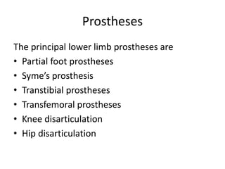
  • 1. Prostheses The principal lower limb prostheses are • Partial foot prostheses • Syme’s prosthesis • Transtibial prostheses • Transfemoral prostheses • Knee disarticulation • Hip disarticulation
  • 2. Partial Foot Prostheses • Purpose 1. Restore foot function particularly in walking 2. Simulate the shape of the missing foot segment • Indications 1. Loss of one or more toes 2. Transmetatarsal amputation 3. Amputation or disarticulation through tarsals
  • 3. Partial Foot Prostheses • Loss of one or more toes  Pad the toe section to improve appearance of the upper section of the shoe  Arch support to maintain alignment of amputated foot. • Transmetatarsal amputation  Plastic socket to protect amputated ends of the metatarsals  Rigid plate restores the foot length  Toe filler  Rocker bar at the bottom of the prosthesis to aid late stance.
  • 4. Partial foot prostheses • Amputation or disarticulation through tarsals Prostheses used in transmetatarsal amputation augmented with a plastic calf shell which is strapped around the leg.
  • 5. Trans-tibial Prostheses Trans-tibial level refers to - • Amputation in which tibia and fibula are transected • Retention of anatomical knee joint • Intact motor and sensory functions. • Indicated in vascular diseases.
  • 6. Parts of Trans-tibial Prostheses 1. Foot-ankle assembly 2. Socket 3. Shank 4. Suspension component
  • 7. Foot-ankle assembly Uses • Restores contour of the foot • Absorbs shock at heel contact • Plantarflexes in early stance • Simulates metatarsophalangeal hyperextension in late stance phase • In neutral position during swing phase
  • 8. Foot-ankle assembly  Types • Non-articulated feet 1. SACH foot (solid ankle cushion heel) 2. SAFE foot (stationary attachment flexible endoskeleton) • Articulated feet 1. Single-Axis feet 2. Multiple-Axis feet
  • 9. SACH FOOT (Solid ankle Cushion Heel)
  • 10. SAFE FOOT ( Solid Ankle Flexible Endoskeleton)
  • 11. Shank  Substitute for human leg  Restores length and shape  Located above foot-ankle assembly and below socket in transtibial prostheses  Types • Exoskeleton shank • Endoskeleton shank
  • 13. Socket Uses • Maximum distribution of load • Assist in venous blood circulation • Provide tactile feedback Patellar tendon bearing (PTB) socket has a prominent indentation over patella tendon. Socket has reliefs and build-ups
  • 14.
  • 15.
  • 16. Structure of Socket • Reliefs-concavities in the socket over areas contacting sensitive structures such as bony prominences. • Located over i. fibular head, ii. tibial crest, iii. tibial condyles and iv. anterior-distal tibia. • Posterior brim is trimmed to provide room for hamstring tendons
  • 17. Structure of Socket • Build-ups- convexities n the socket over areas contacting pressure-tolerant tissues. • Located over i. belly of gastrocnemius, ii. patellar tendon, iii. proximomedial tibia (pes anserinus) and iv. tibial and fibular shafts.
  • 18.
  • 19.
  • 20. Alignment of Socket On the shank in slight flexion and in slight lateral tilt. • Flexion i. enhances loading on patellar tendon, ii. prevent genu recurvatum, iii. resist tendency of amputated limb to slide too deeply into socket, iv. facilitates contraction of quadriceps muscle • Lateral tilt reduces loading on fibular head
  • 21. Types of socket • Lined socket i. Made of polyethylene foam liner, silicone ii. Liner adds or reduces volume of the socket iii. Adds to the bulk of the prosthesis iv. Heat insulator v. Reduces risk of abrasion between socket and skin • Unlined socket i. Made of thermoplastic material ii. Usually given when stump has stabilized in volume iii. Easier to clean iv. Difficult to alter the shape of the socket.
  • 22. Suspension • Prosthesis requires some from suspension to hold it in place while walking or climbing stairs or jumping. • Types of suspension i. Cuff variants ii. Distal attachment iii. Brim variants iv. Thigh corset v. Vacuum-assisted socket system
  • 23.
  • 24.
  • 26.
  • 27. Trans-femoral prosthesis Components • Foot-ankle assembly • Shank • Knee unit • Socket • Suspension device
  • 28. Knee Unit Knee units have four features: • Axis • Friction mechanism • Extension aid • Mechanical stabilizer
  • 29. Knee Unit: AXIS SYSYTEM Two types of knee units i. Single axis ii. Polycentric linkage a) 4 or more pivoting bars b) Provide greater stability c) center of knee rotation in posterior to weight bearing line
  • 30. Knee Unit: FRICTION MECHANISMS • Change the knee swing by modifying speed of knee motion during swing phase • Affect knee swing according to walking speed. • Two factors affecting friction mechanism are i. Time during swing phase when friction affects knee joint ii. medium through which mechanism operates
  • 31. Knee unit:FRICTION MECHANISMS • Constant friction i. Clamp grasping knee joint ii. Amount of friction is unvarying in swing phase iii. Manually adjusted to loosen or tighten • Variable friction i. Amount of friction is variable in swing phase ii. Early swing –high friction iii. Mid-swing –friction diminishes iv. Late swing -friction increases
  • 32. Knee Unit: FRICTION MECHANISM Medium  Sliding friction  Fluid friction - hydraulic friction - pneumatic friction  Microprocessor controls
  • 33. Knee Unit: EXTENSION AID • A mechanism to assist knee extension during latter part of swing phase • Types- i. Elastic webbing ii. Internal extension aid
  • 34. Knee Unit:STABILIZERS • To increase stability of knee unit • Hip motion controls knee action, aided by alignment of knee in relation to other components of prosthesis • Knee joint is aligned posterior to line extending from trochanter to ankle (TKA line) • Types i. Manual lock ii. Friction brakes
  • 35. SOCKET Pressure tolerant areas • Gluteal musculature • Sides of the thigh • And distal end of amputated limb Pressure sensitive areas • Pubic symphysis • Perineum
  • 36.
  • 37. Types of socket Quadrilateral socket i. Post wall-ischial tuberosity + gluteal muscles ii. Ant wall- applies post directed pressure iii. Lat wall- aid in medio-lateral stabilization iv. Med wall Reliefs i. Antero-medial ii. Postero-medial iii. Antero-lateral iv. Postero-lateral Ischial Containment socket i. Contoured adducted trochanter-controlled alignment method. ii. Covers ischial tuberosity and part of ischiopubic ramus to increase stability. iii. To increase frontal stability medial-lateral width is narrow. iv. Lateral wall covers greater trochanter.
  • 38. Suspension Suction suspension i. Refers to pressure differences inside and outside the socket. ii. In suction suspension,(int socket press) < (ext pressure), therefore atm press. causes the socket to remain on the thigh iii. One-way air-release valve enables residual air to be expelled iv. Types of suspension are a) total suction, b) partial suction and c) no suction
  • 40. FIT AND ALIGNMENT • FIT i. Snug fitting to minimize chaffing and maximize control • ALIGNMENT – slight socket flexion i. Facilitates contraction of hip extensors ii. Reduce lumbar lordosis iii. Allows equal step length
  • 41. DIFFERENT TYPES OF LOWER LIMB PROSTHESIS
  • 42. KNEE DISARTICULATION PROSTHESIS Excellent prosthetic control because i. Thigh leverage is maximum ii. Body weight can be borne through distal end of femur iii. Epicondyles provide rotational stability
  • 43. HIP DISARTICULATION PROSTHESIS • Indications i. Amputation above greater trochanter (short transfemoral) ii. Removal of the femoral head from acetabulum (hip disarticulation) iii. Removal of femur and portion of pelvis (transpelvic amputation)
  • 44.

Notas do Editor

  1. 1 retard excessive knee flexion 2 permit knee to swing easily 3 to dampen impact