
Recomendados
Mais conteúdo relacionado
Mais procurados
Mais procurados (20)
Destaque
Destaque (19)
Semelhante a Drug receptors
Semelhante a Drug receptors (20)
Último
Último (20)
Drug receptors
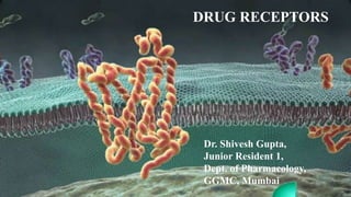
- 1. DRUG RECEPTORS Dr. Shivesh Gupta, Junior Resident 1, Dept. of Pharmacology, GGMC, Mumbai
- 2. Flow of Seminar 1. Introduction 2. History 3. Drug-Receptor Interactions 4. Classification 5. Transducer mechanisms 6. Regulation of receptors 7. Function of receptors 8. Recent advances 9. Summary 10. References
- 3. Introduction • The term drug receptor or drug target denotes the cellular macromolecule or macromolecular complex with which the drug interacts to elicit a cellular response, i.e., a change in cell function. • These are the sensing elements in the system of chemical communications that coordinates the function of all the different cells in the body. GOODMAN, GILMAN, L.BRUTON: THE PHARMACOLOGICAL BASIS OF THERAPEUTICS; 12TH EDITION, NEW YORK: MCGRAW HILL MEDICAL; 2011.
- 4. HISTORY 1. The birth of the receptor concept was the outcome of circumstances in the lives of its two founding fathers, the physiologist John Newport Langley (1852–1925) and the immunologist and bacteriologist Paul Ehrlich (1854– 1915). 2. John N. Langley is known as one of the fathers of the chemical receptor theory, and gave the origin of the concept of "receptive substance“ in 1878. 3. P. Ehrlich designated the term ‘Receptor’ in 1900. K.D TRIPATHI: ESSENTIALS OF MEDICAL PHARMACOLOGY; 7TH EDITION. NEW DELHI: JAYPEE BROTHERS; 2013.
- 5. Theories Proposed Receptor occupancy theory: Clark (1937) propounded a theory of drug action based on occupation of receptors by specific drugs. The interaction between the two molecular species, viz. drug (D ) and receptor (R) to be governed by the law of mass action, and the effect (E) to be a direct function of the drug-receptor complex (DR) formed. K.D TRIPATHI: ESSENTIALS OF MEDICAL PHARMACOLOGY; 7TH EDITION. NEW DELHI: JAYPEE BROTHERS; 2013.
- 6. Contd…. Two – state receptor theory: It was first described by Black and Leff in 1983 as an alternative model of receptor activation. It proposes that ligand binding results in a change in receptor state from an inactive to an active state based on the receptor's conformation. A receptor in its active state will ultimately elicit its biological response. K.D TRIPATHI: ESSENTIALS OF MEDICAL PHARMACOLOGY; 7TH EDITION. NEW DELHI: JAYPEE BROTHERS; 2013.
- 7. Contd…. It is model proposed for explaining the action of agonists, antagonists, partial agonists and inverse agonists. The receptor is believed to exist in two interchangeable states: Ra (active) and Ri (inactive) which are in equilibrium. In the case of majority of receptors, the Ri state is favoured at equilibrium
- 8. Drug-Receptor Interactions The drug receptor interaction is characterized by (1) binding of drug to receptor and (2) generation of a response in a biological system. Affinity: The ability of the drug to bind with the receptor and form D-R complex. Intrinsic activity (IA) or efficacy: The capacity of the drug to induce a functional change in the receptor after binding. K.D TRIPATHI: ESSENTIALS OF MEDICAL PHARMACOLOGY; 7TH EDITION. NEW DELHI: JAYPEE BROTHERS; 2013.
- 9. Contd…. Based on Affinity and Intrinsic Activity, the drug can be labelled as follows: Agonist: An agent which activates a receptor to produce an effect similar to that of the physiological signal molecule. Agonists have both affinity and maximal intrinsic activity (IA = 1). e.g. adrenaline, histamine, morphine. Antagonist: An agent which prevents the action of an agonist on a receptor or the subsequent response, but does not have any effect of its own. Competitive antagonists have affinity but no intrinsic activity (IA = 0), e.g. propranolol, atropine, chlorpheniramine, naloxone. K.D TRIPATHI: ESSENTIALS OF MEDICAL PHARMACOLOGY; 7TH EDITION. NEW DELHI: JAYPEE BROTHERS; 2013.
- 10. Contd…. Inverse agonist: An agent which activates a receptor to produce an effect in the opposite direction to that of the agonist. Inverse agonists have affinity but intrinsic activity with a minus sign (IA between 0 and –1). e.g. DMCM (on benzodiazepine receptor), chlorpheniramine (on H1 histamine receptor). Partial agonist: An agent which activates a receptor to produce submaximal effect but antagonizes the action of a full agonist. Partial agonists have affinity and submaximal intrinsic activity (IA between 0 and 1). e.g. dichloroisoproterenol (on β adrenergic receptor), pentazocine (on μ opioid receptor). K.D TRIPATHI: ESSENTIALS OF MEDICAL PHARMACOLOGY; 7TH EDITION. NEW DELHI: JAYPEE BROTHERS; 2013.
- 11. Contd…. GOODMAN, GILMAN, L.BRUTON: THE PHARMACOLOGICAL BASIS OF THERAPEUTICS; 12TH EDITION, NEW YORK: MCGRAW HILL MEDICAL; 2011.
- 12. Classification The IUPHAR ( International Union of Pharmacological Science) system broadly classifies receptors as: 1. Pharmacological ( Mediator based e.g. Nor Epinephrine, Insulin) 2. Biochemical & Biophysical (Second messenger based e.g. cAMP, PLC) 3. Molecular or Structural (Subunit Composition e.g. 5HT1A) 4. Anatomical ( Tissue e.g. muscle or ganglionic Ach or Cellular e.g. Cell surface or intracellular) IUPHAR/ GUIDE TO PHARMACOLOGY
- 13. Transducer Mechanisms o These are complex multistep processes that provide for amplification and integration of concurrently received extra- and intracellular signals at each step. o Only a handful of transducer pathways are shared by large number of receptors, the cell is able to generate an integrated response reflecting the sum total of diverse signal input. H.P RANG, J.M. RITTER, R.J. FLOWER, G.HENDERSON: RANG & DALE’S PHARMACOLOGY; 8TH EDITION. CHINA: ELSEVIER; 2016.
- 14. Contd…. The transducer mechanisms classifies receptors into 4 major categories: 1. Ligand Gated Ion channel receptor 2. G-Protein Coupled Receptors (GPCRs) 3. Kinase-linked and related receptors 4. Receptors regulating gene expression ( Transcription factors, Nuclear factors) H.P RANG, J.M. RITTER, R.J. FLOWER, G.HENDERSON: RANG & DALE’S PHARMACOLOGY; 8TH EDITION. CHINA: ELSEVIER; 2016
- 15. Ligand-gated ion channels Also known as Ionotropic receptors. These are the receptors on which fast neurotransmitters act. These mediate fast synaptic transmission, on a millisecond time scale, in the nervous system and at the somatic neuromuscular junction. Typical example of this is Nicotinic Acetylcholine receptor at NM junction. Other examples include GABAA , 5HT3 receptors, Glycine receptors, IP3 receptors, Ionotropic Glutamate receptors, Purinergic PAX, etc. H.P RANG, J.M. RITTER, R.J. FLOWER, G.HENDERSON: RANG & DALE’S PHARMACOLOGY; 8TH EDITION. CHINA: ELSEVIER; 2016
- 16. Contd…. These are integral membrane proteins that contain a pore which allows the regulated flow of selected ions across the plasma membrane. Ion flux is passive and driven by the electrochemical gradient for the permeant ions. These channels are open, or gated, by the binding of a neurotransmitter to an orthosteric site(s) that triggers a conformational change that results in the conducting state. Modulation of gating can occur by the binding of endogenous, or exogenous, modulators to allosteric sites. H.P RANG, J.M. RITTER, R.J. FLOWER, G.HENDERSON: RANG & DALE’S PHARMACOLOGY; 8TH EDITION. CHINA: ELSEVIER; 2016
- 17. Molecular structure The nicotinic acetylcholine receptor consists of a pentameric assembly of four subunits, termed α, β, γ and δ, each of molecular weight 40–58 kDa. The pentameric structure (2α, β, γ, δ) possesses two acetylcholine binding sites, each lying at the interface between one of the two α subunits and its neighbour. Both must bind acetylcholine molecules in order for the receptor to be activated. Each subunit contains four membrane-spanning α-helices, inserted into the membrane. H.P RANG, J.M. RITTER, R.J. FLOWER, G.HENDERSON: RANG & DALE’S PHARMACOLOGY; 8TH EDITION. CHINA: ELSEVIER; 2016
- 18. Contd…. Mechanism of Action: The five helices that form the pore are sharply kinked inwards halfway through the membrane, forming a constriction. When two acetylcholine molecules bind to the binding sites, a conformational change occurs in the extracellular part of the receptor. This twists the α subunits, causing the kinked helical segments to swivel out of the way, thus opening the channel. H.P RANG, J.M. RITTER, R.J. FLOWER, G.HENDERSON: RANG & DALE’S PHARMACOLOGY; 8TH EDITION. CHINA: ELSEVIER; 2016
- 19. Contd…. Clinical Significance: Nicotinic Ach receptors and Glutamate receptors are responsible for the majority of synaptic transmission by neurons in CNS as well as in the periphery. So, they help in generation and propagation in nerve impulses. IP3 sensitive Ca2+ channels are responsible for release of Ca2+ from ER and this can be blocked by drugs in treatment of Malignant Hyperthermia. The Sulfonylurea Receptor ‘SUR 1’ regulates the ATP-dependent K+ channels are the prime target of oral hypoglycemic drugs in treatment of Type II DM. GOODMAN, GILMAN, L.BRUTON: THE PHARMACOLOGICAL BASIS OF THERAPEUTICS; 12TH EDITION, NEW YORK: MCGRAW HILL MEDICAL; 2011.
- 20. G-protein coupled receptors (GPCRs): These are also known as Metabotropic or 7- Transmembrane (7TM) receptors. G protein-coupled receptors (GPCRs) are the largest class of membrane proteins in the human genome. Humans express over 800 GPCRs that make up the third largest family of genes in humans. Examples include chemoreceptors in olfaction, taste & vision, Musuranic AchRs, Adrenoceptors, dopamine receptors, GABAB many peptide and purine receptors and many Orphan receptors. GOODMAN, GILMAN, L.BRUTON: THE PHARMACOLOGICAL BASIS OF THERAPEUTICS; 12TH EDITION, NEW YORK: MCGRAW HILL MEDICAL; 2011.
- 21. Contd…. They are membrane receptors that are coupled to intracellular effector systems primarily via a G protein. G proteins are heterotrimeric GTP-binding regulatory proteins. G proteins are signal transducers that convey the information that agonist is bound to the receptor from the receptor to one or more effector proteins. G–protein-regulated effectors include enzymes such as adenylyl cyclase, phospholipase C, cyclic GMP, phosphodiesterase (PDE6), and membrane ion channels selective for Ca2+ and K+. GOODMAN, GILMAN, L.BRUTON: THE PHARMACOLOGICAL BASIS OF THERAPEUTICS; 12TH EDITION, NEW YORK: MCGRAW HILL MEDICAL; 2011.
- 22. Classification of GPCRs On the basis of sequence homology, GPCRs are classified into six classes. These classes and their prototype members were as follows: Class A (rhodopsin-like), Class B (secretin receptor family), Class C (metabotropic glutamate), Class D (fungal mating pheromone receptors), Class E (cyclic AMP receptors) and Class F (frizzled/smoothened). Of these, classes D and E are not found in vertebrates IUPHAR/ GUIDE TO PHARMACOLOGY
- 23. Contd…. H.P RANG, J.M. RITTER, R.J. FLOWER, G.HENDERSON: RANG & DALE’S PHARMACOLOGY; 8TH EDITION. CHINA: ELSEVIER; 2016
- 24. Contd…. An alternative classification scheme "GRAFS" divides GPCRs into five classes: o Glutamate family (class C): eg. metabotropic glutamate receptors, a calcium-sensing receptor and GABAB receptors. o Rhodopsin family (class A): eg. neurotransmitters, peptides and hormones, olfactory receptors, visual pigments, taste type 2 receptors. o Adhesion family: These are phylogenetically realted to class B receptors, from which they differ by possessing large extracellular N-terminal. o Frizzled family: Consists of 10 Frizzled proteins (FZD(1-10)) and Smoothened (SMO). o Secretin family: These are encoded by 15 genes in humans. The ligands for receptors in this family are: glucagon, glucagon-like peptides (GLP-1, GLP-2), GIP, secretin, VIP, pituitary adenylate cyclase-activating polypeptide (PACAP) and GHRH. IUPHAR/ GUIDE TO PHARMACOLOGY
- 25. Molecular structure The first GPCR to be fully characterised was the β adrenoceptor, which was cloned in 1986. The GPCR molecule has 7 α-helical membrane spanning hydrophobic amino acid (AA) segments which run into 3 extracellular and 3 intracellular loops. Two binding sites: agonist binding and G-protein coupling. The third intracellular loop interacts with the G protein. K.D TRIPATHI: ESSENTIALS OF MEDICAL PHARMACOLOGY; 7TH EDITION. NEW DELHI: JAYPEE BROTHERS; 2013.
- 26. G-proteins These comprise a family of membrane-resident proteins whose function is to recognise activated GPCRs and pass on the message to the effector systems that generate a cellular response. They are called G proteins because of their interaction with the guanine nucleotides, GTP and GDP. G proteins consist of three subunits: α, β and γ. Guanine nucleotides bind to the α subunit, which has enzymatic (GTPase) activity, catalysing the conversion of GTP to GDP. The β and γ subunits remain together as a βγ complex. H.P RANG, J.M. RITTER, R.J. FLOWER, G.HENDERSON: RANG & DALE’S PHARMACOLOGY; 8TH EDITION. CHINA: ELSEVIER; 2016
- 27. Contd…. GOODMAN, GILMAN, L.BRUTON: THE PHARMACOLOGICAL BASIS OF THERAPEUTICS; 12TH EDITION, NEW YORK: MCGRAW HILL MEDICAL; 2011. In the basal state of the receptor-heterotrimer complex, the α subunit contains bound GDP and the α-GDP:βγ complex is bound to the unliganded receptor. The G protein family is comprised of 23 α subunits, 7 β subunits, and 12 γ subunits.
- 28. Contd…. A number of G-proteins distinguished by their α subunits have been described. The important ones with their action on the effector are: 1. Gs- Adenylyl cyclase activation, Ca+ channel opening. (Activated by cholera toxin) 2. Gi- Adenylyl cyclase inhibition, K+ channel opening. (Blocked by pertussis toxin) 3. Go- Ca+ channel inhibiton. 4. Gq- Phospholipase C activation. K.D TRIPATHI: ESSENTIALS OF MEDICAL PHARMACOLOGY; 7TH EDITION. NEW DELHI: JAYPEE BROTHERS; 2013.
- 29. Contd…. The following couplers have been associated with different G receptors: In addition, Gs is the coupler for histamine H2, serotonin 5HT4-7 and many more. Gi is utilized by opioid, cannabinoid and some other receptors. K.D TRIPATHI: ESSENTIALS OF MEDICAL PHARMACOLOGY; 7TH EDITION. NEW DELHI: JAYPEE BROTHERS; 2013. Receptor Coupler Muscuranic M2 Gi, Go Muscuranic M1, M3 Gq Dopamine D2 Gi, Go β-adrenergic Gs α1- adrenergic Gq α2- adrenergic Gi, Go
- 30. Mechanism of Action When an agonist binds to a GPCR, there is a conformational change in the receptor. This causes the α subunit to exchange its bound GDP for GTP. Binding of GTP activates the α subunit and causes it to release both the βγ dimer and the receptor. Both the GTP bound α subunit and the βγ heterodimer become active signaling molecules. GOODMAN, GILMAN, L.BRUTON: THE PHARMACOLOGICAL BASIS OF THERAPEUTICS; 12TH EDITION, NEW YORK: MCGRAW HILL MEDICAL; 2011.
- 31. Contd…. GOODMAN, GILMAN, L.BRUTON: THE PHARMACOLOGICAL BASIS OF THERAPEUTICS; 12TH EDITION, NEW YORK: MCGRAW HILL MEDICAL; 2011.
- 32. Contd…. GOODMAN, GILMAN, L.BRUTON: THE PHARMACOLOGICAL BASIS OF THERAPEUTICS; 12TH EDITION, NEW YORK: MCGRAW HILL MEDICAL; 2011
- 33. Effector pathways The main targets for G proteins, through which GPCR control different aspects of cell function are: o Adenylyl cyclase, the enzyme responsible for cAMP formation. o Phospholipase C, the enzyme responsible for inositol phosphate and diacylglycerol (DAG) formation. o Ion channels, particularly calcium and potassium channels. H.P RANG, J.M. RITTER, R.J. FLOWER, G.HENDERSON: RANG & DALE’S PHARMACOLOGY; 8TH EDITION. CHINA: ELSEVIER; 2016
- 34. Contd…. H.P RANG, J.M. RITTER, R.J. FLOWER, G.HENDERSON: RANG & DALE’S PHARMACOLOGY; 8TH EDITION. CHINA: ELSEVIER; 2016 Activation of AC intracellular accumulation of second messenger cAMP Activates Protein Kinase (PKA) Phosphorylates and alters the function of many enzymes, ion channels, transporters, transcription factors and structural proteins Increased contractility/ impulse generation, relaxation, glycogenolysis, lipolysis, secretion of hormones, etc. Adenylyl cyclase/cAMP pathway:
- 35. Contd…. Some examples of role of increased cAMP are: β-adrenoceptor activation: helps in increasing blood glucose. Phosphorylation of voltage gated Ca2+ channels in heart muscle cells increases the force of contraction of the heart. In smooth muscle, cAMP phosphorylates (thereby inactivating), myosin light-chain kinase, required for contraction. This accounts for the smooth muscle relaxation. H.P RANG, J.M. RITTER, R.J. FLOWER, G.HENDERSON: RANG & DALE’S PHARMACOLOGY; 8TH EDITION. CHINA: ELSEVIER; 2016
- 36. Contd…. H.P RANG, J.M. RITTER, R.J. FLOWER, G.HENDERSON: RANG & DALE’S PHARMACOLOGY; 8TH EDITION. CHINA: ELSEVIER; 2016 Activation of phospholipase Cβ Hydrolyses the membrane PIP2 to generate IP3 and DAG IP3 diffuses to cytosol and mobilises calcium from ER whereas, DAG stays in the membrane and recruits and activates PKc. The activated PKc phosphorylates many intracellular proteins and mediated various physiological responses. Phospholipase C : IP3-DAG pathway:
- 37. Contd…. • Another major function of G protein-coupled receptors is to control ion channel function directly. • These do not involve second messengers such as cAMP or inositol phosphates. • Examples: • In Cardiac muscles, mAChRs enhance K+ permeability. • In neurons, inhibitory drugs such as opioid analgesics reduce excitability by opening certain K+ channels or by inhibiting voltage-activated Ca2+ channels and thus reducing neurotransmitter release. H.P RANG, J.M. RITTER, R.J. FLOWER, G.HENDERSON: RANG & DALE’S PHARMACOLOGY; 8TH EDITION. CHINA: ELSEVIER; 2016
- 38. K.D TRIPATHI: ESSENTIALS OF MEDICAL PHARMACOLOGY; 7TH EDITION. NEW DELHI: JAYPEE BROTHERS; 2013. Major functional pathways of GPCR transduction Adenylyl cyclase: cAMP Phospholipase IP3-DAG Channel regulation Increases Decreases Increase Ca2+ Decrease Ca2+ Increase K+ Adrenergic-β Adrenergic-α2 Adrenergic-α1 Adrenergic-β1 (Heart, Sk. Muscle) Dopamine-D2 Adrenergic-α2 Histamine-H2 Muscuranic-M2 Histamine-H1 GABAB Muscuranic-M2 Dopamine-D1 Dopamine-D2 Muscuranic-M1, M3 Opioid- κ Dopamine-D2 Glucagon 5-HT1 5-HT2 Adenosine-A1 5-HT1A FSH & LH GABAB Vasopressin-oxytocin Somtostatin GABAB ACTH Opioid- µ, δ Bradykinin-B2 Opioid- µ, δ TSH Angiotensin-AT1 Angiotensin-AT1 Adenosine-A1 Prostaglandin- EP2 Prostaglandin- EP3 Prostaglandin-FP, EP1 EP3 Prostacyclin-IP Somatostatin Thromboxane- TP Adenosine-A2 Adenosine-A1 Leukotriene BLT, cys LT Cholecystokinin-Gastrin PAF
- 39. Contd…. Clinical Significance: Approx 45% of all pharmaceutical drugs are known to target these GPCRs. Examples include: K.D TRIPATHI: ESSENTIALS OF MEDICAL PHARMACOLOGY; 7TH EDITION. NEW DELHI: JAYPEE BROTHERS; 2013.
- 40. Contd…. K.D TRIPATHI: ESSENTIALS OF MEDICAL PHARMACOLOGY; 7TH EDITION. NEW DELHI: JAYPEE BROTHERS; 2013.
- 41. Contd…. Abnormal G protein signalling can occur due to: a) Bacterial toxins. Eg. Cholera and Pertussis toxin. b) Gene mutations: 1. Gain of function mutation: can lead to development of Congenital Nightblindness (rhodopsin), Familial Precocious puberty (LH), Familial Hypocalcemia (Ca2+ sensing), etc. 2. Loss of function mutation: can lead to development of Retinitis Pigmentosa (rhodopsin), Diabetes Insipidus (V2), Hypocalciuric Hypercalcemia (Ca2+ sensing), etc. c) GPCR Misfoldings: can lead to diseases like Nephrogenic DI (V2R), Hypogonadotrophic Hypogonadism (GnRHR),etc. GOODMAN, GILMAN, L.BRUTON: THE PHARMACOLOGICAL BASIS OF THERAPEUTICS; 12TH EDITION, NEW YORK: MCGRAW HILL MEDICAL; 2011
- 42. Kinase-linked and related receptors • Diverse group of physiological membrane receptors. • They have extracellular ligand binding domains and an intrinsic enzymatic activity on the cytoplasmic surface of the cell. • Most are activated by a wide variety of protein mediators, including growth factors and cytokines, hormones such as insulin and leptin. • Here, the effects are exerted mainly at the level of gene transcription. H.P RANG, J.M. RITTER, R.J. FLOWER, G.HENDERSON: RANG & DALE’S PHARMACOLOGY; 8TH EDITION. CHINA: ELSEVIER; 2016
- 43. Contd…. The main types are as follows: 1. Receptor tyrosine kinases (RTKs) 2. Cytokine receptors ( JAK-STAT Receptor) 3. Receptor serine/threonine kinases 4. Others ( Toll-like Receptors, TNF-α) H.P RANG, J.M. RITTER, R.J. FLOWER, G.HENDERSON: RANG & DALE’S PHARMACOLOGY; 8TH EDITION. CHINA: ELSEVIER; 2016
- 44. Contd…. Receptor tyrosine kinases (RTKs): These molecules consist of single polypeptide chains. Have large, cysteine-rich extracellular domains. Have short transmembrane domains. The intracellular region containing one (or in some cases two) protein tyrosine kinase domains. Examples include receptors for hormones such as insulin, for multiple growth factors such as EGF, PDGF, NGF, FGF, VEGF, and ephrins. GOODMAN, GILMAN, L.BRUTON: THE PHARMACOLOGICAL BASIS OF THERAPEUTICS; 12TH EDITION, NEW YORK: MCGRAW HILL MEDICAL; 2011
- 45. Mechanism of action H.P RANG, J.M. RITTER, R.J. FLOWER, G.HENDERSON: RANG & DALE’S PHARMACOLOGY; 8TH EDITION. CHINA: ELSEVIER; 2016
- 46. Contd…. Cytokine receptors ( JAK-STAT Receptor): • These receptors signal to the nucleus by a more direct manner than the receptor tyrosine kinases. • These receptors have no intrinsic enzymatic activity. • The intracellular domain binds a separate, intracellular tryosine kinase termed a Janus kinase (JAK). • JAKs phosphorylate other proteins termed signal transducers and activators of transcription (STATs). • Examples include receptors for cytokines such as γ-interferon, hormones like growth hormone and prolactin. GOODMAN, GILMAN, L.BRUTON: THE PHARMACOLOGICAL BASIS OF THERAPEUTICS; 12TH EDITION, NEW YORK: MCGRAW HILL MEDICAL; 2011
- 47. Mechanism of action H.P RANG, J.M. RITTER, R.J. FLOWER, G.HENDERSON: RANG & DALE’S PHARMACOLOGY; 8TH EDITION. CHINA: ELSEVIER; 2016
- 48. Contd…. Receptor serine/threonine kinases: This smaller class is similar in structure to RTKs. However, they phosphorylate serine and/or threonine residues rather than tyrosine. The activated receptor on ligand binding, phosphorylates a gene regulatory protein termed a Smad. The main example is the receptor for transforming growth factor (TGF-β). GOODMAN, GILMAN, L.BRUTON: THE PHARMACOLOGICAL BASIS OF THERAPEUTICS; 12TH EDITION, NEW YORK: MCGRAW HILL MEDICAL; 2011
- 49. Contd…. Other receptors include: Toll-Like Receptors: • Signaling related to the innate immune system. • Highly expressed in hematopoeitic cells. • Ligands comprise of a multitude of pathogen products including lipids, peptidoglycans, lipopeptides, and viruses. • Activation of these receptors produces an inflammatory response to the pathogenic microorganisms. Tumor Necrosis Factor α (TNF-α): Eg: Signaling to the NF-κB transcription factors. GOODMAN, GILMAN, L.BRUTON: THE PHARMACOLOGICAL BASIS OF THERAPEUTICS; 12TH EDITION, NEW YORK: MCGRAW HILL MEDICAL; 2011
- 50. Contd…. Clinical Significance: These are key regulators of critical cellular processes, such as proliferation and differentiation, cell survival and metabolism, cell migration and cell cycle control. RTKs are seen as drug targets in many types of cancer and other disease states like inflammation, neurodegeneration, atherosclerosis. Many diseases result from genetic changes or abnormalities that either alter the activity and/or regulation of RTKs. IUPHAR/ GUIDE TO PHARMACOLGY
- 51. Contd…. Few classic examples of Drugs that act at receptors in this diverse family: 1. Insulin for the treatment of diabetes mellitus. 2. Imatinib used to treat chronic myelogenous leukemia and several solid tumors with dysregulated tyrosine kinases. 3. Humanized monoclonal antibodies to TNF-α itself, such as infiximab and adalimumab, are important for the treatment of rheumatoid arthritis and Crohn’s disease. GOODMAN, GILMAN, L.BRUTON: THE PHARMACOLOGICAL BASIS OF THERAPEUTICS; 12TH EDITION, NEW YORK: MCGRAW HILL MEDICAL; 2011
- 52. Receptors regulating gene expression Also called as Nuclear Receptors (NR). NRs can directly interact with DNA, so called ligand activated transcription factors. These transduce signals by modifying gene transcription. Examples are receptors for steroid hormones, glucocorticoids, mineralocorticoids, thyroid hormone, Vit D, RXR, LXR, PPAR, etc. H.P RANG, J.M. RITTER, R.J. FLOWER, G.HENDERSON: RANG & DALE’S PHARMACOLOGY; 8TH EDITION. CHINA: ELSEVIER; 2016
- 53. Contd…. H.P RANG, J.M. RITTER, R.J. FLOWER, G.HENDERSON: RANG & DALE’S PHARMACOLOGY; 8TH EDITION. CHINA: ELSEVIER; 2016 Diagram of a nuclear receptor Molecular Structure: The N-terminal domain displays the most heterogeneity. It harbours the AF1 (activation function 1) site. The core domain of the receptor is highly conserved and consists of the structure responsible for DNA recognition and binding. It is the highly flexible hinge region in the molecule that allows it to dimerise with other NRs. The C-terminal domain contains the ligand-binding module and is specific to each class of receptor.
- 54. Classification H.P RANG, J.M. RITTER, R.J. FLOWER, G.HENDERSON: RANG & DALE’S PHARMACOLOGY; 8TH EDITION. CHINA: ELSEVIER; 2016
- 55. Mechanism ofAction H.P RANG, J.M. RITTER, R.J. FLOWER, G.HENDERSON: RANG & DALE’S PHARMACOLOGY; 8TH EDITION. CHINA: ELSEVIER; 2016
- 56. Contd H.P RANG, J.M. RITTER, R.J. FLOWER, G.HENDERSON: RANG & DALE’S PHARMACOLOGY; 8TH EDITION. CHINA: ELSEVIER; 2016
- 57. Contd…. Clinical Significance: 1. NRs are very important drug targets, being responsible for the biological effects of approximately 10–15% of all prescription drugs. 2. NRs also regulate expression of many drug metabolising enzymes and transporters. 3. Many illnesses are associated with malfunctioning of the NR system, including inflammation, cancer, diabetes, cardiovascular disease, obesity and reproductive disorders. H.P RANG, J.M. RITTER, R.J. FLOWER, G.HENDERSON: RANG & DALE’S PHARMACOLOGY; 8TH EDITION. CHINA: ELSEVIER; 2016
- 58. H.P RANG, J.M. RITTER, R.J. FLOWER, G.HENDERSON: RANG & DALE’S PHARMACOLOGY; 8TH EDITION. CHINA: ELSEVIER; 2016
- 59. Regulation of Receptors Receptors exist in a dynamic state, i.e. their density and efficacy to elicit a response is subject to regulation by the level of on-going activity, feedback from their own signal output and other pathophysiological influences. The mechanisms involved may be unmasking of receptors or their proliferation (up regulation) or accentuation of signal amplification by the transducer. Eg. – Estrogens increase the density of oxytocin receptors on the myometrium, and the sensitivity of uterus to contractile action of oxytocin increases progressively during the third trimester of pregnancy, especially near term. K.D TRIPATHI: ESSENTIALS OF MEDICAL PHARMACOLOGY; 7TH EDITION. NEW DELHI: JAYPEE BROTHERS; 2013.
- 60. Contd…. Conversely, continued / intense receptor stimulation causes desensitization or refractoriness: the receptor becomes less efficient in transducing response to the agonist. The mechanisms by which this is brought about is: Masking or internalization of the receptor or impaired coupling of the transducer to the receptor. Eg. In case of β adrenergic receptors. Decreased synthesis/ increased destruction of receptors (down regulation). Eg. In tyrosine kinase receptors. Eg. Includes patients of bronchial asthma being treated continuously with β adrenergic agonists gradually become less responsive. K.D TRIPATHI: ESSENTIALS OF MEDICAL PHARMACOLOGY; 7TH EDITION. NEW DELHI: JAYPEE BROTHERS; 2013.
- 61. Functions These are: 1. To propagate regulatory signals from outside to inside the effector cell when the molecular species carrying the signal cannot itself penetrate the cell membrane. 2. To amplify the signal. 3. To integrate various extracellular and intracellular regulatory signals. 4. To adapt to short term and long term changes in the regulatory melieu and maintain homeostasis. K.D TRIPATHI: ESSENTIALS OF MEDICAL PHARMACOLOGY; 7TH EDITION. NEW DELHI: JAYPEE BROTHERS; 2013.
- 62. Recent Advances 1. YM-254890 and FR900359: These are related cyclic depsi-peptide natural products. Specifically and potently inhibit the Gq subfamily of G proteins. Is targeted in treatment of CNS disorders. 2. GPR83: Is currently classified as an orphan receptor. Mainly localised in the mouse to the CNS. It has been implicated in behavior, learning, and metabolic regulation. A biologically active peptide, PEN is now identified as a ligand activating GPR83. 1. NISHIMURA A. ET. AL.(2010) STRUCTURAL BASIS FOR THE SPECIFIC INHIBITION OF HETEROTRIMERIC GQ PROTEIN BY A SMALL MOLECULE. PROC NATL ACAD SCI; 107(31): 13666–13671. 2. GOMES I ET AL. (2016). IDENTIFICATION OF GPR83 AS THE RECEPTOR FOR THE NEUROENDOCRINE PEPTIDE PEN.SCI. SIGNAL. 9(425): RA43.
- 63. Contd…. 1. GPR68: G-protein coupled receptor belongs to a proton sensing family detecting acidic pH. A potent GPR86 positive allosteric modulator (PAM) named ogerin is identified. GPR68 plays a role in anxiety. This suggests a potential new drug target in this and related CNS disorders. 2. CSF1R: Inhibition of CSF1R by a tyrosine kinase inhibitor results in the blockade of microglial proliferation on transgenic mouse model of Alzheimer's-like pathology. There was improved memory and behavioural performance as well as prevention of synaptic degeneration. This suggests the therapeutic strategy of modifying CSF1R activation could ameliorate Alzheimer's disease. 1. HUANG XP, KARPIAK J, KROEZE WK ET AL. (2015). ALLOSTERIC LIGANDS FOR THE PHARMACOLOGICALLY DARK RECEPTORS GPR68 AND GPR65 NATURE 527: 477-83. 2. OLMOS-ALONSO A, SCHETTERS ST, SRI S ET AL. (2016). PHARMACOLOGICAL TARGETING OF CSF1R INHIBITS MICROGLIAL PROLIFERATION AND PREVENTS THE PROGRESSION OF ALZHEIMER'S-LIKE PATHOLOGY BRAIN : EPUB JAN 8
- 64. Contd…. 1. Sigma receptor: It has been a ‘receptor-in-waiting’. Revealed as ‘fourth’ opioid receptor. Responds to nociceptin/orphanin. Has functional impact, in particular in the nervous and cardiovascular systems. 2. Sigma-2 Receptors: Receptor activation produced both transient and sustained increases in Ca2+ and cytotoxic effects. Sigma-2 agonists induced apoptosis in drug-resistant cancer cells. Also, enhanced the potency of DNA damaging agents. Sigma-2 receptor agonists may be useful in treatment of drug-resistant cancers. 1. SCHMIDT HR, ZHENG S, GURPINAR E ET AL. (2016). CRYSTAL STRUCTURE OF THE HUMAN Σ1 RECEPTOR. NATURE 532(7600): 527-530. 2. WAYNE D BOWEN, ET AL. SIGMA RECEPTORS: RECENT ADVANCES AND NEW CLINICAL POTENTIALS. PHARMACEUTICA ACTA HELVETIAE, VOL 74, ISSUES 2–3, 2000: 211–218.
- 65. Summary Receptors are molecules which are essential for majority of biochemical and metabolic processes in the body. Extensive research is being done on receptor pharmacology to find out new class of receptors. Newer Drug molecules that target different receptor proteins and alter their physiology are needed to be searched for. Discovery about mechanism of orphan receptors can lead to drug development for the effective treatment of diseases.
- 66. References 1. Goodman, Gilman, L.Bruton: The Pharmacological Basis of THERAPEUTICS; 12th Edition. New York: McGraw Hill Medical; 2011. 2. H.P Rang, J.M. Ritter, R.J. Flower, G.Henderson: RANG & DALE’S Pharmacology; 8th Edition. China: Elsevier; 2016. 3. K.D Tripathi: Essentials of Medical Pharmacology; 7th Edition. New Delhi: Jaypee Brothers; 2013. 4. IUPHAR/BPS. Guide to Pharmacology. www.guidetopharmacology.org/ 5. Nishimura A. et. al.(2010) Structural basis for the specific inhibition of heterotrimeric Gq protein by a small molecule. Proc Natl Acad Sci; 107(31): 13666–13671. 6. Gomes I et al. (2016). Identification of GPR83 as the receptor for the neuroendocrine peptide PEN.Sci. Signal. 9(425): ra43. 7. Huang XP, Karpiak J, Kroeze WK et al. (2015). Allosteric ligands for the pharmacologically dark receptors GPR68 and GPR65 Nature 527: 477-83. 8. Olmos-Alonso A, Schetters ST, Sri S et al. (2016). Pharmacological targeting of CSF1R inhibits microglial proliferation and prevents the progression of Alzheimer's-like pathology Brain : epub jan 8. 9. Schmidt HR, Zheng S, Gurpinar E et al. (2016). Crystal structure of the human σ1 receptor. Nature 532(7600): 527-530. 10. Wayne D Bowen, et al. Sigma receptors: recent advances and new clinical potentials. Pharmaceutica Acta Helvetiae, Vol 74, Issues 2– 3, 2000: 211–218.
- 67. THANK YOU
Notas do Editor
- However, the receptor itself has no other function. The drugs that act on the receptors just modify or alter the rate and magnitude of the response, but don’t create a new response.
- Langley studied mutual antagonism between pilocarpine and atropine and proposed that they both reacted with samr receptive substance. Ehlrich studied about quantitative neutralisation between toxin and antitoxin.
- He stated that mere occupation of receptor by the drug is not enough, the drug must also activate the receptor to generate the response.
- The channel lining contains a series of anionic residues, making the channel selectively permeable to cations, mainly to Na+ and K+, and few to Ca2+ as well. Cation selective receptors are for excitatory ligands like Ach. Anion selective receptors are for inhibitory ligands like Glycine & GABA.
- 5HT3 antagonists like Ondansetron is used to inhibit the Emesis caused by Various drugs. Autoantibodies against nAch is Myasthenia Gravis.
- The α subunit confers specific recognition to both receptors and effectors. The dimer of β and γ subunits confers membrane localization of the G protein heterorimer by prenylation of the γ subunit.
- The βγ diamer has also been shown to activate K+ channels, to inhibit Ca2+ channels and activate PI-3 kinase (PI3K).
- Following activation of one G protein, the receptor is freed to interact with other G proteins. The G protein remains active until the GTP bound to the α subunit is hydrolyzed to GDP by regulators of G protein signalling (RGSs).
- RGS is regulator of G prot Signalling.
- GPCR Kinases (GRK) phosphorylates specific GPCR and cause rapid desensitization of these GPCRs. This phosphorylations cause binding of arrestins that cause uncoupling of G prot from the receptors.
- Rho A/Rho kinase, a system that regulates the activity of many signalling pathways controlling cell growth and proliferation, smooth muscle contraction, etc. Mitogen-activated protein kinase (MAP kinase), a system that controls many cell functions, including cell division.
- Similarly, Receptors linked to Gi rather than Gs inhibit adenylyl cyclase, and thus reduce cAMP formation. Examples include: the M2 receptor of cardiac muscle, α2 smooth muscle and opioid receptors. Cyclic AMP is hydrolysed within cells by phosphodiesterases (PDEs), an important and ubiquitous family of enzymes. Eleven PDE subtypes exist, of which some isoforms of PDE (PDE-3, PDE-4) are selective for cAMP, while PDE-5 is selective for cGMP. Eg. of drugs that inhibit PDEs are Methylxanthines, Milrinone, Sildenafil, etc.
- Here, interaction through the βγ subunits of Gi and Go proteins, appears to be a general mechanism for controlling K+ and Ca2+ channels.
- So basically, they play a major role in controlling cell division, growth, differentiation, inflammation, tissue repair, apoptosis and immune responses.
- Activation of growth factor receptors leads to cell survival, cell proliferation, and differentiation. Activation of the ephrin receptors leads to neuronal angiogenesis, axonal migration, and guidance.
- There are 4 Jaks and 6 stats that combine differently depending upon the signal to produce nuclear transcription.
- Smad migrates to the nucleus, associates with transcription factors and regulates genes leading to morphogenesis and transformation. There are also inhibitory Smads ( smad 6 and 7 )that terminate the signalling.
- Therefore, drugs that modify the dysregulated functions of these RTKs have been developed that block the activation of RTKs directly or indirectly, or inhibit the tyrosine kinase activity directly.
- NRs are not embedded in membranes like GPCRs or ion channels, but are present as freely diffusible molecules in the soluble phase of the cell. Some (steroid receptors) can translocate from the cytoplasm to the nucleus, while others (RXR), probably dwell mainly within the nuclear compartment.
- The core region basically consists of two zinc fingers, i.e. cysteine rich loops in amino acid chain that is kept in this confirmation by zinc ions. AF1 is important for ligand-independent activation. The AF2 region is important in ligand-dependent activation.
- Heterologous desensitization: Some times response to all agonists, which act through different receptors but produce the same overt effect, is decreased by exposure to any one of these agonists. Eg. of agonists Include Histamine and Acetyl choline, both contract intestinal smooth muscles. Homologous desensitization: Here desensitization is limited to agonists of the same receptor that is being repeatedly activated.