SlideShare uma empresa Scribd logo
1 de 29
Baixar para ler offline
HYOID BONE


                                        Hyoid


 The U-shaped bone at the base of the tongue that
 supports the tongue muscles
SPINAL CORD & RIBS
CERVICAL VERTEBREA
The upper part of the spine consist of 7 bones.
The atlis is the bone right below the skull that hold it up and
the axis is the bone tight below that that helps it turn.
THORACIC VERTEBRAE
 This part of the vertebrae consist of 12 bones.
LUMBAR VERTEBRAE
This part of the vertebrae consist of 5 bones.
SACRUM
This contains 5 different bones that over time fuse into one.
COCCYX
• or tailbone, is the triangular bony structure located at the bottom
  of the vertebral column. It is composed of three to five bony
  segments held in place by joints and ligaments.
STERNUM
Also known as the manubrium sterni it is the broad,
upper part of the sternum.
TRUE RIBS
They are the first 7 ribs in the body. They are called
true ribs because they are actually connected to the
sternum directly.
FALSE RIBS
These are the 3 ribs right under the true ribs. They are
called false because they are no directly connected the
sternum, instead they are connected to a cartilage.
FLOATING RIBS
These are the last 2 ribs right below the rib cage. They are
called floating ribs because they are not connected to the
sternum or another ribs at all. They are just connected to the
spine and stick out.
CLAVICLE


           6
SCAPULA


          7
HUMERUS


          6
RADIUS AND ULNA


                  6
CARPALS AND METACARPALS AND PHALANGES

•   There are eight carpal bones in the base of each hand. There are five metacarpals in each hand.
    There are 16 bones for phalanges. From closet to the center of the hand going outward it goes from
    proximal, middle, and distal. There is no first middle phalange only proximal and distal.


                                                                                           1
The upper and lower extremities of humans offer many interesting points of
  comparison and of contrast. They and their individual components are
 homologous—i.e., of a common origin and patterned on the same basic
 plan. A long evolutionary history and profound changes in the function of
     these two pairs of extremities have led, however, to considerable
                       differences between them. (3)
•   lower extremity-
     •   Coxal (Pelvic Girdle)- What bones make up the Acetabelum? Howcan an anthropologist
         tell the difference between a male and female?
           • Ilium- Iliac crest, ASIS, AIIS, PSIS, PIIS, Greater Sciatic Notch
           • Ishium- ishial tuberosity
           • Pubis- Pubic Tubercle
     •   Femur- head, neck, greater trochanter, Lesser trochanter, condyles, epicondyles, trochlea
     •   Patella
     •   Tibia- condyles, Tibial Tuberosity, Tibial Plateau, Medial Malleolus
     •   Fibula- Head, Lateral malleolus
     •   Tarsals- Talus, Calcaneus, Navicular, (1,2,3) Cuneiforms, Cuboid
     •   Metatarsals- Long Bones Numbered I through V
     •   Phalanges- Proximal, Medial, Distal I through V
COXAL PELVIC GIRDLE
 The ilium / pubis and ischium bones make up the Acetabelum.

Male                                            Female
•narrower, heart-shaped pelvic inlet            •open, circular pelvic inlet
•narrower sciatic notch                         •broader sciatic notch
•narrower angle where the two pubic bones in    •wider angle where the two pubic bones meet in
front                                           front
                                                •more outwardly flared hip bones


  This chart helps to explain how anthropologists can determine weather pelvic bones are
  male of female. (1)
Female pelvic
                   bone


Male pelvic bone             (1)
ISHIUM- ISHIAL TUBEROSITY
ISHIUM
• is a bone in pelvis: the lowest and rearmost of the three bones that
   make up each half of the pelvis
PUBIS
• lower front of hipbone: the bone that forms the lower front section of
   the hipbone in humans and is one of a pair joined at the front of the
   pelvis.
ILIUM
• Ilium the uppermost of the three bones composing each half of the pelvis(3)
FEMUR
•   of thigh: relating to, in, or
    involving the thigh or
    femur
                (3)
PATELLA
•   middle joint of human leg: the joint of the human leg between the thigh and the lower leg,
    where the femur and the tibia meet, covered in front by the kneecap patella
                                                                                 (4)


                                                                          patella
TIBIA
•   inner bone of lower leg: the inner and larger of the two bones in the lower leg, extending
    from the knee to the ankle bone alongside the fibula (5)


                                                         tibia
FIBULA
•   human leg bone: the outer and narrower of the two bones in the human lower leg between
    the knee and ankle(6)


                                                                    fibula
TARSALS
•   human leg bone: the outer and narrower of the two bones in the human lower leg between
    the knee and ankle(7)


                                                                    tarsals
METATARSALS
  • Metatarsals- Long Bones Numbered I through V
  • of foot bones: belonging or relating to the bones between the toes and ankle(6)


                                                                  metatarsals
PHALANGES
•   Phalanges- Proximal, Medial, Distal I through V
•   finger and toe bone: a finger or toe bone of a human being or vertebrate animal(6)


                                                              phalanges

Mais conteúdo relacionado

Mais procurados

Mais procurados (20)

Lec 2 anatomy
Lec 2 anatomyLec 2 anatomy
Lec 2 anatomy
 
Sacrum
SacrumSacrum
Sacrum
 
Bones of hand and anatomical spaceas
Bones of hand and anatomical spaceasBones of hand and anatomical spaceas
Bones of hand and anatomical spaceas
 
General anatomy of muscle
General anatomy of muscleGeneral anatomy of muscle
General anatomy of muscle
 
Muscle anatomy
Muscle anatomy Muscle anatomy
Muscle anatomy
 
Lecture 7
Lecture 7Lecture 7
Lecture 7
 
Osteology of abdomen
Osteology of abdomenOsteology of abdomen
Osteology of abdomen
 
Psoas major
Psoas majorPsoas major
Psoas major
 
2 anatomy of ls coccyx and sacrum
2 anatomy of ls coccyx and sacrum2 anatomy of ls coccyx and sacrum
2 anatomy of ls coccyx and sacrum
 
Purvi shah hip bone anatomy ppt
Purvi shah hip bone anatomy pptPurvi shah hip bone anatomy ppt
Purvi shah hip bone anatomy ppt
 
Lecture 9
Lecture 9Lecture 9
Lecture 9
 
Skeletal System 2
Skeletal System 2Skeletal System 2
Skeletal System 2
 
Tibiofibular joints
Tibiofibular jointsTibiofibular joints
Tibiofibular joints
 
The femur
The femurThe femur
The femur
 
Lec 4 patella
Lec 4 patellaLec 4 patella
Lec 4 patella
 
Bone of fore_limb
Bone of fore_limbBone of fore_limb
Bone of fore_limb
 
Lecture 9 (2)
Lecture 9 (2)Lecture 9 (2)
Lecture 9 (2)
 
Development of Diaphragm, Skull, Vertebrae & Limbs (Special Embryology)
Development of Diaphragm, Skull, Vertebrae & Limbs (Special Embryology)Development of Diaphragm, Skull, Vertebrae & Limbs (Special Embryology)
Development of Diaphragm, Skull, Vertebrae & Limbs (Special Embryology)
 
Pectoral muscles
Pectoral musclesPectoral muscles
Pectoral muscles
 
Bony pelvis
Bony pelvisBony pelvis
Bony pelvis
 

Semelhante a Skeletal disection ppt

The lower extremity1
The lower extremity1The lower extremity1
The lower extremity1mc1726bn
 
The lower extremity1
The lower extremity1The lower extremity1
The lower extremity1mc1726bn
 
skeletal system
skeletal systemskeletal system
skeletal systemDevu67
 
Final apendicular skeleton (lower limbs).
Final apendicular skeleton (lower limbs).Final apendicular skeleton (lower limbs).
Final apendicular skeleton (lower limbs).Shahzadshams shams
 
Final apendicular skeleton (lower limbs).
Final apendicular skeleton (lower limbs).Final apendicular skeleton (lower limbs).
Final apendicular skeleton (lower limbs).Shahzadshams shams
 
Carpal, Meta Carpal, Tarsal Meta Tarsal & Phalanges By M Thiru murugan.pptx
Carpal, Meta Carpal, Tarsal Meta Tarsal & Phalanges By M Thiru murugan.pptxCarpal, Meta Carpal, Tarsal Meta Tarsal & Phalanges By M Thiru murugan.pptx
Carpal, Meta Carpal, Tarsal Meta Tarsal & Phalanges By M Thiru murugan.pptxthiru murugan
 
2 Osteology_of_the_lower_limb[1].1111111pptx
2 Osteology_of_the_lower_limb[1].1111111pptx2 Osteology_of_the_lower_limb[1].1111111pptx
2 Osteology_of_the_lower_limb[1].1111111pptxmarrahmohamed33
 
Msu3780 2016 human_body_systems__1._skeletal
Msu3780 2016 human_body_systems__1._skeletalMsu3780 2016 human_body_systems__1._skeletal
Msu3780 2016 human_body_systems__1._skeletalWillemndeutapo
 
ANATOMICAL FAETURES OF BONES FOR NURSING STUDENTS .pptx
ANATOMICAL FAETURES OF BONES  FOR NURSING STUDENTS .pptxANATOMICAL FAETURES OF BONES  FOR NURSING STUDENTS .pptx
ANATOMICAL FAETURES OF BONES FOR NURSING STUDENTS .pptxWINCY THIRUMURUGAN
 
ANATOMY OF THE HUMAN SKELETON POWERPOINT
ANATOMY OF THE HUMAN SKELETON POWERPOINTANATOMY OF THE HUMAN SKELETON POWERPOINT
ANATOMY OF THE HUMAN SKELETON POWERPOINTmhixgoodie
 
skeletel system part 2.pptx
skeletel system part 2.pptxskeletel system part 2.pptx
skeletel system part 2.pptxabdiazizhamud1
 
Appendicular Skeleton.ppt
Appendicular Skeleton.pptAppendicular Skeleton.ppt
Appendicular Skeleton.pptlovekeshSingh12
 
Anthropology ppt 1.pdf
Anthropology ppt 1.pdfAnthropology ppt 1.pdf
Anthropology ppt 1.pdfSuchita Rawat
 
Skeletal System.pdf
Skeletal System.pdfSkeletal System.pdf
Skeletal System.pdfKeval81
 
THE SKELETAL SYSTEM ppt.pptx
THE SKELETAL SYSTEM ppt.pptxTHE SKELETAL SYSTEM ppt.pptx
THE SKELETAL SYSTEM ppt.pptxAbhishek Borkar15
 
Anatomy and PhysiologySkeletal system II
 Anatomy and PhysiologySkeletal system II Anatomy and PhysiologySkeletal system II
Anatomy and PhysiologySkeletal system IImrhunterspage
 
Basic ap chapter 8 powerpoint 2017
Basic ap chapter 8 powerpoint 2017Basic ap chapter 8 powerpoint 2017
Basic ap chapter 8 powerpoint 2017Kathy Richards
 

Semelhante a Skeletal disection ppt (20)

The lower extremity1
The lower extremity1The lower extremity1
The lower extremity1
 
The lower extremity1
The lower extremity1The lower extremity1
The lower extremity1
 
skeletal system
skeletal systemskeletal system
skeletal system
 
Final apendicular skeleton (lower limbs).
Final apendicular skeleton (lower limbs).Final apendicular skeleton (lower limbs).
Final apendicular skeleton (lower limbs).
 
Final apendicular skeleton (lower limbs).
Final apendicular skeleton (lower limbs).Final apendicular skeleton (lower limbs).
Final apendicular skeleton (lower limbs).
 
Carpal, Meta Carpal, Tarsal Meta Tarsal & Phalanges By M Thiru murugan.pptx
Carpal, Meta Carpal, Tarsal Meta Tarsal & Phalanges By M Thiru murugan.pptxCarpal, Meta Carpal, Tarsal Meta Tarsal & Phalanges By M Thiru murugan.pptx
Carpal, Meta Carpal, Tarsal Meta Tarsal & Phalanges By M Thiru murugan.pptx
 
2 Osteology_of_the_lower_limb[1].1111111pptx
2 Osteology_of_the_lower_limb[1].1111111pptx2 Osteology_of_the_lower_limb[1].1111111pptx
2 Osteology_of_the_lower_limb[1].1111111pptx
 
Msu3780 2016 human_body_systems__1._skeletal
Msu3780 2016 human_body_systems__1._skeletalMsu3780 2016 human_body_systems__1._skeletal
Msu3780 2016 human_body_systems__1._skeletal
 
Osteology Hind Limb.pdf
Osteology Hind Limb.pdfOsteology Hind Limb.pdf
Osteology Hind Limb.pdf
 
ANATOMICAL FAETURES OF BONES FOR NURSING STUDENTS .pptx
ANATOMICAL FAETURES OF BONES  FOR NURSING STUDENTS .pptxANATOMICAL FAETURES OF BONES  FOR NURSING STUDENTS .pptx
ANATOMICAL FAETURES OF BONES FOR NURSING STUDENTS .pptx
 
ANATOMY OF THE HUMAN SKELETON POWERPOINT
ANATOMY OF THE HUMAN SKELETON POWERPOINTANATOMY OF THE HUMAN SKELETON POWERPOINT
ANATOMY OF THE HUMAN SKELETON POWERPOINT
 
skeletel system part 2.pptx
skeletel system part 2.pptxskeletel system part 2.pptx
skeletel system part 2.pptx
 
Appendicular Skeleton.ppt
Appendicular Skeleton.pptAppendicular Skeleton.ppt
Appendicular Skeleton.ppt
 
Anthropology ppt 1.pdf
Anthropology ppt 1.pdfAnthropology ppt 1.pdf
Anthropology ppt 1.pdf
 
Skeletal System.pdf
Skeletal System.pdfSkeletal System.pdf
Skeletal System.pdf
 
Femur
FemurFemur
Femur
 
Femur
FemurFemur
Femur
 
THE SKELETAL SYSTEM ppt.pptx
THE SKELETAL SYSTEM ppt.pptxTHE SKELETAL SYSTEM ppt.pptx
THE SKELETAL SYSTEM ppt.pptx
 
Anatomy and PhysiologySkeletal system II
 Anatomy and PhysiologySkeletal system II Anatomy and PhysiologySkeletal system II
Anatomy and PhysiologySkeletal system II
 
Basic ap chapter 8 powerpoint 2017
Basic ap chapter 8 powerpoint 2017Basic ap chapter 8 powerpoint 2017
Basic ap chapter 8 powerpoint 2017
 

Skeletal disection ppt

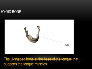
  • 1. HYOID BONE Hyoid The U-shaped bone at the base of the tongue that supports the tongue muscles
  • 3. CERVICAL VERTEBREA The upper part of the spine consist of 7 bones. The atlis is the bone right below the skull that hold it up and the axis is the bone tight below that that helps it turn.
  • 4. THORACIC VERTEBRAE This part of the vertebrae consist of 12 bones.
  • 5. LUMBAR VERTEBRAE This part of the vertebrae consist of 5 bones.
  • 6. SACRUM This contains 5 different bones that over time fuse into one.
  • 7. COCCYX • or tailbone, is the triangular bony structure located at the bottom of the vertebral column. It is composed of three to five bony segments held in place by joints and ligaments.
  • 8. STERNUM Also known as the manubrium sterni it is the broad, upper part of the sternum.
  • 9. TRUE RIBS They are the first 7 ribs in the body. They are called true ribs because they are actually connected to the sternum directly.
  • 10. FALSE RIBS These are the 3 ribs right under the true ribs. They are called false because they are no directly connected the sternum, instead they are connected to a cartilage.
  • 11. FLOATING RIBS These are the last 2 ribs right below the rib cage. They are called floating ribs because they are not connected to the sternum or another ribs at all. They are just connected to the spine and stick out.
  • 12.
  • 13. CLAVICLE 6
  • 14. SCAPULA 7
  • 15. HUMERUS 6
  • 17. CARPALS AND METACARPALS AND PHALANGES • There are eight carpal bones in the base of each hand. There are five metacarpals in each hand. There are 16 bones for phalanges. From closet to the center of the hand going outward it goes from proximal, middle, and distal. There is no first middle phalange only proximal and distal. 1
  • 18. The upper and lower extremities of humans offer many interesting points of comparison and of contrast. They and their individual components are homologous—i.e., of a common origin and patterned on the same basic plan. A long evolutionary history and profound changes in the function of these two pairs of extremities have led, however, to considerable differences between them. (3)
  • 19. lower extremity- • Coxal (Pelvic Girdle)- What bones make up the Acetabelum? Howcan an anthropologist tell the difference between a male and female? • Ilium- Iliac crest, ASIS, AIIS, PSIS, PIIS, Greater Sciatic Notch • Ishium- ishial tuberosity • Pubis- Pubic Tubercle • Femur- head, neck, greater trochanter, Lesser trochanter, condyles, epicondyles, trochlea • Patella • Tibia- condyles, Tibial Tuberosity, Tibial Plateau, Medial Malleolus • Fibula- Head, Lateral malleolus • Tarsals- Talus, Calcaneus, Navicular, (1,2,3) Cuneiforms, Cuboid • Metatarsals- Long Bones Numbered I through V • Phalanges- Proximal, Medial, Distal I through V
  • 20. COXAL PELVIC GIRDLE The ilium / pubis and ischium bones make up the Acetabelum. Male Female •narrower, heart-shaped pelvic inlet •open, circular pelvic inlet •narrower sciatic notch •broader sciatic notch •narrower angle where the two pubic bones in •wider angle where the two pubic bones meet in front front •more outwardly flared hip bones This chart helps to explain how anthropologists can determine weather pelvic bones are male of female. (1)
  • 21. Female pelvic bone Male pelvic bone (1)
  • 22. ISHIUM- ISHIAL TUBEROSITY ISHIUM • is a bone in pelvis: the lowest and rearmost of the three bones that make up each half of the pelvis PUBIS • lower front of hipbone: the bone that forms the lower front section of the hipbone in humans and is one of a pair joined at the front of the pelvis. ILIUM • Ilium the uppermost of the three bones composing each half of the pelvis(3)
  • 23. FEMUR • of thigh: relating to, in, or involving the thigh or femur (3)
  • 24. PATELLA • middle joint of human leg: the joint of the human leg between the thigh and the lower leg, where the femur and the tibia meet, covered in front by the kneecap patella (4) patella
  • 25. TIBIA • inner bone of lower leg: the inner and larger of the two bones in the lower leg, extending from the knee to the ankle bone alongside the fibula (5) tibia
  • 26. FIBULA • human leg bone: the outer and narrower of the two bones in the human lower leg between the knee and ankle(6) fibula
  • 27. TARSALS • human leg bone: the outer and narrower of the two bones in the human lower leg between the knee and ankle(7) tarsals
  • 28. METATARSALS • Metatarsals- Long Bones Numbered I through V • of foot bones: belonging or relating to the bones between the toes and ankle(6) metatarsals
  • 29. PHALANGES • Phalanges- Proximal, Medial, Distal I through V • finger and toe bone: a finger or toe bone of a human being or vertebrate animal(6) phalanges