SlideShare uma empresa Scribd logo
1 de 22
MICROTOME
A microtome is a cutting tool used to produce extremely thin slices of material known as sections. Important in science,
microtomes are used in microscopy, allowing for the preparation of samples for observation under transmitted light or
electron radiation.
The most common applications of
microtomes are:
• Traditional Histology Technique:
• Cryosectioning Technique:
• Electron Microscopy Technique:
• Botanical Microtomy Technique:
(Hard materials like wood require a sledge microtome.
These microtomes have heavier blades and cannot cut as thin as a regular
microtome.)
Parts of MICROTOME
There are different microtomes, but they all consist of three main parts:
1.Base (microtome body)
2.Knife attachment and blade
3. Material or tissue holder
Some microtomes have more features, like a desk clamp to hold the machine in
place. Some devices are manually used, while others are automatic, so the specific
features would differ in these microtomes.
Various types of microtomes are available.
• Rotary Mictrotome
• Sledge Microtome
• Cryomicrotome
• Cyrostat
• Ultramicrotome
• Vibrating microtome
• Saw microtome
• Laser microtome
Rotary Mictrotome
• It is most commonly used microtome. This device operates with a
staged rotary action such that the actual cutting is part of the rotary
motion. In a rotary microtome, the knife is typically fixed in a
horizontal position.
• The typical cut thickness for a rotary microtome is between 1 and 60
μm.
• For hard materials, such as a sample embedded in a synthetic resin,
this design of microtome can allow for good “Semi-thin” sections with
a thickness of as low as 0.5 μm.
Advantages of Rotary Mictrotome
• The machine is heavy, so it is stable and does not vibrate during
cutting.
• Serial sections can be obtained.
• Cutting angle and knife angle can be adjusted.
• It may also be used for cutting celloidin embedded sections with the
help of special holder to set the knife.
Sledge Microtome
• Sample is placed into a fixed holder (shuttle), the sledge placed upon
a linear bearing, a design that allows for the microtome to readily cut
many coarse sections.
• Applications for this design of microtome are of the preparation of
large samples, such as those embedded in paraffin for biological
preparations.
• Typical cut thickness achievable on a sledge microtome is between is
10 and 60 micron.
Freezing microtome
• For the cutting of frozen samples, many rotary microtomes can be
adapted to cut in a liquid nitrogen chamber, in a so-called
cryomicrotome setup.
• The reduced temperature allows for the hardness of the sample to be
increased, such as by undergoing a glass transition, which allows for
the preparation of semi- thin samples.
• However the sample temperature and the knife temperature must be
controlled in order to optimise the resultant sample thickness
Cryostat
• The introduction of fluorescent antibody staining techniques by Coons, Creech
and Jones in 1941 led to a need for thin section(3-5 microns) of fresh frozen
tissue free of ice crystal defect.
• [So there must be quick frozen at a very low temp , and section cut without
allowing the tissue to thaw.
• Cryostat is primarily used for cutting sections of frozen tissueFrozen sections
were originally produced for histological techniques, but were later used to
demonstrate soluble substance and the diagnosis or urgent biopsy specimens.
• Specimens are frozen and cut at 4-8 um thickness in an cryo-microtome using
an anti-roll plate
Ultramicrotome
• A ribbon of ultrathin sections prepared by room temperature
ultramicrotomy, floating on water in the boat of a diamond knife used
to cut the sections.
• The knife blade is the edge at the upper end of the trough of water.
• It can allow for the preparation of extremely thin sections
• These extremely thin cuts are important for use with transmission
electron microscope (TEM) and Serial Block-Face Scanning Electron
Microscopy (SBFSEM), and are sometimes also important for light-
optical microscopy.
Vibrating microtome
• The vibrating microtome operates by cutting using a vibrating blade,
allowing the resultant cut to be made with less pressure than would
be required for a stationary blade.
• The vibrating microtome is usually used for difficult biological
samples.
• The cut thickness is usually around 30-500 μm for live tissue and 10-
500 μm for fixed tissue.
Saw microtome
• The saw microtome is especially for hard materials such as teeth or
bones.
• The microtome of this type has a recessed rotating saw, which slices
through the sample.
• The minimal cut thickness is approximately 30 μm, and can be made
for comparatively large samples.
Laser microtome
• The laser microtome is an instrument for contact free slicing. Prior
preparation of the sample through embedding, freezing or chemical
fixation is not required, thereby minimizing the artifacts from
preparation methods.
• Alternately this design of microtome can also be used for very hard
materials, such as bones or teeth as well as some ceramics.
Dependent upon the properties of the sample material, the thickness
achievable is between 10 and 100 μm.
Laser microtome
• The device operates using a cutting action of an infra-red laser.
• As the laser emits a radiation in the near infra-red, in this wavelength
regime the laser can interact with biological materials.
• Through the non-linear interaction of the optical penetration in the
focal region a material separation in a process known as photo-
disruption is introduced.
Laser microtome
• By limiting the laser pulse durations to the femtoseconds range, the
energy expended at the target region is precisely controlled, thereby
limiting the interaction zone of the cut to under a micrometre.
• External to this zone the ultra- short beam application time
introduces minimal to no thermal damage to the remainder of the
sample.
MICROTOME KNIFE
• It is the important instrument used to cut uniform thin serial sections
of the tissue. Various types of knives are used with different
microtomes. For routine purpose wedge (C type) knife is used. It is
plain on both sides. The size varies from 100 mm to 350 mm in
length.
• Microtome knives are made of good quality of high carbon or steel
which is tempered at the tip. Hardness of knife is essential to obtain
good tissue sections.
Types of microtome knives
1The Heiffor knife (used on rocking microtomes with a fixed handle)
2Larger knives with detachable handle ranging from (8- 24 cm).8cm for freezing microtomes and 24 cm for
base sledge microtomes.
3HEEL-Angle formed by the cutting edge and end of the knife nearest the handle.
4TOE-Angle formed by the cutting edge and end of the knife farthest from the handle
Sharpening of microtome knife
• To achieve good sections knife should be very sharp.
• The knife is put in the knife back to sharpen.
• Knife can be sharpened manually or by the use of automatic machine.
Honing
• This is done to remove nicks and irregularity from the knife edge.
Coarse and fine honing is done using different abrasives.
• Consider following these six easy steps to sharpen a knife with a
honing steel:
1.Adapt to the circumstances. You should sharpen a serrated knife
and a paring knife using different methods. ...
2.Determine dullness. ...
3.Drag across the sharpening steel. ...
4.Repeat the process. ...
5.Scrap any shavings. ...
6.Use the correct angle.
Stropping
• The purpose of stropping is to remove the “burr” formed during
honing and to polish cutting edge.

Mais conteúdo relacionado

Mais procurados

Fixatives in Histopathology
Fixatives in HistopathologyFixatives in Histopathology
Fixatives in Histopathology
Ishwar9
 
Fixatives used in histopathology
Fixatives used in histopathologyFixatives used in histopathology
Fixatives used in histopathology
Hitendra Prajapati
 

Mais procurados (20)

Fixatives in Histopathology
Fixatives in HistopathologyFixatives in Histopathology
Fixatives in Histopathology
 
Microtome
Microtome Microtome
Microtome
 
1. Microtome
1. Microtome1. Microtome
1. Microtome
 
Principles, Methods and Types of Fixatives
Principles, Methods and Types of FixativesPrinciples, Methods and Types of Fixatives
Principles, Methods and Types of Fixatives
 
Fixation of tissues
Fixation of tissuesFixation of tissues
Fixation of tissues
 
Fixation
Fixation Fixation
Fixation
 
Freezing microtome_ cryostat.pptx
Freezing microtome_ cryostat.pptxFreezing microtome_ cryostat.pptx
Freezing microtome_ cryostat.pptx
 
microtomy .ppt
microtomy .pptmicrotomy .ppt
microtomy .ppt
 
Microtomy
MicrotomyMicrotomy
Microtomy
 
Histopathology - CRYOSTAT
Histopathology - CRYOSTATHistopathology - CRYOSTAT
Histopathology - CRYOSTAT
 
Fixation
FixationFixation
Fixation
 
Embedding
EmbeddingEmbedding
Embedding
 
Section Cutting
Section CuttingSection Cutting
Section Cutting
 
Microtomy - Preparation of Histological Slides
Microtomy - Preparation of Histological SlidesMicrotomy - Preparation of Histological Slides
Microtomy - Preparation of Histological Slides
 
ULTRAMICROTOME
ULTRAMICROTOMEULTRAMICROTOME
ULTRAMICROTOME
 
Histology & Cytology Instruments
Histology & Cytology Instruments Histology & Cytology Instruments
Histology & Cytology Instruments
 
Microtomy
MicrotomyMicrotomy
Microtomy
 
Fixatives used in histopathology
Fixatives used in histopathologyFixatives used in histopathology
Fixatives used in histopathology
 
Fixatives
Fixatives Fixatives
Fixatives
 
Mounting media
Mounting mediaMounting media
Mounting media
 

Semelhante a MICROTOME and it's TYPES

Semelhante a MICROTOME and it's TYPES (20)

Microtomes, Section Cutting and Sharpening of razor
Microtomes, Section Cutting and Sharpening of razorMicrotomes, Section Cutting and Sharpening of razor
Microtomes, Section Cutting and Sharpening of razor
 
Microtome
MicrotomeMicrotome
Microtome
 
Microtomy
MicrotomyMicrotomy
Microtomy
 
Microtome
MicrotomeMicrotome
Microtome
 
microtomy-161016082109.pdf
microtomy-161016082109.pdfmicrotomy-161016082109.pdf
microtomy-161016082109.pdf
 
Microtomy.pptx
Microtomy.pptxMicrotomy.pptx
Microtomy.pptx
 
microtomy.pptx
microtomy.pptxmicrotomy.pptx
microtomy.pptx
 
Microtomy and Paraffin Section Preparation ( PDFDrive ).pdf
Microtomy and Paraffin Section Preparation ( PDFDrive ).pdfMicrotomy and Paraffin Section Preparation ( PDFDrive ).pdf
Microtomy and Paraffin Section Preparation ( PDFDrive ).pdf
 
Botany:Microtome And Their Types
Botany:Microtome And Their TypesBotany:Microtome And Their Types
Botany:Microtome And Their Types
 
Beam modifying devices
Beam modifying devices Beam modifying devices
Beam modifying devices
 
Cutting Machines
Cutting MachinesCutting Machines
Cutting Machines
 
Rotary microtome seminar.pptx
Rotary microtome seminar.pptxRotary microtome seminar.pptx
Rotary microtome seminar.pptx
 
4. MICROTOMY.pptx
4. MICROTOMY.pptx4. MICROTOMY.pptx
4. MICROTOMY.pptx
 
Cutting
CuttingCutting
Cutting
 
2. Microtomy Bio Techniques.pptx
2. Microtomy Bio Techniques.pptx2. Microtomy Bio Techniques.pptx
2. Microtomy Bio Techniques.pptx
 
Laser cutting machine | Metal sheet & tube laser Cutter‎, Amtec inc.
Laser cutting machine | Metal sheet & tube laser Cutter‎, Amtec inc.Laser cutting machine | Metal sheet & tube laser Cutter‎, Amtec inc.
Laser cutting machine | Metal sheet & tube laser Cutter‎, Amtec inc.
 
Dhruv barot production technology
Dhruv barot   production technologyDhruv barot   production technology
Dhruv barot production technology
 
Beam modification devices
Beam modification devicesBeam modification devices
Beam modification devices
 
Micro and nano manufacturing
Micro and nano manufacturingMicro and nano manufacturing
Micro and nano manufacturing
 
Micro and nano manufacturing
Micro and nano manufacturingMicro and nano manufacturing
Micro and nano manufacturing
 

Mais de ShahzebHUSSAIN5 (12)

hormones
hormoneshormones
hormones
 
lipotropic factors.pptx
lipotropic factors.pptxlipotropic factors.pptx
lipotropic factors.pptx
 
kidney disease.pptx
kidney disease.pptxkidney disease.pptx
kidney disease.pptx
 
Immunohistochemistry
Immunohistochemistry Immunohistochemistry
Immunohistochemistry
 
PAS STAIN.pptx
PAS STAIN.pptxPAS STAIN.pptx
PAS STAIN.pptx
 
Stains - -staining.pptx
Stains - -staining.pptxStains - -staining.pptx
Stains - -staining.pptx
 
Gastric Function Test.pptx
Gastric Function Test.pptxGastric Function Test.pptx
Gastric Function Test.pptx
 
General_Properties_of_fungi.pptx
General_Properties_of_fungi.pptxGeneral_Properties_of_fungi.pptx
General_Properties_of_fungi.pptx
 
Health problem in infants and school going children
Health problem in infants and school going children Health problem in infants and school going children
Health problem in infants and school going children
 
haemoglobin derivatives and disorder
haemoglobin derivatives and disorder haemoglobin derivatives and disorder
haemoglobin derivatives and disorder
 
cardiovascular system disease there Gross appearances and morphological chang...
cardiovascular system disease there Gross appearances and morphological chang...cardiovascular system disease there Gross appearances and morphological chang...
cardiovascular system disease there Gross appearances and morphological chang...
 
porphyrins chemistry and metabolism.pptx
porphyrins chemistry and metabolism.pptxporphyrins chemistry and metabolism.pptx
porphyrins chemistry and metabolism.pptx
 

Último

Último (20)

Application orientated numerical on hev.ppt
Application orientated numerical on hev.pptApplication orientated numerical on hev.ppt
Application orientated numerical on hev.ppt
 
General Principles of Intellectual Property: Concepts of Intellectual Proper...
General Principles of Intellectual Property: Concepts of Intellectual  Proper...General Principles of Intellectual Property: Concepts of Intellectual  Proper...
General Principles of Intellectual Property: Concepts of Intellectual Proper...
 
SOC 101 Demonstration of Learning Presentation
SOC 101 Demonstration of Learning PresentationSOC 101 Demonstration of Learning Presentation
SOC 101 Demonstration of Learning Presentation
 
How to setup Pycharm environment for Odoo 17.pptx
How to setup Pycharm environment for Odoo 17.pptxHow to setup Pycharm environment for Odoo 17.pptx
How to setup Pycharm environment for Odoo 17.pptx
 
Accessible Digital Futures project (20/03/2024)
Accessible Digital Futures project (20/03/2024)Accessible Digital Futures project (20/03/2024)
Accessible Digital Futures project (20/03/2024)
 
ICT role in 21st century education and it's challenges.
ICT role in 21st century education and it's challenges.ICT role in 21st century education and it's challenges.
ICT role in 21st century education and it's challenges.
 
2024-NATIONAL-LEARNING-CAMP-AND-OTHER.pptx
2024-NATIONAL-LEARNING-CAMP-AND-OTHER.pptx2024-NATIONAL-LEARNING-CAMP-AND-OTHER.pptx
2024-NATIONAL-LEARNING-CAMP-AND-OTHER.pptx
 
How to Give a Domain for a Field in Odoo 17
How to Give a Domain for a Field in Odoo 17How to Give a Domain for a Field in Odoo 17
How to Give a Domain for a Field in Odoo 17
 
Basic Civil Engineering first year Notes- Chapter 4 Building.pptx
Basic Civil Engineering first year Notes- Chapter 4 Building.pptxBasic Civil Engineering first year Notes- Chapter 4 Building.pptx
Basic Civil Engineering first year Notes- Chapter 4 Building.pptx
 
Plant propagation: Sexual and Asexual propapagation.pptx
Plant propagation: Sexual and Asexual propapagation.pptxPlant propagation: Sexual and Asexual propapagation.pptx
Plant propagation: Sexual and Asexual propapagation.pptx
 
HMCS Vancouver Pre-Deployment Brief - May 2024 (Web Version).pptx
HMCS Vancouver Pre-Deployment Brief - May 2024 (Web Version).pptxHMCS Vancouver Pre-Deployment Brief - May 2024 (Web Version).pptx
HMCS Vancouver Pre-Deployment Brief - May 2024 (Web Version).pptx
 
On National Teacher Day, meet the 2024-25 Kenan Fellows
On National Teacher Day, meet the 2024-25 Kenan FellowsOn National Teacher Day, meet the 2024-25 Kenan Fellows
On National Teacher Day, meet the 2024-25 Kenan Fellows
 
UGC NET Paper 1 Mathematical Reasoning & Aptitude.pdf
UGC NET Paper 1 Mathematical Reasoning & Aptitude.pdfUGC NET Paper 1 Mathematical Reasoning & Aptitude.pdf
UGC NET Paper 1 Mathematical Reasoning & Aptitude.pdf
 
Sociology 101 Demonstration of Learning Exhibit
Sociology 101 Demonstration of Learning ExhibitSociology 101 Demonstration of Learning Exhibit
Sociology 101 Demonstration of Learning Exhibit
 
COMMUNICATING NEGATIVE NEWS - APPROACHES .pptx
COMMUNICATING NEGATIVE NEWS - APPROACHES .pptxCOMMUNICATING NEGATIVE NEWS - APPROACHES .pptx
COMMUNICATING NEGATIVE NEWS - APPROACHES .pptx
 
Food safety_Challenges food safety laboratories_.pdf
Food safety_Challenges food safety laboratories_.pdfFood safety_Challenges food safety laboratories_.pdf
Food safety_Challenges food safety laboratories_.pdf
 
Python Notes for mca i year students osmania university.docx
Python Notes for mca i year students osmania university.docxPython Notes for mca i year students osmania university.docx
Python Notes for mca i year students osmania university.docx
 
HMCS Max Bernays Pre-Deployment Brief (May 2024).pptx
HMCS Max Bernays Pre-Deployment Brief (May 2024).pptxHMCS Max Bernays Pre-Deployment Brief (May 2024).pptx
HMCS Max Bernays Pre-Deployment Brief (May 2024).pptx
 
How to Add New Custom Addons Path in Odoo 17
How to Add New Custom Addons Path in Odoo 17How to Add New Custom Addons Path in Odoo 17
How to Add New Custom Addons Path in Odoo 17
 
How to Create and Manage Wizard in Odoo 17
How to Create and Manage Wizard in Odoo 17How to Create and Manage Wizard in Odoo 17
How to Create and Manage Wizard in Odoo 17
 

MICROTOME and it's TYPES

  • 1. MICROTOME A microtome is a cutting tool used to produce extremely thin slices of material known as sections. Important in science, microtomes are used in microscopy, allowing for the preparation of samples for observation under transmitted light or electron radiation.
  • 2. The most common applications of microtomes are: • Traditional Histology Technique: • Cryosectioning Technique: • Electron Microscopy Technique: • Botanical Microtomy Technique: (Hard materials like wood require a sledge microtome. These microtomes have heavier blades and cannot cut as thin as a regular microtome.)
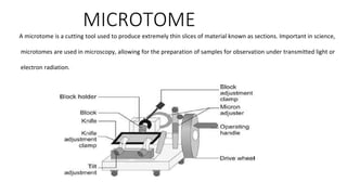
  • 3. Parts of MICROTOME There are different microtomes, but they all consist of three main parts: 1.Base (microtome body) 2.Knife attachment and blade 3. Material or tissue holder Some microtomes have more features, like a desk clamp to hold the machine in place. Some devices are manually used, while others are automatic, so the specific features would differ in these microtomes.
  • 4. Various types of microtomes are available. • Rotary Mictrotome • Sledge Microtome • Cryomicrotome • Cyrostat • Ultramicrotome • Vibrating microtome • Saw microtome • Laser microtome
  • 5. Rotary Mictrotome • It is most commonly used microtome. This device operates with a staged rotary action such that the actual cutting is part of the rotary motion. In a rotary microtome, the knife is typically fixed in a horizontal position.
  • 6. • The typical cut thickness for a rotary microtome is between 1 and 60 μm. • For hard materials, such as a sample embedded in a synthetic resin, this design of microtome can allow for good “Semi-thin” sections with a thickness of as low as 0.5 μm.
  • 7. Advantages of Rotary Mictrotome • The machine is heavy, so it is stable and does not vibrate during cutting. • Serial sections can be obtained. • Cutting angle and knife angle can be adjusted. • It may also be used for cutting celloidin embedded sections with the help of special holder to set the knife.
  • 8. Sledge Microtome • Sample is placed into a fixed holder (shuttle), the sledge placed upon a linear bearing, a design that allows for the microtome to readily cut many coarse sections. • Applications for this design of microtome are of the preparation of large samples, such as those embedded in paraffin for biological preparations. • Typical cut thickness achievable on a sledge microtome is between is 10 and 60 micron.
  • 9. Freezing microtome • For the cutting of frozen samples, many rotary microtomes can be adapted to cut in a liquid nitrogen chamber, in a so-called cryomicrotome setup. • The reduced temperature allows for the hardness of the sample to be increased, such as by undergoing a glass transition, which allows for the preparation of semi- thin samples. • However the sample temperature and the knife temperature must be controlled in order to optimise the resultant sample thickness
  • 10. Cryostat • The introduction of fluorescent antibody staining techniques by Coons, Creech and Jones in 1941 led to a need for thin section(3-5 microns) of fresh frozen tissue free of ice crystal defect. • [So there must be quick frozen at a very low temp , and section cut without allowing the tissue to thaw. • Cryostat is primarily used for cutting sections of frozen tissueFrozen sections were originally produced for histological techniques, but were later used to demonstrate soluble substance and the diagnosis or urgent biopsy specimens. • Specimens are frozen and cut at 4-8 um thickness in an cryo-microtome using an anti-roll plate
  • 11. Ultramicrotome • A ribbon of ultrathin sections prepared by room temperature ultramicrotomy, floating on water in the boat of a diamond knife used to cut the sections. • The knife blade is the edge at the upper end of the trough of water. • It can allow for the preparation of extremely thin sections • These extremely thin cuts are important for use with transmission electron microscope (TEM) and Serial Block-Face Scanning Electron Microscopy (SBFSEM), and are sometimes also important for light- optical microscopy.
  • 12. Vibrating microtome • The vibrating microtome operates by cutting using a vibrating blade, allowing the resultant cut to be made with less pressure than would be required for a stationary blade. • The vibrating microtome is usually used for difficult biological samples. • The cut thickness is usually around 30-500 μm for live tissue and 10- 500 μm for fixed tissue.
  • 13. Saw microtome • The saw microtome is especially for hard materials such as teeth or bones. • The microtome of this type has a recessed rotating saw, which slices through the sample. • The minimal cut thickness is approximately 30 μm, and can be made for comparatively large samples.
  • 14. Laser microtome • The laser microtome is an instrument for contact free slicing. Prior preparation of the sample through embedding, freezing or chemical fixation is not required, thereby minimizing the artifacts from preparation methods. • Alternately this design of microtome can also be used for very hard materials, such as bones or teeth as well as some ceramics. Dependent upon the properties of the sample material, the thickness achievable is between 10 and 100 μm.
  • 15. Laser microtome • The device operates using a cutting action of an infra-red laser. • As the laser emits a radiation in the near infra-red, in this wavelength regime the laser can interact with biological materials. • Through the non-linear interaction of the optical penetration in the focal region a material separation in a process known as photo- disruption is introduced.
  • 16. Laser microtome • By limiting the laser pulse durations to the femtoseconds range, the energy expended at the target region is precisely controlled, thereby limiting the interaction zone of the cut to under a micrometre. • External to this zone the ultra- short beam application time introduces minimal to no thermal damage to the remainder of the sample.
  • 17. MICROTOME KNIFE • It is the important instrument used to cut uniform thin serial sections of the tissue. Various types of knives are used with different microtomes. For routine purpose wedge (C type) knife is used. It is plain on both sides. The size varies from 100 mm to 350 mm in length. • Microtome knives are made of good quality of high carbon or steel which is tempered at the tip. Hardness of knife is essential to obtain good tissue sections.
  • 18. Types of microtome knives 1The Heiffor knife (used on rocking microtomes with a fixed handle) 2Larger knives with detachable handle ranging from (8- 24 cm).8cm for freezing microtomes and 24 cm for base sledge microtomes. 3HEEL-Angle formed by the cutting edge and end of the knife nearest the handle. 4TOE-Angle formed by the cutting edge and end of the knife farthest from the handle
  • 19.
  • 20. Sharpening of microtome knife • To achieve good sections knife should be very sharp. • The knife is put in the knife back to sharpen. • Knife can be sharpened manually or by the use of automatic machine.
  • 21. Honing • This is done to remove nicks and irregularity from the knife edge. Coarse and fine honing is done using different abrasives. • Consider following these six easy steps to sharpen a knife with a honing steel: 1.Adapt to the circumstances. You should sharpen a serrated knife and a paring knife using different methods. ... 2.Determine dullness. ... 3.Drag across the sharpening steel. ... 4.Repeat the process. ... 5.Scrap any shavings. ... 6.Use the correct angle.
  • 22. Stropping • The purpose of stropping is to remove the “burr” formed during honing and to polish cutting edge.