Gall bladder & biliary tract anomalies and variants
•
219 gostaram•39,029 visualizações

GALL BLADDER & BILIARY TRACT - ANATOMY & VARIANTS

Recomendados
Recomendados
Mais conteúdo relacionado
Mais procurados
Mais procurados (20)
Destaque
Destaque (20)
Semelhante a Gall bladder & biliary tract anomalies and variants
Semelhante a Gall bladder & biliary tract anomalies and variants (20)
Último
Último (20)
Gall bladder & biliary tract anomalies and variants
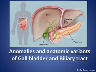
- 1. Anomalies and anatomic variants of Gall bladder and Biliary tract Dr. M Sanal kumar
- 2. Normal Anatomy Gallbladder The gallbladder is an elliptical organ located in a fossa on the undersurface of the liver between the right and the left lobes. Although size and shape vary, the relaxed gallbladder is approximately 10 cm long and 3 to 5 cm in diameter. Normal capacity is approximately 50 mL. The normal gallbladder wall is 2 to 3 mm thick, and the mucosa is composed of simple columnar epithelium. The gallbladder is usually apposed to the liver surface by parietal peritoneum.
- 4. The gallbladder is divided into 4 parts— fundus, body, infundibulum, and neck. The fundus is the rounded distal tip, which may project below the anterior inferior liver edge. The body is the midportion of the gallbladder, which may be in contact with the duodenum and hepatic flexure. The infundibulum (Hartmann pouch) is the focally enlarged segment between the body and the neck. The neck of the gallbladder lies between the body and the cystic duct and points toward the porta hepatis.
- 7. Agenesis Of The Gallbladder Agenesis of the gallbladder is caused by failure of development of the caudal division of the primitive hepatic diverticulum or failure of vacuolization after the solid phase of embryonic development. Other congenital anomalies are present in two thirds of these patients, including congenital heart lesions, polysplenia, imperforate anus, absence of one or more bones, and rectovaginal fistula. The surgical incidence of gall bladder agenesis is approx. 0.02%. Agenesis of the gallbladder (AGB) is a very rare congenital anomaly. Till July 2010, a total of 413 cases have been reported in the literature Nearly 2/3 of adult patients with AGB have biliary tract symptoms & extrahepatic biliary calculi are reported in 25% -50% of pts.
- 8. There are two main expressions: 1) AGB without cystic duct remnant and 2) AGB with cystic remnant . Ultrasound or CT may suggest the diagnosis, but it is usually diagnosed at surgery when the gallbladderis not found at cholangiography. Intraoperative ultrasound may be helpful in establishing the diagnosis and excluding a completely intrahepatic gallbladder.
- 9. Duplication Of The Gallbladder (Vesica fellea duplex) Gallbladder duplication occurs in about 1 in 4000 people. This anomaly is caused by incomplete revacuolization of the primitive gallbladder, resulting in a persistent longitudinal septum that divides the gallbladder lengthwise / occurrence of separate cystic buds. To establish the diagnosis, two separate gallbladder cavities, each with its own cystic duct, must be present. These duplicated cystic ducts may enter the common duct separately or form a Y-configuration before a common entrance. Most reported cases of gallbladder duplication have a clinical picture of cholecystitis with cholelithiasis in at least one of the gallbladders.
- 11. Wandering Gallbladder When the gallbladder has an unusually long mesentery, it can “wander” or “float.” The gallbladder may “disappear” into the pelvis on upright radiographs or wander in front of the spine or to the left of the abdomen. Rarely, the gallbladder can herniate through the foramen of Winslow into the lesser sac. The herniation can be intermittent and may be responsible for abdominal pain. The mesentery is sufficiently long to permit torsion in 4.5% of the population.
- 12. Three unusual anatomic situations give rise to torsion of the gallbladder, and they all produce twisting of an unusually mobile gallbladder on a pedicle: (1) a gallbladder that is completely free of mesenteric or peritoneal investments except for its cystic duct and artery, (2) a long gallbladder mesentery sufficient to allow twisting, and (3) the presence of large stones in the gallbladder fundus that cause lengthening and torsion of the gallbladder mesentery. Kyphosis, vigorous gallbladder peristalsis have also been implicated as other predisposing or contributing factors.
- 13. Most cases of gallbladder torsion occur in women (F/M ratio of 3:1). The usual preoperative diagnosis is acute cholecystitis. Gangrene develops in more than 50% of cases and is extremely common when the pain has been present for more than 48 hours. On cross-sectional imaging, the gallbladder is distended and may have an unusual location and show mural thickening.
- 14. Anomalies Of Gallbladder Shape Phrygian Cap Diverticula Multiseptate Gallbladder
- 15. Phrygian Cap Phrygian cap is the most common abnormality of gallbladder shape, occurring in 1% to 6% of the population. Named after the headgear worn by ancient Greek slaves as a sign of liberation. This deformity is characterized by a fold or septum of the gallbladder between the body and fundus. Two variations of this anomaly have been described. Retroserosal /concealed type,the GB is smoothly invested by peritoneum, and the mucosal fold that projects into lumen may not be visible externally. Serosal or visible type, the peritoneum follows the bend in the fundus, then reflects on itself as the fundus overlies the body.
- 18. Multiseptate Gallbladder The multiseptate gallbladder is a solitary gallbladder characterized by multiple septa of various sizes internally and a faintly bosselated surface externally. The gallbladder is usually normal in size and position, and the chambers communicate with one another by one or more orifices from fundus to cystic duct. These septations lead to stasis of bile and gallstone formation. On ultrasound studies, multiple communicating septations and locules are seen bridging the gallbladder lumen. The sonographic differential diagnoses are desquamated gallbladder mucosa and hyperplastic cholecystoses.
- 20. Diverticula Gallbladder diverticula are rare and usually clinically silent. They can occur anywhere in the gallbladder and are usually single and vary greatly in size. Congenital diverticula are true diverticula and contain all the mural layers, as opposed to the pseudodiverticula of adenomyomatosis, which have little or no smooth muscle in their walls. Acquired traction diverticula from adjacent adhesions or duodenal disease must be also be excluded.
- 22. The bilobed gallbladder, isextremely rare congenital anomaly in man. This malformation is represented by a structure having two separate fundic cavities, united at their bases and joined to the ductus choledochus by a single cystic duct. It is differentiated fromdouble/accessory GB by presence of independent cystic ducts, draining individual fundic cavities of the latter type. Bilobed gall bladder ( Vesica fellea divisa)
- 23. Ectopic Gallbladder The gallbladder can be located in a variety of anomalous positions. Intrahepatic, suprahepatic, retrohepatic, supradiaphragmatic, and retroperitoneal. A. Intrahepatic. B. Left sided. C. Transverse. D. Retrodisplaced.
- 24. In patients with an intrahepatic gallbladder, the GB is completely surrounded by hepatic parenchyma. Intrahepatic GB usually presents little difficulty imaging, but may complicate clinical diagnosis of acute cholecystitis because of paucity of peritoneal signs resulting from long distance between GB & peritoneum. This anomaly also makes cholecystectomy more difficult. On sulfur colloid scans,the intrahepatic GB presents as a cold hepatic defect. In patients with cirrhosis, small or absent right lobes, or chronic obstructive pulmo - nary disease, the gallbladder together with the colon is often interposed between the liver and the diaphragm. Left-sidedgallbladders may occur in situs inversus or as an isolated finding.
- 26. Retro hepatic GB Suprahepatic GB
- 27. Cystic Duct The gallbladder is attached to the common bile duct (CBD) via the cystic duct, which is usually 2 to 4 cm long and contains tortuous folds, the spiral valves of Heister . Diameter 2-3 mm. The cystic duct usually joins the common hepatic duct (CHD) from the right lateral aspect approximately halfway between the porta hepatis and the ampulla of Vater to form the CBD. The point at which the cystic duct joins the CHD is variable, from high in the upper extrahepatic bile duct or one of the intrahepatic ducts (more often the right) to low at the ampulla. The cystic duct usually runs parallel to the CHD at least for a short distance and may insert either anteriorly or posteriorly or spiral around to insert on the medial aspect.
- 32. Bile Ducts Intrahepatic Ducts The liver is divided into right and left lobes on the basis of portal vein anatomy and biliary drainage. The right lobe is divided into anterior and posterior segments, and the left lobe is divided into medial and lateral segments by the fissure of the ligamentum teres. Small branching interlobar bile ducts merge into larger ducts until the major left and right hepatic ducts are formed. A left medial segment duct and a left lateral segment duct normally join to form the main left hepatic duct.
- 33. The left lateral section is divided into superior (segment II) and inferior(segment III) segments. Union of ducts of segment II and III behind the umbilical part of left portal vein form the left hepatic duct (LHD) which then receives the duct from segment IV. Average length of the LHD is 1.7 cm and diameter is 3.0 mm (1.08). The right hepatic duct branches near its take off from the CHD. In approximately 60% of patients, the right hepatic duct has a dorsocaudal branch, with a characteristic hook-like configuration proximally, draining the posterior segment of the right lobe, and a ventrocranial branch, draining the anterior segment of the right lobe. The right anterior sectoral duct (RASD) drains segments V and VIII and the right posterior sectoral duct (RPSD) drains segments VI and VII. The RPSD passes horizontally and generally curves round the RASD to join its medial side to form the right hepatic duct (RHD). Average length of RHD is 0.9 cm and diameter is 2.6 mm.
- 37. The bile ducts generally follow the internal hepatic segmental anatomy; however, marked variation in the branching pattern is common. Anomalies of the biliary system are found in 2.4% of autopsies, 28% of surgical dissections, and 5% to 13% of operative cholangiograms.
- 43. Extrahepatic Ducts The right and left hepatic ducts emerge from the liver and unite to form the 3- to 4-cm-long CHD, which then joins ystic duct to form the CBD. The union of the right and left main hepatic ducts is usually just outside the liver but may be lower, resulting in a shorter CHD or CBD. The CBD averages 6 to 7 cm in length and is usually divided into suprapancreatic, intra-pancreatic, and ampullary segments. In approximately 70% of patients, the CBD courses through the pancreatic head; in a smaller percentage, the CBD is located in a groove on the posterior surface of the pancreas. The CBD enters the posteriormedial aspect of the second portion of the duodenum through an oblique, 1- to 2-cm-long intramural tunnel terminating at the papilla of Vater.
- 44. The exact union of the CBD and the pancreatic duct at the ampulla varies. Most commonly, the two ducts join in the duodenal wall and have a short common channel. Occasionally, separate orifices are present at the ampulla, or the ducts unite, forming a long common channel before enter ing the duodenal wall . The sphincter of Oddi surrounds the common channel and the choledochal sphincter (sphincter of Boyden) surrounds the CBD from its entrance into the duodenal wall to its junction with the pancreatic duct.
- 45. Choledochal Cysts Choledochal cysts are congenital cystic dilatations of any portion of extra hepatic bile ducts, most commonly the main portion of CBD. It is postulated that this condition begins with an anomalous junction of the common bile duct and pancreatic duct proximal to the duodenal papilla. Higher pressure in pancreatic duct combined + an absent ductal sphincter allows free reflux of enzymes into biliary tree, weakening wall of the common bile duct. Normal Abnormal
- 46. Diagnosis of a choledochal cyst is made on the basis of disproportional dilatation of the extrahepatic bile ducts after excluding the possibility of a tumor, stone, or inflammation as the cause of the dilatation. The estimated incidence of choledochal cysts in Western countries varies between 1 in 100,000 and 1 in 150,000 individuals. The rate of incidence is higher in Asia and occurs more frequently in women (M: F- 4:1).60% of patients present before age 10, although choledochal cysts can present from birth to old age. This anomaly is associated with increased incidence of GB anomalies, biliary anomalies ( stenosis /atresia), and congenital hepatic fibrosis. Complications of choledochal cysts in adults include rupture with bile peritonitis, secondary infection (cholangitis), biliary cirrhosis and portal hypertension, calculus formation, portal vein thrombosis, liver abscess, hemorrhage, and malignant transformation into cholangiocarcinoma.
- 50. Newborns and infants present with obstructive jaundice. Older children and adults may have the classic triad of right upper quadrant pain, intermittent jaundice, and a palpable right upper quadrant mass. In adult patients, a choledochal cyst is often first diagnosed on cross- sectional imaging. CT and ultrasound demonstrate a fluid-filled structure beneath the porta hepatis separate from the gallbladder that communicates with the hepatic ducts. An abrupt change in the caliber of the ducts occurs at the site of the cysts. Intrahepatic ductal dilatation may be present as well.
- 54. Caroli’s Disease Caroli’s disease, also known as communicating cavernous ectasia, is characterized by multifocal segmental saccular dilatation of the intrahepatic bile ducts, a predisposition to biliary calculi and cholangitis, and an association with various forms of cystic renal disease. It is an autosomal recessive disease secondary to the ductal plate malformation. It is associated with polycystic kidney disease, medullary sponge kidney and medullary cystic disease. Caroli’s disease usually manifests in adulthood; however, it can be seen in newborns and infants. Adult patients present with recurrent attacks of cholangitis and crampy right upper quadrant pain with occasional fever and mild jaundice. Infants and children may present with hematemesis caused by portal hypertension from hepatic fibrosis.
- 55. Complications of Caroli’s disease include stone formation (95%) within the dilated intra-hepatic ducts, recurrent cholangitis, and liver abscess. There is also a 100-fold increase in incidence of bile duct carcinoma, occurring in 7% of patients. Caroli’s disease is best demonstrated by cholangiography which shows saccular dilatations of the intrahepatic ducts, stones, strictures, and communicating hepatic abscesses.
- 56. Ultrasound May show dilated intrahepatic bile ducts (IHBD). intraductal bridging: echogenic septa traversing the dilatedbile duct lumen. smallportal venous branches partially/completely surroundedbydilatedbile ducts. intraductal calculi. CT multiple hypodense rounded areas which are inseperable from the dilated intrahepatic bile ducts “centraldot” sign: enhancing dots within the dilated intrahepatic bile duct s, these intraluminal dots correspond to intraluminal portal veins
- 57. MRCP with three-dimensional display is an accurate method for demonstrating Caroli’s disease because the luminal contents of the bile ducts appear hyperintense in contrast to the portal vein, which usually appears as signal void. Cystic expansions of the intrahepatic biliary tract are depicted as oval- shaped structures in continuity with the biliary tract.
- 59. Biliary atresia Biliary atresia is a congenital biliary disorder, which is characterised by an absence or severe deficiency of the extra-hepatic biliary tree . It is one of the most common causes of neonatal cholestasis, often causing cirrhosis immediately and leading to death and accounts for over half of children who undergo liver transplantation. Incidence- 1 in 10,000-15,000 newborn infants. There is a recognised male predilection. Luminal obstruction of the extrahepatic bile duct with a fibrous ductal remnant is the pathology.
- 60. Infants with biliary atresia may appear normal & healthy at birth. Most often, symptoms develop between 2wks-2mnths, and may include : Jaundice Dark yellow or brown urine Pale or clay-colored (acholic) stools Hepatomegaly Affected neonates have associated congenital defects, including situs inversus, polysplenia, malrotation, intestinal atresia, and cardiac anomalies.
- 61. Type I, the common bile duct is obliterated while the proximal bile ducts are patent. Type II, atresia of the hepatic duct is seen, with cystic bile ducts found at the porta hepatis. Type IIa, the cystic and common bile ducts are patent, whereas in type IIb, the cystic, common bile duct, and hepatic ducts are obliterated. Type III atresia refers to discontinuity of the right and the left hepatic ducts to the level of the porta hepatis. This form of biliary atresia is common, accounting for more than 90% of cases. Kasai classification
- 63. Ultrasound Echogenic triangular cord sign Tubular echogenic cord of fibrous tissue seen in the porta hepatis at ultrasonography and is relatively specific in diagnosis of biliary atresia. It is defined as more than 4 mm thickness of echogenic anterior wall of right portal vein (EARPV) measured on a longitudinal ultrasound scan. Gallbladder ghost triad Atretic gallbladder, length less than 19 mm Irregular or lobular contour Lack of smooth/complete echogenic mucosal lining with indistinct wall Larger hepatic arterial calibre
- 64. Positive of triangular cord sign = thickness of EARPV > 4 mm on a longitudinal scan 5.1mm 5.4mm
- 65. Length= 1.6mm Irregular or lobular contour Lack of smooth/complete echogenic mucosal lining with indistinct wall Gall bladder ghost triad
- 66. Nuclear medicine (hepatobiliary (HIDA) scan) Cases of biliary atresia typically demonstrate relatively good hepatic uptake with no evidence of excretion into the bowel at 24 hours. Pretreatment with phenobarbital (5 mg/kg/day for 5 days) to increase biliary secretion by stimulating hepatic enzymes is frequently helpful to minimize the possibility of a false-positive study in a patient with a patent biliary system but poor excretion. Good hepatic uptake, but no excretion into bowel even after 24hrs.
- 67. Magnetic Resonance Cholangiography MRCP is a relatively new technique for neonatal imaging. Findings in infants with biliary atresia include incomplete visualization of the extrahepatic biliary system and periportal high signal intensity on T2-weighted magnetic resonance imaging (MRI) scans (which may represent cystic dilatation of fetal bile ducts with surrounding fibrosis).
- 68. Thank you..
Notas do Editor
- Diagrams depicting types of gallbladder variants. A, Gallbladder diverticulum. B, Hourglass gallbladder (left), retroserosal (middle), and serosal phrygian cap (right). C, Floating gallbladder. D, Bilobed gallbladder. E, V- and Y-shaped duplications.
- Suprahepatic- hypoplasia of right lobe Retrohepatic - posterior and inferior to right hepatic lobe
- Normal and aberrant sectoral ductal anatomy. (A) Typical ductal anatomy, (B) triple confluence, (C) Ectopic drainage of a right sectoral duct into the common hepatic duct (C1, right anterior duct draining into the common hepatic duct; C2, right posterior duct draining into the common hepatic duct), (D) ectopic drainage of a right sectoral duct into the left hepatic ductal system (D1, right posterior sectoral duct draining into the left hepatic ductal system; D2, right anterior sectoral duct draining into the left hepatic ductal system, (E) absence of the hepatic duct confluence, (F) absence of right hepatic duct and ectopic drainage of the right posterior duct into the cystic duct.
- Diagrams depicting types of commonly encountered biliary variants in decreasing order of their occurrence. A, Normal pattern. B, Right posterior hepatic duct enters the left hepatic duct (19% of normal population). C, Trifurcation (11% of the normal population). D, Right anterior hepatic duct enters the left hepatic duct. E, Low formation of common hepatic duct. F, Right hepatic duct enters cystic duct.
- Drawing illustrates the sphincter of Oddi complex (arrow) encompassing the distal CBD and pancreatic duct. B. Drawing illustrates a long common channel (>15 mm). Note that the sphincter of Oddi does not reach the confluence (arrow) of the ducts.
- The EARPV was 5.4 mm thick on this longitudinal US scan, which shows the TC sign (cursors) as a thick, tubular, echogenic area along the anterior aspect of the right portal vein (long arrow). The right hepatic artery (short arrow) is encased within the EARPV.
- hepatobiliary iminodiacetic acid (HIDA), paraisopropyl iminodiacetic acid (PIPIDA), or diisopropyl iminodiacetic acid (DISIDA)