SlideShare uma empresa Scribd logo
1 de 21
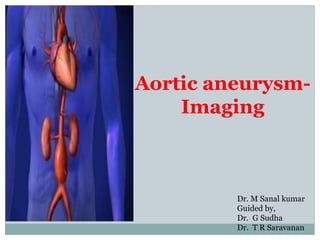
Aortic aneurysm-
Imaging
Dr. M Sanal kumar
Guided by,
Dr. G Sudha
Dr. T R Saravanan
Abdominal aortic aneurysms (AAA) are focal dilatations of the
abdominal aorta that are 50% greater than the proximal normal
segment or that is greater than 3 cm in maximum diameter.
Epidemiology
Its prevalence increases with age.
Males much more commonly affected than females (with a
male:female ratio of 4:1).
Clinical presentation
Most AAAs are asymptomatic unless they
leak or rupture.
Unruptured aneurysms may uncommonly
cause abdominal or back pain, or a
pulsatile mass, if large.
Ruptured aneurysms present with severe
abdominal or back pain, hypotension and
shock.
Anatomy
The aorta passes through the diaphragm at the level of the T12 vertebral body.
It lies slightly to the left of the midline and bifurcates at the level of L4 vertebral body.
The surface anatomy landmarks corresponding to these two points are the xiphoid process
and the umbilicus.
The length of the abdominal aorta is about 13 cm (6 inches). Most scanning of the aorta
will therefore take place in the short distance between the sternum and the umbilicus.
Immediately below the diaphragm, the celiac trunk is the first major vessel to arise from
the aorta in the midline anteriorly.
This short (usually less than 1 cm) vessel can often be seen sonographically in the
transverse plane, dividing in a “wide Y”. The fork on the patient’s right is the common
hepatic artery, heading to the porta hepatis; the fork on the patient’s left, is the splenic
artery. This sonographic view is known as the “seagull sign”.
About 1 cm inferior to the celiac trunk, arises the superior mesenteric artery
(SMA). Measurements of the proximal aorta to use as a comparison with distal
measurements are made at this level.
One centimeter below the SMA, the renal arteries arise on either side.. Thus, these
three major vessels occur within about 3 centimeters of the diaphragm.
90% of all AAA’s will occur distal to this point.
Most AAAs begin below the renal arteries and end above the iliac
arteries.
The size, shape, and extent of AAAs vary considerably.
Like aneurysms of the thoracic aorta, AAAs may be broadly described
as either fusiform (circumferential) or saccular (more localized).
Causes
Atherosclerosis (most common)
Inflammatory abdominal aortic aneurysm
Chronic aortic dissection
Vasculitis, e.g. Takayasu arteritis
Connective tissue disorders, e.g.
Marfan syndrome
Ehlers-Danlos syndrome
Mycotic aneurysm
Traumatic pseudoaneurysm
Anastomotic pseudoaneurysm
The natural history of abdominal aortic aneurysms (AAA) is that of
slow expansion and rupture with devastating consequences.
The risk of rupture is proportional to the size of the aneurysm and
the rate of growth.
Differing rates of rupture for a given aneurysm size have been
reported in the literature but the general consensus is that
aneurysms greater than 5.0 cm in women and 5.5 to 6.0 cm in men
carry a significantly increased risk of rupture and should be
treated.
Furthermore, aneurysms that expand greater than 10 mm per year
are also at significant risk of rupture and are considered for
treatment even when less than 5.0 cm.
Ultrasound
Ultrasound assessment is simple, safe and inexpensive.
It has a reported sensitivity of 95% and specificity close to 100%.
It is usually the preferred choice for monitoring of small aneurysms.
Technique for ultrasound scanning of the aorta
1) Orientation. Start in the transverse plane (pointer to “9 o’clock”), high in the
epigastrium, using the liver as a sonic “window”. Identify the vertebral body (a
dark, rounded shape, with dense shadow).
2) Identify the aorta on the patient’s left, and the IVC (patient’s right) “above” the
vertebral body on the ultrasound image.
3) In real time obtain transverse images of the aorta from the celiac to the
bifurcation.
4) Obtain views of the iliacs if possible.
5) Rotate the probe’s pointer clockwise from the "9 o' clock" to the “12 o’clock”
position for sagittal views from the celiac to the bifurcation.
6) Attempt to obtain:
1) at least 3 transverse views, labeled, “high”, “middle”, “low”, with
calipers. One view should show the maximal aortic diameter.
2) Sagittal view(s) from the celiac to the bifurcation
Aorta is visualized first in short axis and then in long axis. Large (>7 cm) abdominal
aortic aneurysm with mural thrombus and hypoechoic areas is noted outside aorta,
which may represent rupture. Colorflow Doppler illustrates turbulent flow within lumen.
Using Sonography to Monitor the Growth of an
Abdominal Aortic Aneurysm
The accepted method of monitoring abdominal aortic aneurysm is as follows:
If aneurysm measures < 4cm in diameter, the patient is scanned once each year.
If aneurysm measures between 4cm- 5cm in diameter, the patient is scanned
every 6 months.
If aneurysm measures between 5cm - 5.5cm in diameter, the patient is scanned
every 3 months.
Once the aneurysm reaches 5.5cm in diameter, the patient is scheduled for
surgical repair.
It is important to repair the aneurysm before the diameter reaches 6cm because of
increased risk of rupture.
If an aneurysm is growing rapidly, repair be scheduled sooner to avoid rupture.
The mortality rate from a ruptured AAA is high (59-83%) of
patients succumb to death before they make it to hospital or
undergo surgery. The operative mortality rate for those who make
it to surgery tends to be around 40%.
Pearls and Pitfalls
•Obtain measurements of the aorta from outer wall to outer wall. Since
aneurysms will often contain a thrombus, one may accidentally mistake the
inner rim of the thrombus for the aortic wall. Doing this will lead a falsely
decreased measurement of the true aortic diameter.
•Avoid oblique or angled cuts if possible, especially with a tortuous aorta,
which will exaggerate the true aortic diameter.
•Transverse views are needed because many AAAs have larger transverse than
AP diameter.
•A small aneurysm does not preclude rupture: Any symptoms consistent with
rupture in a patient with an aortic diameter greater than 3.0 cm should have
this diagnosis (or alternative vascular catastrophes) ruled out.
•Scanning should be systematically performed in real-time from the
diaphragmatic hiatus to the bifurcation in order to avoid missing small,
localized saccular aneurysms.
Aortic aneurysm imaging

Mais conteúdo relacionado

Mais procurados

Presentation1, radiological imaging of thoracic aortic aneurysm.
Presentation1, radiological imaging of thoracic aortic aneurysm.Presentation1, radiological imaging of thoracic aortic aneurysm.
Presentation1, radiological imaging of thoracic aortic aneurysm.
Abdellah Nazeer
 
Doppler ultrasound of lower limb arteries
Doppler ultrasound of lower limb arteriesDoppler ultrasound of lower limb arteries
Doppler ultrasound of lower limb arteries
Samir Haffar
 
Cystic liver lesions - An ultrasound perspective
Cystic liver lesions - An ultrasound perspectiveCystic liver lesions - An ultrasound perspective
Cystic liver lesions - An ultrasound perspective
Samir Haffar
 
Doppler ultrasound of carotid arteries
Doppler ultrasound of carotid arteriesDoppler ultrasound of carotid arteries
Doppler ultrasound of carotid arteries
Samir Haffar
 
Doppler ultrasound of A-V access for hemodialysis
Doppler ultrasound of A-V access for hemodialysisDoppler ultrasound of A-V access for hemodialysis
Doppler ultrasound of A-V access for hemodialysis
Samir Haffar
 
Presentation1.pptx, ultrasound examination of the liver and gall bladder.
Presentation1.pptx, ultrasound examination of the liver and gall bladder.Presentation1.pptx, ultrasound examination of the liver and gall bladder.
Presentation1.pptx, ultrasound examination of the liver and gall bladder.
Abdellah Nazeer
 

Mais procurados (20)

Aortic dissection 01
Aortic dissection 01Aortic dissection 01
Aortic dissection 01
 
Doppler ultrasound of acute scrotum
Doppler ultrasound of acute scrotumDoppler ultrasound of acute scrotum
Doppler ultrasound of acute scrotum
 
Presentation1, radiological imaging of thoracic aortic aneurysm.
Presentation1, radiological imaging of thoracic aortic aneurysm.Presentation1, radiological imaging of thoracic aortic aneurysm.
Presentation1, radiological imaging of thoracic aortic aneurysm.
 
Role of medical imaging in management of arteriovenous fistula Dr. Muhammad B...
Role of medical imaging in management of arteriovenous fistula Dr. Muhammad B...Role of medical imaging in management of arteriovenous fistula Dr. Muhammad B...
Role of medical imaging in management of arteriovenous fistula Dr. Muhammad B...
 
Doppler ultrasound of lower limb arteries
Doppler ultrasound of lower limb arteriesDoppler ultrasound of lower limb arteries
Doppler ultrasound of lower limb arteries
 
Cystic liver lesions - An ultrasound perspective
Cystic liver lesions - An ultrasound perspectiveCystic liver lesions - An ultrasound perspective
Cystic liver lesions - An ultrasound perspective
 
Peripheral arterial doppler
Peripheral  arterial dopplerPeripheral  arterial doppler
Peripheral arterial doppler
 
Doppler ultrasound in peripheral arterial disease
Doppler ultrasound in peripheral arterial diseaseDoppler ultrasound in peripheral arterial disease
Doppler ultrasound in peripheral arterial disease
 
Radiological imaging of mediastinal masses
Radiological imaging of mediastinal massesRadiological imaging of mediastinal masses
Radiological imaging of mediastinal masses
 
Doppler ultrasound of carotid arteries
Doppler ultrasound of carotid arteriesDoppler ultrasound of carotid arteries
Doppler ultrasound of carotid arteries
 
USG of Aorta and Coeliac axis
USG of Aorta and Coeliac axisUSG of Aorta and Coeliac axis
USG of Aorta and Coeliac axis
 
Venous Doppler upper limb
Venous Doppler upper limb Venous Doppler upper limb
Venous Doppler upper limb
 
Retroperitoneal masses radiology
Retroperitoneal masses radiologyRetroperitoneal masses radiology
Retroperitoneal masses radiology
 
Imaging of aortic pathologies
Imaging of aortic pathologies Imaging of aortic pathologies
Imaging of aortic pathologies
 
Imaging in stroke
Imaging in stroke Imaging in stroke
Imaging in stroke
 
Doppler ultrasound of visceral arteries
Doppler ultrasound of visceral arteriesDoppler ultrasound of visceral arteries
Doppler ultrasound of visceral arteries
 
Renal doppler usg
Renal doppler usgRenal doppler usg
Renal doppler usg
 
Doppler of the portal system
Doppler of the portal systemDoppler of the portal system
Doppler of the portal system
 
Doppler ultrasound of A-V access for hemodialysis
Doppler ultrasound of A-V access for hemodialysisDoppler ultrasound of A-V access for hemodialysis
Doppler ultrasound of A-V access for hemodialysis
 
Presentation1.pptx, ultrasound examination of the liver and gall bladder.
Presentation1.pptx, ultrasound examination of the liver and gall bladder.Presentation1.pptx, ultrasound examination of the liver and gall bladder.
Presentation1.pptx, ultrasound examination of the liver and gall bladder.
 

Semelhante a Aortic aneurysm imaging

24 US Abominal Aorta evaluation via emergency ultrasound
24 US Abominal Aorta evaluation via emergency ultrasound24 US Abominal Aorta evaluation via emergency ultrasound
24 US Abominal Aorta evaluation via emergency ultrasound
SiriC5
 
ANUERYSMS ,AV FISTULAS ,ARTERISTIS ,RAYNAUDS DISEASE -1.pptx
ANUERYSMS ,AV FISTULAS ,ARTERISTIS ,RAYNAUDS DISEASE -1.pptxANUERYSMS ,AV FISTULAS ,ARTERISTIS ,RAYNAUDS DISEASE -1.pptx
ANUERYSMS ,AV FISTULAS ,ARTERISTIS ,RAYNAUDS DISEASE -1.pptx
musayansa
 

Semelhante a Aortic aneurysm imaging (20)

Radiological approach to aortic aneurysm and acute diseases
Radiological approach to aortic aneurysm and acute diseasesRadiological approach to aortic aneurysm and acute diseases
Radiological approach to aortic aneurysm and acute diseases
 
Abdominal aortic aneurysm
Abdominal aortic aneurysmAbdominal aortic aneurysm
Abdominal aortic aneurysm
 
Abdominal aorta.pptx
Abdominal aorta.pptxAbdominal aorta.pptx
Abdominal aorta.pptx
 
24 USAAA.ppt
24 USAAA.ppt24 USAAA.ppt
24 USAAA.ppt
 
24 US Abominal Aorta evaluation via emergency ultrasound
24 US Abominal Aorta evaluation via emergency ultrasound24 US Abominal Aorta evaluation via emergency ultrasound
24 US Abominal Aorta evaluation via emergency ultrasound
 
ABDOMINAL AORTIC ANEURYSM- EPIGASTRIC LUMPS- Abdominal Lumps.pptx
ABDOMINAL AORTIC ANEURYSM- EPIGASTRIC LUMPS- Abdominal Lumps.pptxABDOMINAL AORTIC ANEURYSM- EPIGASTRIC LUMPS- Abdominal Lumps.pptx
ABDOMINAL AORTIC ANEURYSM- EPIGASTRIC LUMPS- Abdominal Lumps.pptx
 
ANUERYSMS ,AV FISTULAS ,ARTERISTIS ,RAYNAUDS DISEASE -1.pptx
ANUERYSMS ,AV FISTULAS ,ARTERISTIS ,RAYNAUDS DISEASE -1.pptxANUERYSMS ,AV FISTULAS ,ARTERISTIS ,RAYNAUDS DISEASE -1.pptx
ANUERYSMS ,AV FISTULAS ,ARTERISTIS ,RAYNAUDS DISEASE -1.pptx
 
Aneurysm
AneurysmAneurysm
Aneurysm
 
aneurysm-210530112039.pdf
aneurysm-210530112039.pdfaneurysm-210530112039.pdf
aneurysm-210530112039.pdf
 
Thoracic aortic aneurysm
Thoracic aortic aneurysmThoracic aortic aneurysm
Thoracic aortic aneurysm
 
Spectrum Of Ct Findings In Rupture And Impendinging Rupture Of AAA
Spectrum Of Ct Findings In Rupture And Impendinging Rupture Of AAASpectrum Of Ct Findings In Rupture And Impendinging Rupture Of AAA
Spectrum Of Ct Findings In Rupture And Impendinging Rupture Of AAA
 
Endovascular Surgery of Abdominal Aorta
Endovascular Surgery of Abdominal AortaEndovascular Surgery of Abdominal Aorta
Endovascular Surgery of Abdominal Aorta
 
Ruptured aortic aneurysms
Ruptured aortic aneurysmsRuptured aortic aneurysms
Ruptured aortic aneurysms
 
artrial disorder managent and nursing care plan
artrial disorder managent and nursing care plan artrial disorder managent and nursing care plan
artrial disorder managent and nursing care plan
 
Aaa
AaaAaa
Aaa
 
AAA
AAAAAA
AAA
 
Aortic aneurysm
Aortic aneurysmAortic aneurysm
Aortic aneurysm
 
Aortic aneurysm final
Aortic aneurysm finalAortic aneurysm final
Aortic aneurysm final
 
thoracic aneurysm
thoracic aneurysmthoracic aneurysm
thoracic aneurysm
 
Aneurysm & THORACIC ANEURYSM
Aneurysm & THORACIC ANEURYSMAneurysm & THORACIC ANEURYSM
Aneurysm & THORACIC ANEURYSM
 

Último

Russian Call Girls Lucknow Just Call 👉👉7877925207 Top Class Call Girl Service...
Russian Call Girls Lucknow Just Call 👉👉7877925207 Top Class Call Girl Service...Russian Call Girls Lucknow Just Call 👉👉7877925207 Top Class Call Girl Service...
Russian Call Girls Lucknow Just Call 👉👉7877925207 Top Class Call Girl Service...
adilkhan87451
 
Call Girl In Pune 👉 Just CALL ME: 9352988975 💋 Call Out Call Both With High p...
Call Girl In Pune 👉 Just CALL ME: 9352988975 💋 Call Out Call Both With High p...Call Girl In Pune 👉 Just CALL ME: 9352988975 💋 Call Out Call Both With High p...
Call Girl In Pune 👉 Just CALL ME: 9352988975 💋 Call Out Call Both With High p...
chetankumar9855
 
Dehradun Call Girls Service {8854095900} ❤️VVIP ROCKY Call Girl in Dehradun U...
Dehradun Call Girls Service {8854095900} ❤️VVIP ROCKY Call Girl in Dehradun U...Dehradun Call Girls Service {8854095900} ❤️VVIP ROCKY Call Girl in Dehradun U...
Dehradun Call Girls Service {8854095900} ❤️VVIP ROCKY Call Girl in Dehradun U...
Sheetaleventcompany
 

Último (20)

Russian Call Girls Lucknow Just Call 👉👉7877925207 Top Class Call Girl Service...
Russian Call Girls Lucknow Just Call 👉👉7877925207 Top Class Call Girl Service...Russian Call Girls Lucknow Just Call 👉👉7877925207 Top Class Call Girl Service...
Russian Call Girls Lucknow Just Call 👉👉7877925207 Top Class Call Girl Service...
 
Call Girls Mumbai Just Call 8250077686 Top Class Call Girl Service Available
Call Girls Mumbai Just Call 8250077686 Top Class Call Girl Service AvailableCall Girls Mumbai Just Call 8250077686 Top Class Call Girl Service Available
Call Girls Mumbai Just Call 8250077686 Top Class Call Girl Service Available
 
Saket * Call Girls in Delhi - Phone 9711199012 Escorts Service at 6k to 50k a...
Saket * Call Girls in Delhi - Phone 9711199012 Escorts Service at 6k to 50k a...Saket * Call Girls in Delhi - Phone 9711199012 Escorts Service at 6k to 50k a...
Saket * Call Girls in Delhi - Phone 9711199012 Escorts Service at 6k to 50k a...
 
Top Rated Pune Call Girls (DIPAL) ⟟ 8250077686 ⟟ Call Me For Genuine Sex Serv...
Top Rated Pune Call Girls (DIPAL) ⟟ 8250077686 ⟟ Call Me For Genuine Sex Serv...Top Rated Pune Call Girls (DIPAL) ⟟ 8250077686 ⟟ Call Me For Genuine Sex Serv...
Top Rated Pune Call Girls (DIPAL) ⟟ 8250077686 ⟟ Call Me For Genuine Sex Serv...
 
Low Rate Call Girls Bangalore {7304373326} ❤️VVIP NISHA Call Girls in Bangalo...
Low Rate Call Girls Bangalore {7304373326} ❤️VVIP NISHA Call Girls in Bangalo...Low Rate Call Girls Bangalore {7304373326} ❤️VVIP NISHA Call Girls in Bangalo...
Low Rate Call Girls Bangalore {7304373326} ❤️VVIP NISHA Call Girls in Bangalo...
 
Jogeshwari ! Call Girls Service Mumbai - 450+ Call Girl Cash Payment 90042684...
Jogeshwari ! Call Girls Service Mumbai - 450+ Call Girl Cash Payment 90042684...Jogeshwari ! Call Girls Service Mumbai - 450+ Call Girl Cash Payment 90042684...
Jogeshwari ! Call Girls Service Mumbai - 450+ Call Girl Cash Payment 90042684...
 
Call Girls in Delhi Triveni Complex Escort Service(🔝))/WhatsApp 97111⇛47426
Call Girls in Delhi Triveni Complex Escort Service(🔝))/WhatsApp 97111⇛47426Call Girls in Delhi Triveni Complex Escort Service(🔝))/WhatsApp 97111⇛47426
Call Girls in Delhi Triveni Complex Escort Service(🔝))/WhatsApp 97111⇛47426
 
Call Girl In Pune 👉 Just CALL ME: 9352988975 💋 Call Out Call Both With High p...
Call Girl In Pune 👉 Just CALL ME: 9352988975 💋 Call Out Call Both With High p...Call Girl In Pune 👉 Just CALL ME: 9352988975 💋 Call Out Call Both With High p...
Call Girl In Pune 👉 Just CALL ME: 9352988975 💋 Call Out Call Both With High p...
 
Call Girls Jaipur Just Call 9521753030 Top Class Call Girl Service Available
Call Girls Jaipur Just Call 9521753030 Top Class Call Girl Service AvailableCall Girls Jaipur Just Call 9521753030 Top Class Call Girl Service Available
Call Girls Jaipur Just Call 9521753030 Top Class Call Girl Service Available
 
Call Girls Service Jaipur {9521753030} ❤️VVIP RIDDHI Call Girl in Jaipur Raja...
Call Girls Service Jaipur {9521753030} ❤️VVIP RIDDHI Call Girl in Jaipur Raja...Call Girls Service Jaipur {9521753030} ❤️VVIP RIDDHI Call Girl in Jaipur Raja...
Call Girls Service Jaipur {9521753030} ❤️VVIP RIDDHI Call Girl in Jaipur Raja...
 
Mumbai ] (Call Girls) in Mumbai 10k @ I'm VIP Independent Escorts Girls 98333...
Mumbai ] (Call Girls) in Mumbai 10k @ I'm VIP Independent Escorts Girls 98333...Mumbai ] (Call Girls) in Mumbai 10k @ I'm VIP Independent Escorts Girls 98333...
Mumbai ] (Call Girls) in Mumbai 10k @ I'm VIP Independent Escorts Girls 98333...
 
Call Girls Service Jaipur {9521753030 } ❤️VVIP BHAWNA Call Girl in Jaipur Raj...
Call Girls Service Jaipur {9521753030 } ❤️VVIP BHAWNA Call Girl in Jaipur Raj...Call Girls Service Jaipur {9521753030 } ❤️VVIP BHAWNA Call Girl in Jaipur Raj...
Call Girls Service Jaipur {9521753030 } ❤️VVIP BHAWNA Call Girl in Jaipur Raj...
 
Premium Call Girls In Jaipur {8445551418} ❤️VVIP SEEMA Call Girl in Jaipur Ra...
Premium Call Girls In Jaipur {8445551418} ❤️VVIP SEEMA Call Girl in Jaipur Ra...Premium Call Girls In Jaipur {8445551418} ❤️VVIP SEEMA Call Girl in Jaipur Ra...
Premium Call Girls In Jaipur {8445551418} ❤️VVIP SEEMA Call Girl in Jaipur Ra...
 
💕SONAM KUMAR💕Premium Call Girls Jaipur ↘️9257276172 ↙️One Night Stand With Lo...
💕SONAM KUMAR💕Premium Call Girls Jaipur ↘️9257276172 ↙️One Night Stand With Lo...💕SONAM KUMAR💕Premium Call Girls Jaipur ↘️9257276172 ↙️One Night Stand With Lo...
💕SONAM KUMAR💕Premium Call Girls Jaipur ↘️9257276172 ↙️One Night Stand With Lo...
 
Most Beautiful Call Girl in Bangalore Contact on Whatsapp
Most Beautiful Call Girl in Bangalore Contact on WhatsappMost Beautiful Call Girl in Bangalore Contact on Whatsapp
Most Beautiful Call Girl in Bangalore Contact on Whatsapp
 
Dehradun Call Girls Service {8854095900} ❤️VVIP ROCKY Call Girl in Dehradun U...
Dehradun Call Girls Service {8854095900} ❤️VVIP ROCKY Call Girl in Dehradun U...Dehradun Call Girls Service {8854095900} ❤️VVIP ROCKY Call Girl in Dehradun U...
Dehradun Call Girls Service {8854095900} ❤️VVIP ROCKY Call Girl in Dehradun U...
 
Call Girls Madurai Just Call 9630942363 Top Class Call Girl Service Available
Call Girls Madurai Just Call 9630942363 Top Class Call Girl Service AvailableCall Girls Madurai Just Call 9630942363 Top Class Call Girl Service Available
Call Girls Madurai Just Call 9630942363 Top Class Call Girl Service Available
 
Andheri East ) Call Girls in Mumbai Phone No 9004268417 Elite Escort Service ...
Andheri East ) Call Girls in Mumbai Phone No 9004268417 Elite Escort Service ...Andheri East ) Call Girls in Mumbai Phone No 9004268417 Elite Escort Service ...
Andheri East ) Call Girls in Mumbai Phone No 9004268417 Elite Escort Service ...
 
Call Girls Ahmedabad Just Call 9630942363 Top Class Call Girl Service Available
Call Girls Ahmedabad Just Call 9630942363 Top Class Call Girl Service AvailableCall Girls Ahmedabad Just Call 9630942363 Top Class Call Girl Service Available
Call Girls Ahmedabad Just Call 9630942363 Top Class Call Girl Service Available
 
VIP Hyderabad Call Girls Bahadurpally 7877925207 ₹5000 To 25K With AC Room 💚😋
VIP Hyderabad Call Girls Bahadurpally 7877925207 ₹5000 To 25K With AC Room 💚😋VIP Hyderabad Call Girls Bahadurpally 7877925207 ₹5000 To 25K With AC Room 💚😋
VIP Hyderabad Call Girls Bahadurpally 7877925207 ₹5000 To 25K With AC Room 💚😋
 

Aortic aneurysm imaging

  • 1. Aortic aneurysm- Imaging Dr. M Sanal kumar Guided by, Dr. G Sudha Dr. T R Saravanan
  • 2. Abdominal aortic aneurysms (AAA) are focal dilatations of the abdominal aorta that are 50% greater than the proximal normal segment or that is greater than 3 cm in maximum diameter. Epidemiology Its prevalence increases with age. Males much more commonly affected than females (with a male:female ratio of 4:1).
  • 3. Clinical presentation Most AAAs are asymptomatic unless they leak or rupture. Unruptured aneurysms may uncommonly cause abdominal or back pain, or a pulsatile mass, if large. Ruptured aneurysms present with severe abdominal or back pain, hypotension and shock.
  • 4.
  • 5. Anatomy The aorta passes through the diaphragm at the level of the T12 vertebral body. It lies slightly to the left of the midline and bifurcates at the level of L4 vertebral body. The surface anatomy landmarks corresponding to these two points are the xiphoid process and the umbilicus. The length of the abdominal aorta is about 13 cm (6 inches). Most scanning of the aorta will therefore take place in the short distance between the sternum and the umbilicus. Immediately below the diaphragm, the celiac trunk is the first major vessel to arise from the aorta in the midline anteriorly. This short (usually less than 1 cm) vessel can often be seen sonographically in the transverse plane, dividing in a “wide Y”. The fork on the patient’s right is the common hepatic artery, heading to the porta hepatis; the fork on the patient’s left, is the splenic artery. This sonographic view is known as the “seagull sign”.
  • 6. About 1 cm inferior to the celiac trunk, arises the superior mesenteric artery (SMA). Measurements of the proximal aorta to use as a comparison with distal measurements are made at this level. One centimeter below the SMA, the renal arteries arise on either side.. Thus, these three major vessels occur within about 3 centimeters of the diaphragm. 90% of all AAA’s will occur distal to this point.
  • 7. Most AAAs begin below the renal arteries and end above the iliac arteries. The size, shape, and extent of AAAs vary considerably. Like aneurysms of the thoracic aorta, AAAs may be broadly described as either fusiform (circumferential) or saccular (more localized).
  • 8.
  • 9. Causes Atherosclerosis (most common) Inflammatory abdominal aortic aneurysm Chronic aortic dissection Vasculitis, e.g. Takayasu arteritis Connective tissue disorders, e.g. Marfan syndrome Ehlers-Danlos syndrome Mycotic aneurysm Traumatic pseudoaneurysm Anastomotic pseudoaneurysm
  • 10. The natural history of abdominal aortic aneurysms (AAA) is that of slow expansion and rupture with devastating consequences. The risk of rupture is proportional to the size of the aneurysm and the rate of growth. Differing rates of rupture for a given aneurysm size have been reported in the literature but the general consensus is that aneurysms greater than 5.0 cm in women and 5.5 to 6.0 cm in men carry a significantly increased risk of rupture and should be treated. Furthermore, aneurysms that expand greater than 10 mm per year are also at significant risk of rupture and are considered for treatment even when less than 5.0 cm.
  • 11. Ultrasound Ultrasound assessment is simple, safe and inexpensive. It has a reported sensitivity of 95% and specificity close to 100%. It is usually the preferred choice for monitoring of small aneurysms.
  • 12. Technique for ultrasound scanning of the aorta 1) Orientation. Start in the transverse plane (pointer to “9 o’clock”), high in the epigastrium, using the liver as a sonic “window”. Identify the vertebral body (a dark, rounded shape, with dense shadow). 2) Identify the aorta on the patient’s left, and the IVC (patient’s right) “above” the vertebral body on the ultrasound image. 3) In real time obtain transverse images of the aorta from the celiac to the bifurcation. 4) Obtain views of the iliacs if possible. 5) Rotate the probe’s pointer clockwise from the "9 o' clock" to the “12 o’clock” position for sagittal views from the celiac to the bifurcation. 6) Attempt to obtain: 1) at least 3 transverse views, labeled, “high”, “middle”, “low”, with calipers. One view should show the maximal aortic diameter. 2) Sagittal view(s) from the celiac to the bifurcation
  • 13.
  • 14.
  • 15.
  • 16.
  • 17. Aorta is visualized first in short axis and then in long axis. Large (>7 cm) abdominal aortic aneurysm with mural thrombus and hypoechoic areas is noted outside aorta, which may represent rupture. Colorflow Doppler illustrates turbulent flow within lumen.
  • 18. Using Sonography to Monitor the Growth of an Abdominal Aortic Aneurysm The accepted method of monitoring abdominal aortic aneurysm is as follows: If aneurysm measures < 4cm in diameter, the patient is scanned once each year. If aneurysm measures between 4cm- 5cm in diameter, the patient is scanned every 6 months. If aneurysm measures between 5cm - 5.5cm in diameter, the patient is scanned every 3 months. Once the aneurysm reaches 5.5cm in diameter, the patient is scheduled for surgical repair. It is important to repair the aneurysm before the diameter reaches 6cm because of increased risk of rupture. If an aneurysm is growing rapidly, repair be scheduled sooner to avoid rupture.
  • 19. The mortality rate from a ruptured AAA is high (59-83%) of patients succumb to death before they make it to hospital or undergo surgery. The operative mortality rate for those who make it to surgery tends to be around 40%.
  • 20. Pearls and Pitfalls •Obtain measurements of the aorta from outer wall to outer wall. Since aneurysms will often contain a thrombus, one may accidentally mistake the inner rim of the thrombus for the aortic wall. Doing this will lead a falsely decreased measurement of the true aortic diameter. •Avoid oblique or angled cuts if possible, especially with a tortuous aorta, which will exaggerate the true aortic diameter. •Transverse views are needed because many AAAs have larger transverse than AP diameter. •A small aneurysm does not preclude rupture: Any symptoms consistent with rupture in a patient with an aortic diameter greater than 3.0 cm should have this diagnosis (or alternative vascular catastrophes) ruled out. •Scanning should be systematically performed in real-time from the diaphragmatic hiatus to the bifurcation in order to avoid missing small, localized saccular aneurysms.